. 2003 Apr 1;23(7):2557-63.
doi: 10.1523/JNEUROSCI.23-07-02557.2003.

Neuronal cell death is preceded by cell cycle events at all stages of Alzheimer's disease

Affiliations

Neuronal cell death is preceded by cell cycle events at all stages of Alzheimer's disease

Yan Yang et al. J Neurosci. .

Abstract

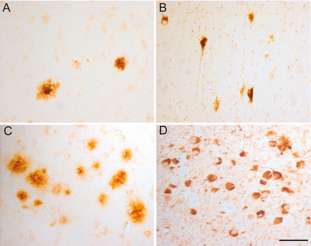
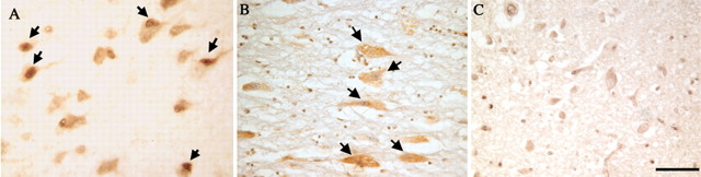
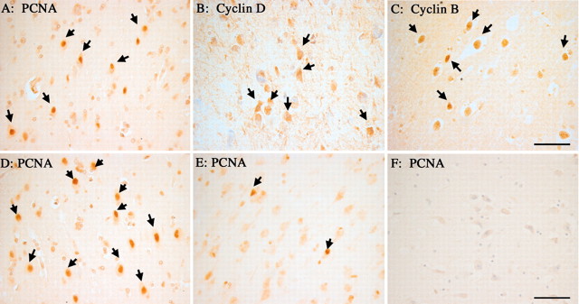
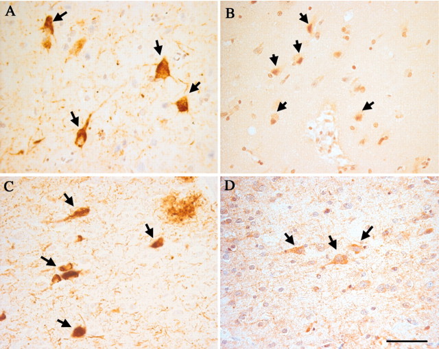
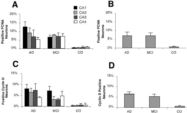
Cell cycle events play a major role in the loss of neurons in advanced Alzheimer's disease (AD). It is currently unknown, however, whether the same is true for the neuronal losses in early disease stages. To explore this issue we analyzed brain autopsy material from individuals clinically categorized with mild cognitive impairment (MCI), many if not most of whom will progress to AD. Immunocytochemistry for three cell cycle-related proteins, proliferating cell nuclear antigen, cyclin D, and cyclin B, was performed on sections from hippocampus, basal nucleus of Meynert, and entorhinal cortex. The results obtained from MCI cases were compared with material from individuals diagnosed with AD and those without cognitive impairment. In both hippocampus and basal nucleus, there was a significant percentage of cell cycle immunopositive neurons in the MCI cases. These percentages were similar to those found in the AD cases but significantly higher than non-cognitively impaired controls. In entorhinal cortex, the density of cell cycle-positive neurons was greater in MCI than in AD. However, we observed large variations in the percentages of immunopositive neurons from individual to individual. These findings lend support to the hypothesis that both the mechanism of cell loss (a cell cycle-induced death) and the rate of cell loss (a slow atrophy over several months) are identical at all stages of the AD disease process. The implication of the findings for human clinical trials is discussed.


Figures

Fig. 1.
Fig. 1.
Histopathology of the hippocampus varies among the different MCI cases. In the hippocampus of the mildest MCI case, the 6E11 antibody recognizes only an occasional β-amyloid plaque (A). Similarly, the mouse monoclonal AT-8 antibody that recognizes phosphorylated tau protein detects relatively few tangle-bearing neurons (B). In other MCI cases the extent of pathology was more significant. In some cases the level of 6E11-positive plaques (C) and AT-8-positive tangles (D) approached levels that might be expected in advanced Alzheimer's disease. Scale bar, 25 μm.
Fig. 2.
Fig. 2.
Immunocytochemistry reveals the expression of various cell cycle proteins in pyramidal neurons of the hippocampus. In both MCI (A) and AD (B), immunostaining for cyclin D reveals that a fraction of the hippocampal pyramidal cells express proteins that are normally found only in actively dividing cells. In contrast, age-matched controls (C) show little if any evidence for cyclin D expression. A second mitotic marker, the DNA polymerase subunit PCNA, is also found in neurons of the hippocampus in MCI cases (D) as well as in material from subjects who died with mild to moderate AD (E). Little evidence for reexpression of PCNA is detected in control material (F). Arrows indicate neurons stained positive for the two cell cycle markers. Scale bars, 25 μm.
Fig. 3.
Fig. 3.
Hippocampal pyramidal cells stained for the cell cycle marker cyclin B1. Cells shown are from MCI (A), AD (B), and a representative field from a normal, nondemented individual (C); the latter illustrates the lack of cyclin B1 staining. Arrows point to the cyclin B1-positive neurons in MCI and AD. Scale bar, 25 μm.
Fig. 4.
Fig. 4.
Nucleus basalis of Meynert immunostained for the cell cycle marker PCNA. In this second region where substantial neurodegeneration is known to occur in Alzheimer's disease, there is significant evidence for ectopic expression of cell cycle proteins in neurons that should be permanently postmitotic. Regions shown are from a representative MCI case (A), an AD case (B), and an age-matched control (C). Arrows indicate immunopositive cells. Scale bar, 25 μm.
Fig. 5.
Fig. 5.
Expression of cell cycle proteins in neurons of the entorhinal cortex in MCI cases. All of the same markers found in the hippocampus are also observed in this region of temporal cortex. PCNA is found in some neuronal nuclei of Layer V of the entorhinal cortex (A). As illustrated here for neurons in layer II, neurons were also found that expressed cyclin D (B) and cyclin B1 (C). Among the different MCI cases, there was variation in the levels of cell cycle protein expression. In some cases, positive cells appeared in clusters consisting of many neurons (D), whereas in other cases only isolated immunopositive neurons were found (E). As in hippocampus, little or no PCNA staining is found in age-matched controls (F). Scale bars, 25 μm.
Fig. 6.
Fig. 6.
Spatial association of hyperphosphorylated tau protein and cell cycle markers in the MCI entorhinal cortex. In this series of near-neighbor sections, the monoclonal AT-8 antibody reveals the location of hyperphosphorylated tau protein in intraneuronal tangles in layer V (A) and layer II (C). In nearby sections, immunostaining for cyclin B1 (B) and cyclin D (D) shows that the cell cycle-positive neurons are closely associated with these NFT-bearing neurons. Scale bar, 25 μm.
Fig. 7.
Fig. 7.
Percentages of immunopositive neurons stained with cell cycle markers in the hippocampus of MCI, AD, and control. Cells were scored for the presence of cell cycle proteins as described in Materials and Methods. Because the anatomical position of the sections scored varied from case to case, all results were expressed as “percent positive” neurons. The results for PCNA (A, B) and cyclin D (C, D) are shown separately. For each case an attempt was made to count percentages in four CA fields separately (A, C). There will be inaccuracies in these estimates as discussed in Results. The percentages of the entire hippocampal pyramidal cell population are also reported (B, D). In A andC, the percentage for CA1 is shown inblack, CA2 is shown in dark gray, CA3 is shown in light gray, and CA4 is shown inwhite. Values shown are means of the percentages calculated in each case; the error bars represent SEs.

References

    1. al-Ubaidi MR, Hollyfield JG, Overbeek PA, Baehr W. Photoreceptor degeneration induced by the expression of simian virus 40 large tumor antigen in the retina of transgenic mice. Proc Natl Acad Sci USA. 1992;89:1194–1198. - PMC - PubMed
    1. Arendt T, Rodel L, Gartner U, Holzer M. Expression of the cyclin-dependent kinase inhibitor p16 in Alzheimer's disease. NeuroReport. 1996;7:3047–3049. - PubMed
    1. Arendt T, Holzer M, Gartner U. Neuronal expression of cyclin dependent kinase inhibitors of the INK4 family in Alzheimer's disease. J Neural Transm. 1998;105:949–960. - PubMed
    1. Arnold S, Hyman B, J F. The topographical and neuroanatomical distribution of neurofibrillary tangles and neuritic plaques in cerebral cortex of patients with Alzheimer's disease. Cereb Cortex. 1991;1:103–116. - PubMed
    1. Baldin V, Lukas J, Marcote MJ, Pagano M, Draetta G. Cyclin D1 is a nuclear protein required for cell cycle progression in G1. Genes Dev. 1993;7:812–821. - PubMed

Publication types

MeSH terms