. 2003 Apr 1;23(7):2665-74.
doi: 10.1523/JNEUROSCI.23-07-02665.2003.

A cell surface receptor complex for fibrillar beta-amyloid mediates microglial activation

Affiliations

A cell surface receptor complex for fibrillar beta-amyloid mediates microglial activation

Maria E Bamberger et al. J Neurosci. .

Abstract

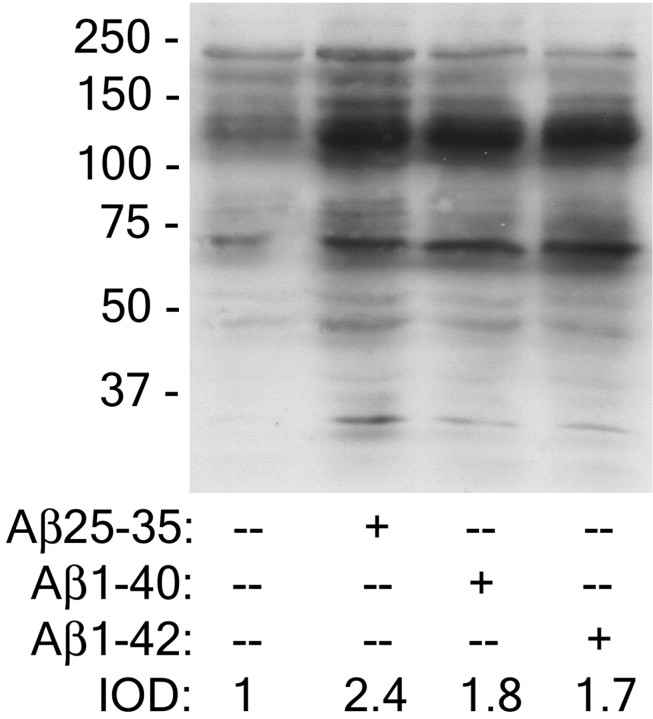
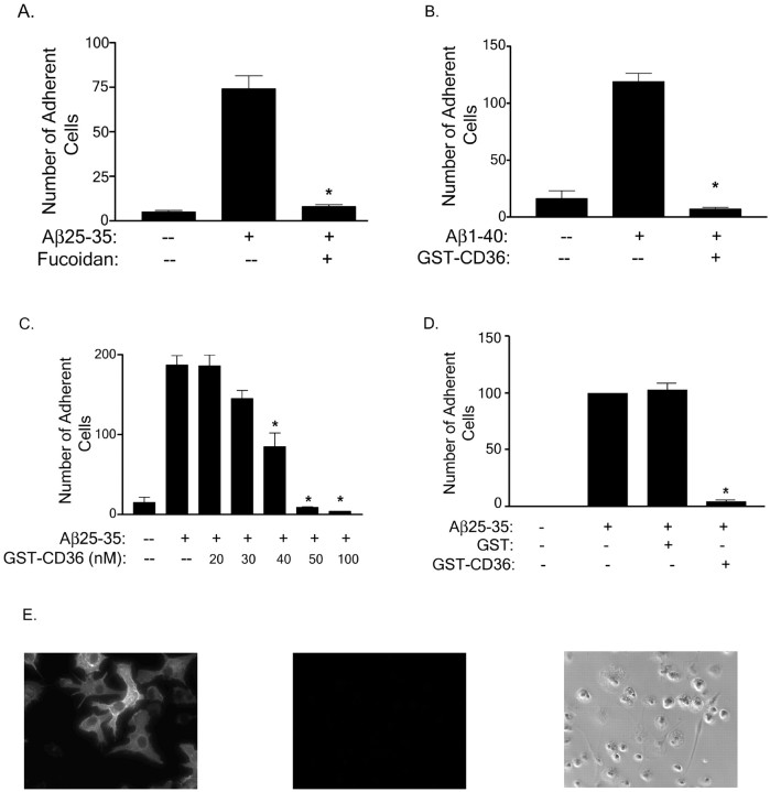
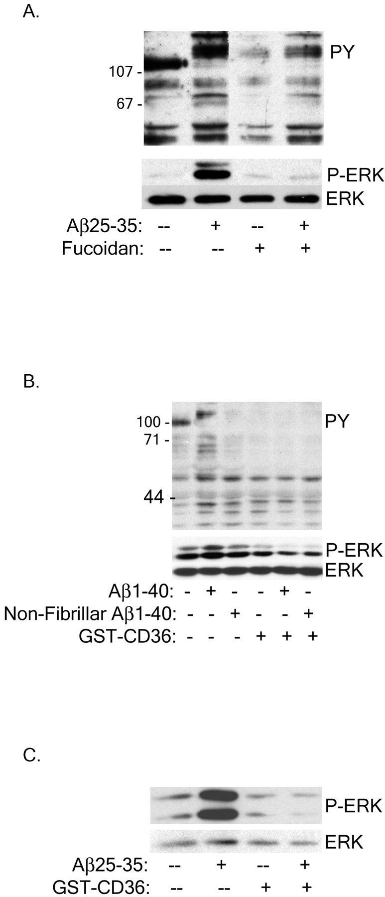
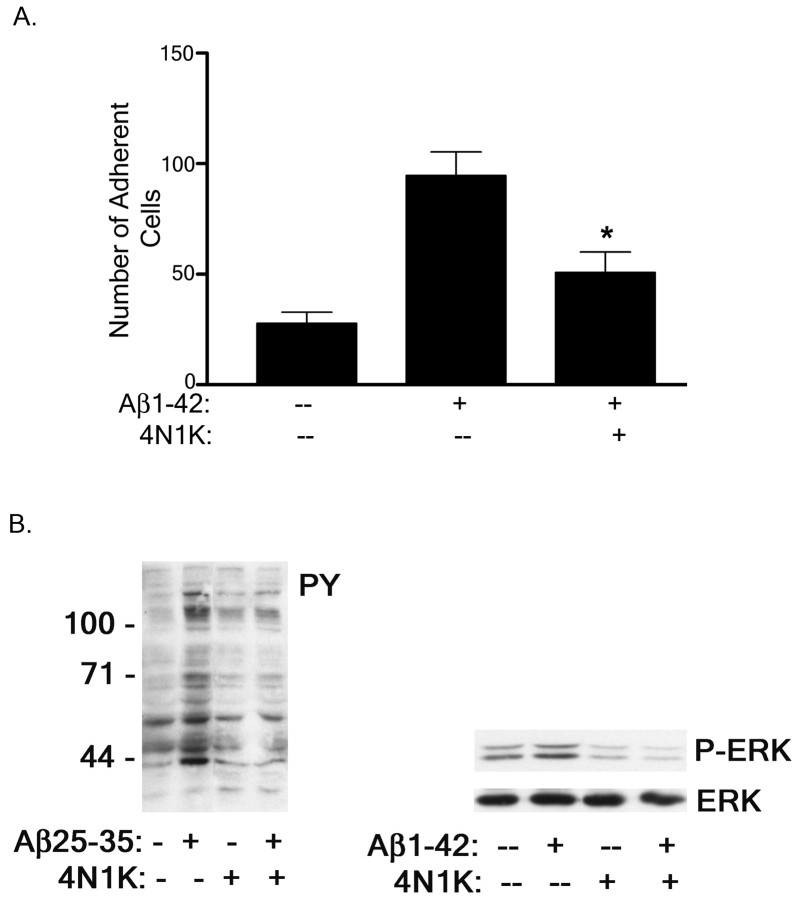
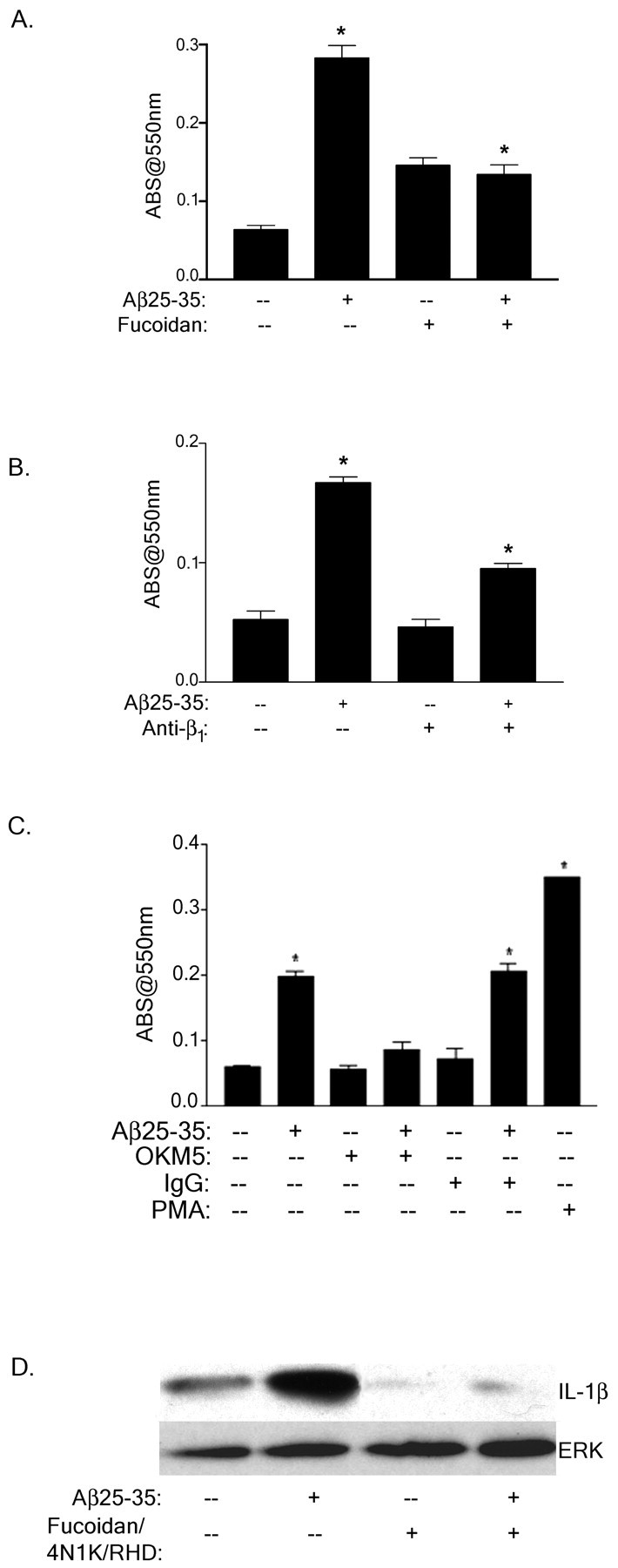
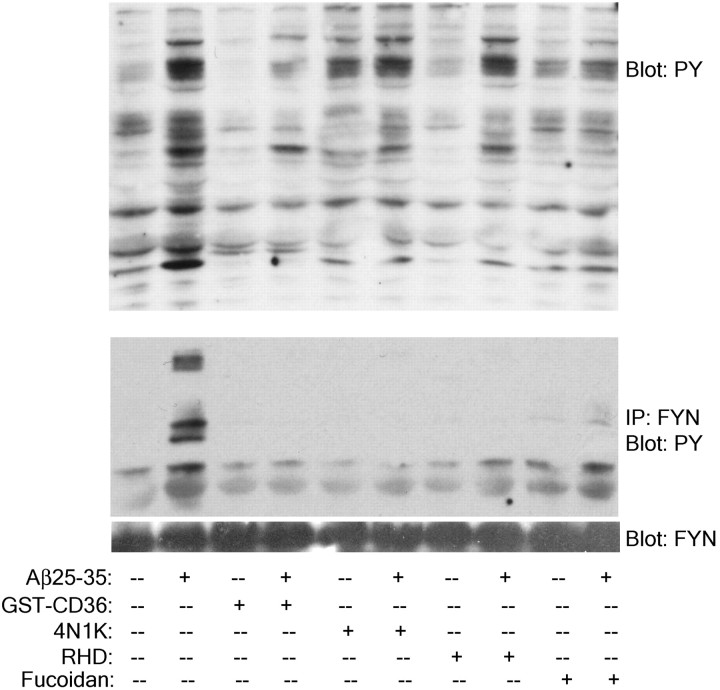
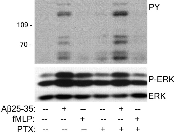
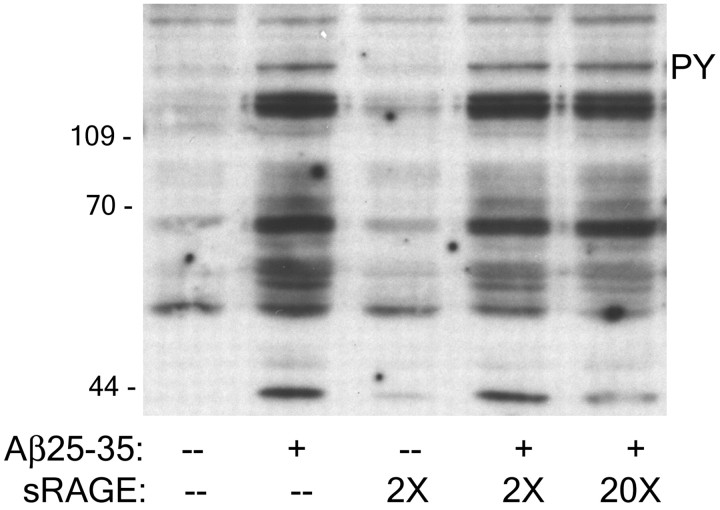
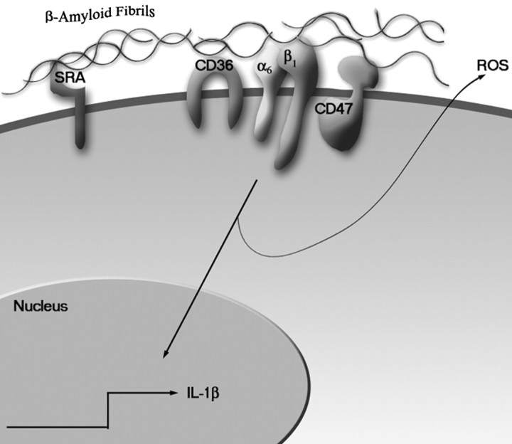
Senile plaques found in the Alzheimer's disease brain are foci of local inflammatory reactions mediated by plaque-associated microglia. The interaction of microglia with compacted deposits of beta-amyloid (Abeta) fibrils results in the stimulation of intracellular Tyr kinase-based signaling cascades and cellular activation, leading to the secretion of proinflammatory molecules. This study identifies a cell surface receptor complex that mediates the binding of microglia to Abeta fibrils and the subsequent activation of intracellular signaling pathways leading to a proinflammatory response. The receptor complex includes the B-class scavenger receptor CD36, the integrin-associated protein/CD47, and the alpha(6)beta(1)-integrin. Antagonists of scavenger receptors, CD36, CD47, and alpha(6)beta(1) inhibited the adhesion of THP-1 monocytes to Abeta fibrils. In addition, peptide competitors of Abeta fibril interactions with CD36, scavenger receptors, CD47, and the alpha(6)beta(1)-integrin inhibited Abeta stimulation of Tyr kinase-based signaling cascades in both THP-1 monocytes and murine microglia as well as interleukin 1beta production. A scavenger receptor antagonist and antibodies specific for CD36 and the beta(1)-integrin subunit also inhibited the Abeta-stimulated generation of reactive oxygen species. Importantly, the principal components of this receptor complex are shared with those for other fibrillar proteins and thus represent general elements through which myeloid lineage cells recognize complex fibrillar proteins. Identification of the cell surface molecules that interact with Abeta fibrils and mediate their activation of intracellular signaling cascades represents a potential intervention point in the treatment of Alzheimer's disease.


Figures

Fig. 1.
Fig. 1.
Fibrillar Aβ25–35, 1–40, and 1–42 peptides stimulate comparable increases in protein-Tyr phosphorylation in THP-1 monocytes. THP-1 monocytes were stimulated with fibrillar Aβ25–35, 1–40, and 1–42 peptides for 3 min. Cell lysates were analyzed by Western blot analysis using the anti-phospho-Tyr antibody 4G10. The integrated optical density (IOD) ratio of the protein-Tyr phosphorylation signal stimulated by the different Aβ ligands was quantitated.
Fig. 2.
Fig. 2.
Scavenger receptors are expressed on microglia and are involved in cellular adhesion to fibrillar Aβ. THP-1 monocytes were preincubated in serum-free RPMI 1640 medium with 300 μg/ml fucoidan (A) or 100 nmGST-CD36-(93–120) (B), and the cell suspension was applied to the fibrillar Aβ (2 μg) bound to a glass slide. Dose-dependent inhibition of adhesion of THP-1 monocytes to Aβ25–35 fibrils in the presence of 20–100 nm GST-CD36-(93–120) (C) and 50 nm GST or GST-CD36 (D) was evaluated. Cells were treated as described in (A, B). The number of adherent cells per square millimeter was recorded. The data shown represent the mean ± SEM of triplicate determinations. (*p < 0.001)E, Primary murine microglia were stained with an anti-CD36 antibody (left panel) or control Ig (middle panel) as described in Materials and Methods or visualized by phase microscopy (right panel).
Fig. 3.
Fig. 3.
Scavenger receptors mediate Aβ stimulation of intracellular Tyr signaling cascades in THP-1 monocytes and microglia. THP-1 monocytes were preincubated with 300 μg/ml fucoidan (A) or 100 nm GST-CD36-(93–120) (B) for 30 min before stimulation with fibrillar or nonfibrillar Aβ. C, Primary murine microglia were treated with 100 nm GST-CD36-(93–120) before Aβ stimulation. Cell lysates were analyzed by Western blot using the anti-phospho-Tyr antibody 4G10 (PY) or an anti-phospho-ERK antibody (P-ERK). P-ERK blots were stripped and reprobed with an anti-ERK antibody (ERK) as a protein-loading control.
Fig. 4.
Fig. 4.
The α6β1-integrin is required for Aβ binding and stimulation of THP-1 monocytes.A, THP-1 monocytes were preincubated with 100 μg/ml RHD peptide for 30 min before fibrillar or nonfibrillar Aβ1–40 stimulation. Cell lysates were analyzed by Western blot using the anti-phospho-Tyr antibody 4G10 (PY) or an anti-phospho-ERK antibody (P-ERK).B, Primary murine microglia were pretreated with RHD peptide as described in A but stimulated with fibrillar Aβ25–35 and analyzed by P-ERK Western blot analysis of cell lysates. Blots were stripped and reprobed with anti-ERK antibody (ERK) as a protein-loading control.C–E, THP-1 monocytes were preincubated with antibodies (40 μg/ml) to the β1 (C), α6 (D), and α1,2,4,5,v (E) integrins as described in A and stimulated with fibrillar Aβ25–35. Cell lysates were evaluated by Western blot analysis, using the 4G10, P-ERK, and ERK antibodies. F, G, Adhesion of THP-1 cells to fibrillar Aβ evaluated by preincubation in serum-free RPMI 1640 medium with antibodies to the α1–6, v- and β1-integrin subunits (40 μg/ml;F) or anti-β2-integrin subunit (40 μg/ml; G) and added to 2 μg of fibrillar Aβ bound to a glass slide. The number of adherent cells per square millimeter was recorded. The data shown represent the mean ± SEM of triplicate determinations (*p < 0.05;F).
Fig. 5.
Fig. 5.
Inhibition of CD47 interactions with Aβ fibrils blocks Aβ-mediated THP-1 monocyte activation. A, THP-1 monocytes were preincubated in serum-free RPMI 1640 medium with 100 μm CD47 antagonist peptide 4N1K and added to 2 μg of fibrillar Aβ bound to a glass slide. The number of adherent cells per square millimeter was recorded. The data shown represent the mean ± SEM of triplicate determinations (*p < 0.01).B, THP-1 monocytes were preincubated with 100 μm 4N1K for 30 min before fibrillar Aβ25–35 or Aβ1–42 stimulation. Cell lysates were analyzed by Western blot using the anti-phospho-Tyr antibody 4G10 (PY) or an anti-phospho-ERK antibody (P-ERK). The P-ERK blot was stripped and reprobed with an anti-ERK antibody (ERK) as a protein-loading control.
Fig. 6.
Fig. 6.
The Aβ receptor complex mediates the stimulation of ROS and IL-1β production. A–C, THP-1 monocytes were incubated with fucoidan (300 μg/ml; A), anti-β1 (40 μg/ml; B), isotype-specific IgG (C) phorbol 12-myristate 13-acetate (PMA, C) or anti-CD36 (OKM5, 40 μg/ml; C) in serum-free RPMI containing NBT and stimulated with fibrillar Aβ25–35 for 25 min at 37°C. Superoxide anion generation was measured by the change in absorbance at 550 nm. The data shown represent the mean ± SEM of triplicate determinations (*p < 0.001;A–C). D, Mature IL-1β production in Aβ-stimulated THP-1 monocytes was evaluated in the presence of 300 μg/ml fucoidan, 100 μm 4N1K, and 100 μg/ml RHD peptide. Cell lysates were resolved by 12% SDS-PAGE, transferred to polyvinylidene difluoride, and probed with the anti-IL-1β antibody. The blot was stripped and reprobed with an anti-ERK antibody (ERK) as a protein-loading control.
Fig. 7.
Fig. 7.
Members of the Aβ receptor complex are required for Aβ stimulation of Fyn phosphorylation. THP-1 monocytes were preincubated in serum free RPMI 1640 medium with 300 μg/ml fucoidan, 100 nm GST-CD36-(93–120), 100 μm 4N1K, or 100 μg/ml RHD for 30 min before stimulation with fibrillar Aβ. Cell lysates were immunoprecipitated with the anti-Fyn JD3 antibody. Cell lysates and immunoprecipitates were resolved by 7.5% SDS-PAGE and analyzed by Western blot using the anti-phospho-Tyr antibody 4G10 (PY). Blots were stripped and reprobed with an anti-Fyn JD3 antibody (FYN) as a protein-loading control.
Fig. 8.
Fig. 8.
A Gi-linked receptor is not involved in Aβ-stimulated intracellular signaling. THP-1 monocytes were preincubated with 200 ng/ml pertussis toxin (PTX) for 30 min at 37°C and stimulated with Aβ25–35 or 1 μm fMLP for 3 min. Western blots of cellular lysates were probed with the anti-phospho-Tyr antibody 4G10 (PY) or with the anti-phospho-ERK antibody (P-ERK). Blots were stripped and reprobed with the anti-ERK antibody (ERK).
Fig. 9.
Fig. 9.
RAGE is not involved in Aβ-mediated intracellular signaling. THP-1 monocytes were incubated for 30 min at 37°C and stimulated with Aβ25–35, 2 or 20× sRAGE, or both for 3 min. Western blots of cellular lysates were probed with the anti-phospho-Tyr antibody 4G10 (PY).
Fig. 10.
Fig. 10.
Model of Aβ fibril interaction with a microglial cell surface receptor complex resulting in activation of intracellular signal transduction cascades.

References

    1. Akiyama H, Barger S, Barnum S, Bradt B, Bauer J, Cole GM, Cooper NR, Eikelenboom P, Emmerling M, Fiebich BL, Finch CE, Frautschy S, Griffin WS, Hampel H, Hull M, Landreth G, Lue L, Mrak R, Mackenzie IR, McGeer PL, O'Banion MK, Pachter J, Pasinetti G, Plata-Salaman C, Rogers J, Rydel R, Shen Y, Streit W, Strohmeyer R, Tooyoma I, Van Muiswinkel FL, Veerhuis R, Walker D, Webster S, Wegrzyniak B, Wenk G, Wyss-Coray T. Inflammation and Alzheimer's disease. Neurobiol Aging. 2000;21:383–421. - PMC - PubMed
    1. Araujo DM, Cotman CW. Beta-amyloid stimulates glial cells in vitro to produce growth factors that accumulate in senile plaques in Alzheimer's disease. Brain Res. 1992;569:141–145. - PubMed
    1. Babic I, Schallhorn A, Lindberg FP, Jirik FR. SHPS-1 induces aggregation of Ba/F3 pro-B cells via an interaction with CD47. J Immunol. 2000;164:3652–3658. - PubMed
    1. Bianca VD, Dusi S, Bianchini E, Dal Pra I, Rossi F. beta-amyloid activates the O-2 forming NADPH oxidase in microglia, monocytes, and neutrophils: a possible inflammatory mechanism of neuronal damage in Alzheimer's disease. J Biol Chem. 1999;274:15493–15499. - PubMed
    1. Boland K, Behrens M, Choi D, Manias K, Perlmutter DH. The serpin-enzyme complex receptor recognizes soluble, nontoxic amyloid-β peptide but not aggregated, cytotoxic amyloid-β peptide. J Biol Chem. 1996;271:18032–18044. - PubMed

Publication types

MeSH terms

Substances