. 2003 Apr 1;23(7):2686-95.
doi: 10.1523/JNEUROSCI.23-07-02686.2003.

Dopamine D1-class receptors selectively modulate a slowly inactivating potassium current in rat medial prefrontal cortex pyramidal neurons

Affiliations

Dopamine D1-class receptors selectively modulate a slowly inactivating potassium current in rat medial prefrontal cortex pyramidal neurons

Yan Dong et al. J Neurosci. .

Abstract

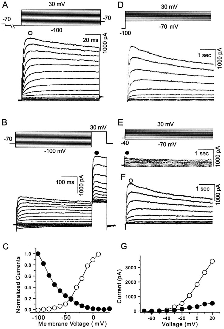
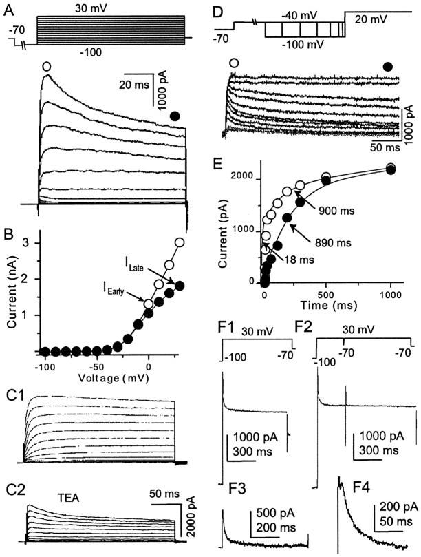
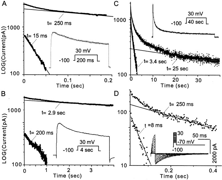
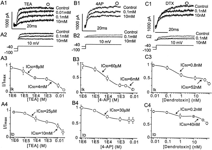
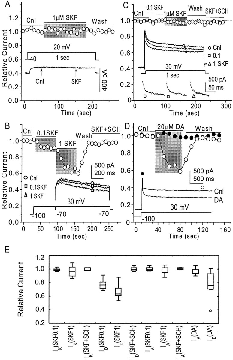
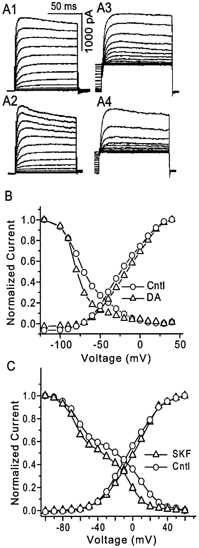
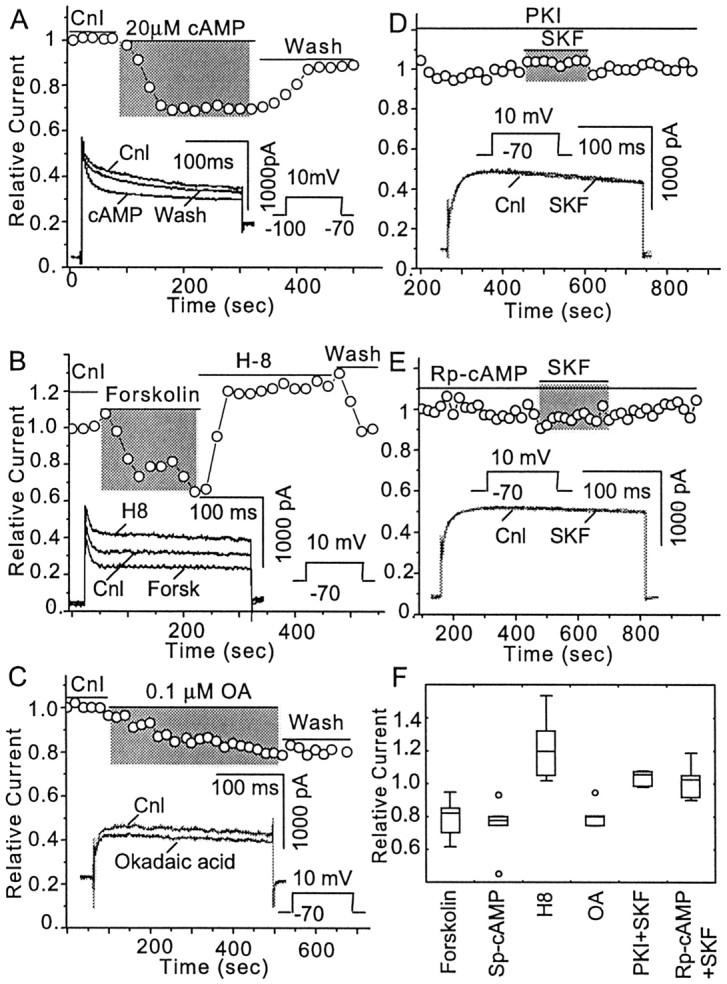
The dopamine (DA) innervation of medial prefrontal cortex (mPFC) regulates cognitive activity in a complex manner. Alterations of DA function, particularly via the DA D1 receptor class (D1R), are implicated in both schizophrenia and drug addiction, yet the precise roles of DA in modulating mPFC excitability remain unclear. We focused on DA modulation of voltage-gated K(+) current (VGKC) in acutely dissociated rat mPFC pyramidal neurons. We defined three components of the whole-cell VGKC according to biophysical and pharmacological properties. The A-type current (I(A)), with rapid activation and inactivation kinetics, was completely inactivated by prolonged holding of the membrane potential at -40 mV and was sensitive to the K(+) channel blocker 4-aminopyridine (4-AP) but not tetraethylammonium (TEA) or dendrotoxin (DTX). The slowly inactivating K(+) current (I(D)), with rapid activation but relatively slow inactivation, was the major contributor to VGKC and was completely inactivated at -40 mV and sensitive to TEA and DTX but less so to 4-AP. The very slowly inactivating K(+) current (I(K)) was elicited by command steps to more depolarized potentials from a prolonged holding potential of -40 mV and was sensitive to all three blockers. Stimulation of DA D2 receptors failed to alter any component of whole-cell VGKC. Stimulation of DA D1Rs selectively suppressed I(D), an effect mimicked by the adenylyl cyclase activator forskolin, the active cAMP analog Sp-cAMP, and the protein phosphatase inhibitor okadaic acid. Inhibition of protein kinase A (PKA) with either PKI or Rp-cAMP abolished D1R modulation. Thus, the DA D1R/cAMP/PKA signaling pathway mediates modulation of I(D) by DA in rat mPFC pyramidal neurons.


Figures

Fig. 1.
Fig. 1.
Whole-cell VGKCs in acutely dissociated mPFC pyramidal neurons. A, After holding the membrane potential at −70 mV and administering a 2 sec prepulse to −100 mV, test protocols from −100 to 30 mV with 10 mV increments elicited the whole-cell currents (n = 16 of 20).B, Whole-cell VGKCs could be partially inactivated by a depolarizing prepulse. After a holding potential at −70 mV and a 600 msec prepulse from −100 to 30 mV (10 mV increments), 100 msec test steps to 30 mV elicited currents showing a (pseudo) steady-state inactivation (n = 6). C, Current was measured at the time point indicated as an open orfilled circle in A and B. The activation and inactivation observed in A andB were plotted as I–V curves.D, Whole-cell VGKC was elicited by a similar activation protocol but with longer time course (4 sec) (n = 5). E, After the holding potential at −40 mV, the same test steps as in D elicited currents showing little inactivation. This current is operationally termedIK (n = 5).F, A slowly inactivating current was obtained by subtraction of traces in E from D. This slowly inactivating component is operationally termedID. G,I–V curves of IK andID indicate that both currents begin to activate at approximately −40 mV (n = 5). Currents were measured at the time points indicated byopen or filled circles inE and F.
Fig. 2.
Fig. 2.
A-type K+ component of whole-cell VGKC. A, A rapid inactivating component could be observed in a small population of neurons (n = 4 of 20) using the protocol described in Figure 1A. The rapidly inactivating component became more obvious at more depolarized steps. Because both activation and inactivation of this component are rapid and consistent with A-type current, it is operationally called IA.B, Activation curves for the early and later components are plotted with the values measured at the time points indicated byfilled and open circles, respectively.C1 and C2 show the activation of the same neuron before and after TEA perfusion, respectively. In most neurons, whole-cell VGKC activation protocols alone failed to show obviousIA. However, after perfusion with 10 mm TEA, IA was revealed (n = 4). D, Recovery protocol shows that IA deinactivates more rapidly than ID. After the holding potential at −70 mV, a prepulse to −40 mV inactivated most of the VGKC. A second hyperpolarizing prepulse to −100 mV recovered the K+ channels from the inactive state. The time course for this hyperpolarization prepulse varied from 10 msec to 1 sec with a nonlinear increment. IA could be recovered with a brief hyperpolarization step but contributes <25% of the whole-cell current (n = 4). E, The recovery time and the current amplitudes (measured at the time points indicated with open and filled circles in D) were plotted. The relationship of the early component (IA+ ID) could be fit with two exponentials (τ1 = 18 msec; τ2 = 890 msec). The relationship of the late component could be fit with one exponential (τ = 900 msec) (n = 5). From F1 toF4, IA was isolated biophysically (n = 4). F1, A neuron with obvious IA component was selected. By running a regular protocol, the whole-cell VGKC was elicited. This current containedIA, ID, and IK. F2, The same testing step as in F1 after a 200 msec depolarization prepulse elicited a K+trace. Because the prepulse had inactivatedIA, the following trace contained onlyID andIK. F3, TheIA component was isolated after subtraction of the trace in F2 from F2. The trace was shown in F4 in a higher magnification.
Fig. 3.
Fig. 3.
Multiple components of whole-cell VGKC revealed by inactivation kinetics. A, A cell with obviousIA was selected for this example. Whole-cell VGKC was elicited during a 200 msec step to 30 mV from −100 mV, as shown in the inset. The current amplitude was digitized and put in a LOG coordinate. The late part, but not the early part, of the current displayed a linear relationship with a time constant of 250 msec. This linear relationship was then extrapolated forward and subtracted from the early part of the digitized current, which resulted in another linear relationship with a time constant of 15 msec. This exponential peeling of decay within the time course demonstrated two components (n = 7).B, Whole-cell VGKC was elicited during a longer (4 sec) step to 30 mV from −100 mV, as shown in the inset. Exponential peeling of the components of decay of this trace revealed two time constants of 200 msec and 2.9 sec (n = 6).C, Whole-cell VGKC was elicited during a 40 sec step to 30 mV from −100 mV, as shown in the inset. Exponential peeling of the components of decay of this trace revealed two time constants of 3.4 and 25 sec (n = 3).D, A protocol as shown in the inset was used to elicit the K+ tail current. Exponential fitting revealed two components in the tail current (using the 40 mV step as the example) with time constants of 8 and 43 msec (n = 4). Taken together, these data support four distinct inactivation time constants, one consistent withIA, one consistent withIK, and two as distinct components ofID.
Fig. 4.
Fig. 4.
Pharmacological analysis also revealed multiple components of the whole-cell VGKC. A modified step to 10 mV elicited whole-cell VGKC when the holding potential was −100 mV but elicitedIK when the holding potential was −40 mV. ID was obtained with subtraction of IK from whole-cell VGKC.A1 and A2 show thatID andIK were blocked by different concentrations of TEA. A3, A4, The dose–response curves for TEA blockade of bothIK (A3) andID (A4) are best fit with two Langmuir isotherms, suggesting two TEA-sensitive components of these currents. B1 and B2show that ID andIK are blocked by different concentrations of 4-AP. B3, B4, The dose–response curve for 4-AP block ofIK (B3) is best fit by two Langmuir isotherms, suggesting at least two 4-AP-sensitive components of this current, whereas ID(B4) appears to possess only one 4-AP-sensitive component. C1 and C2 show thatID andIK are blocked by different concentrations of DTX. C3, C4, The dose–response curves for DTX blockade ofIK (C3) andID are best fit by two Langmuir isotherms, suggesting at least two DTX-sensitive components of these currents. For all graphs, three to six cells are represented at each data point.
Fig. 5.
Fig. 5.
Dopamine D1 receptor-mediated inhibition of VGKC.A, Activation of D1 receptors by the selective agonist SKF 81297 (0.1 and 1 μm) failed to alterIK (n = 5). Theinset shows two traces ofIK in the presence and absence of SKF 81297 (0.1 μm), clearly indicating the lack of effect of D1 receptor stimulation on this current. B, Activation of D1 receptors with SKF 81297 produced a dose-dependent suppression of ID(n = 25), which was prevented by the D1 receptor antagonist SCH 23390 (n = 5). Inset traces clearly show the dose-dependent modulation.C, Activation of D1 receptors by SKF 81297 (1 μm) failed to alterIA (n = 5).Inset traces show the overall effect of SKF 81297 and the effect on the isolated IA. D, The late component of VGKC (mostlyID) was measured at the open circle in inset; peak VGKC (mostlyIA) was measured at the beginning of the current, as indicated by the filled circle ininset. Perfusion of DA (20 μm) failed to alter peak VGKC (mostly IA) (n = 5; filled circles) but significantly inhibited the late component of VGKC (mostlyID) (n = 6;open circles). E, The effects of DA and SKF 81297 on the various components of VGKC are summarized in a box–whisker plot. Concentrations of 0.1 μm SKF 81297 (n = 5), 1 μm SKF 81297 (n = 5), or 0.1 μm SKF 81297 with 1 μm SCH 23390 (n = 3) failed to alter the amplitude of IK. Perfusion of 0.1 μm SKF 81297 produces a 19 ± 3% inhibition of ID(n = 15). Perfusion of 1 μm SKF 81297 produces a 34 ± 17% inhibition ofID (n = 10). Perfusion of 0.1 μm SKF 81297 together with 1 μm SCH 23390 does not alter the amplitude ofID(IRelative = 0.96 ± 0.03;n = 5). Perfusion of 0.1 (n = 3) or 1 μm SKF 81297 (n = 5), or SKF 81297 (0.1 μm) with SCH 23390 (1 μm) (n = 3), does not alter the amplitude of IA. Perfusion of DA does not alter IA(IRelative = 0.93 ± 0.09;n = 5) but inhibitsID(IRelative = 0.78 ± 0.06;n = 6.).
Fig. 6.
Fig. 6.
The effects of dopamine (DA) and SKF 81297 on the VGKC I–V relationship.A1, A2, The activation protocol (as in Fig. 1A) elicited VGKC before and during perfusion of DA (20 μm). A3,A4, The inactivation protocol (as in Fig.1B) elicited VGKC in the same pyramidal mPFC neuron before and during perfusion of 20 μm DA.B, The I–V curve for whole-cell VGKC from the neuron shown in A is plotted. DA clearly produces a hyperpolarizing shift in the inactivation curve of VGKC in mPFC neurons (n = 3 of 3). C, A similar shift of the inactivation curve was observed during perfusion of the D1 receptor agonist SKF 81297 (0.1 μm). The inactivation of these neurons could be best fit with two Boltzmann equations, suggesting that two components with distinct inactivation kinetics are involved in the whole-cell VGKC.
Fig. 7.
Fig. 7.
The adenylyl cyclase/cAMP/protein kinase A (PKA) signaling system mediates DA D1 receptor inhibition ofID. A, Sp-cAMP (20 μm), a membrane-permeable cAMP analog that activates protein kinase A directly, inhibitedID (n = 6) but notIA. B, Forskolin (10 μm), a membrane-permeable stimulator of adenylyl cyclase, also inhibited ID(n = 6). Perfusion of the same neuron with H8 (10 μm), a membrane-permeable PKA inhibitor, increasedID (n = 5).C, The membrane-permeable protein phosphatase inhibitor okadaic acid (100 nm) inhibitedID (n = 4).D, The PKI subunit (1 U/ml) was included in the pipette solution and diffused into the cell when the whole-cell configuration was formed. Once the cAMP/PKA pathway was fully blocked by PKI, indicated as the VGKC reached steady state, the inhibitory effect of SKF 81297 (1 μm) was also blocked (n = 4). E, Perfusion of Rp-cAMP (50 μm), a membrane-permeable cAMP analog with strong inhibitory effects on PKA (n = 5), also blocked the inhibition by SKF 81297 (1 μm). F, This plot summarizes the contribution of each component of the AC/cAMP/PKA pathway to D1R-mediated modulation of VGKC. Perfusion of Sp-Br-cAMP suppresses VGKC by 26 ± 6% (n = 5). Perfusion of forskolin suppresses VGKC by 22 ± 4% (n = 6), whereas perfusion of H8 enhances VGKC by 21 ± 5%. Perfusion of okadaic acid suppresses VGKC by 17 ± 4% (n = 4). VGKCs become resistant to the stimulation of D1R when the cell is treated with either PKI (IRelative = 1.04 ± 0.05;n = 4) or Rp-cAMP (IRelative = 1.02 ± 0.05;n = 5).

References

    1. Bekkers JM, Delaney AJ. Modulation of excitability by α-dendrotoxin-sensitive potassium channels in neocortical pyramidal neurons. J Neurosci. 2001;21:6553–6560. - PMC - PubMed
    1. Berger B, Gaspar P, Verney C. Dopaminergic innervation of the cerebral cortex: unexpected differences between rodents and primates. Trends Neurosci. 1991;14:21–27. - PubMed
    1. Bjorklund A, Divac I, Lindvall O. Regional distribution of catecholamines in monkey cerebral cortex, evidence for a dopaminergic innervation of the primate prefrontal cortex. Neurosci Lett. 1978;7:115–119. - PubMed
    1. Cantrell AR, Smith RD, Goldin AL, Scheuer T, Catterall WA. Dopaminergic modulation of sodium current in hippocampal neurons via cAMP-dependent phosphorylation of specific sites in the sodium channel α subunit. J Neurosci. 1997;17:7330–7338. - PMC - PubMed
    1. Cantrell AR, Scheuer T, Catterall WA. Voltage-dependent neuromodulation of Na+ channels by D1-like dopamine receptors in rat hippocampal neurons. J Neurosci. 1999;19:5301–5310. - PMC - PubMed

Publication types

LinkOut - more resources