. 2003 Apr 1;23(7):2751-8.
doi: 10.1523/JNEUROSCI.23-07-02751.2003.

Vasoactive intestinal polypeptide and pituitary adenylate cyclase-activating polypeptide activate hyperpolarization-activated cationic current and depolarize thalamocortical neurons in vitro

Affiliations

Vasoactive intestinal polypeptide and pituitary adenylate cyclase-activating polypeptide activate hyperpolarization-activated cationic current and depolarize thalamocortical neurons in vitro

Qian-Quan Sun et al. J Neurosci. .

Abstract

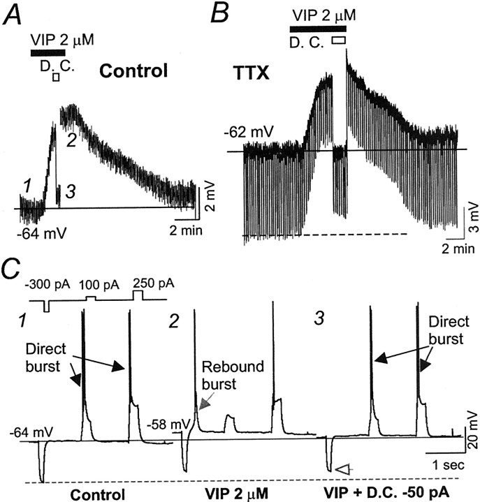
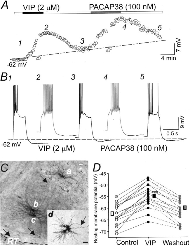
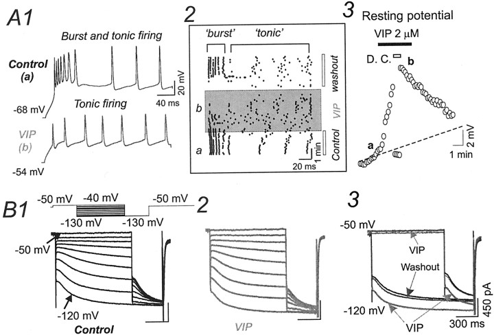
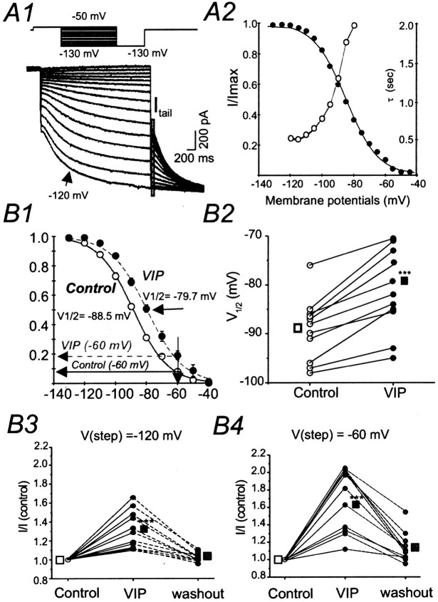
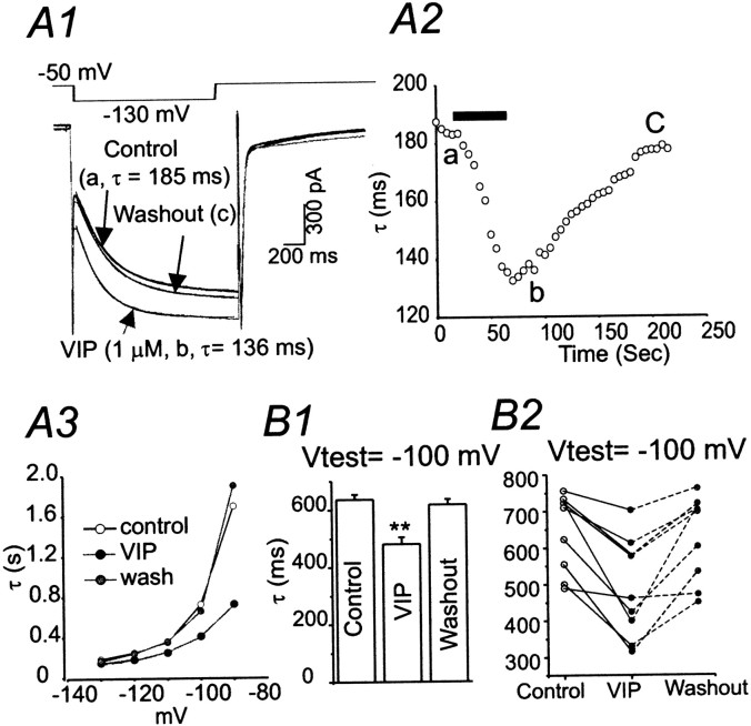
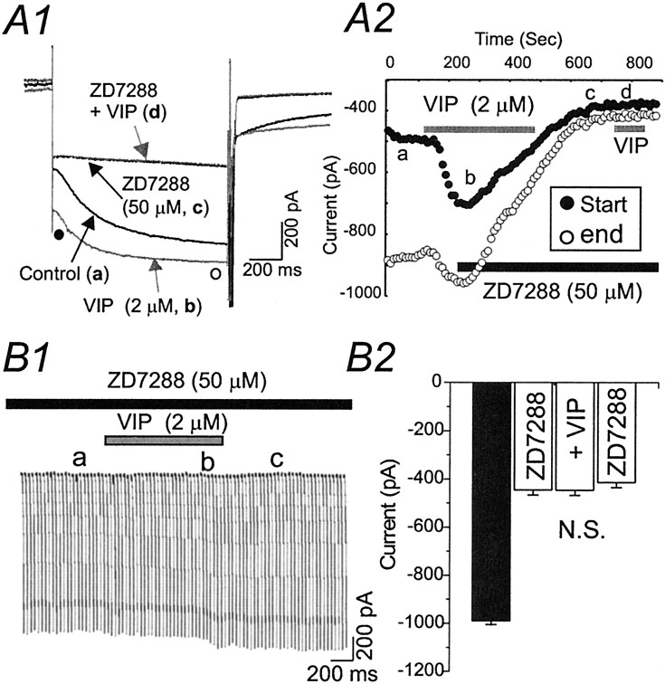
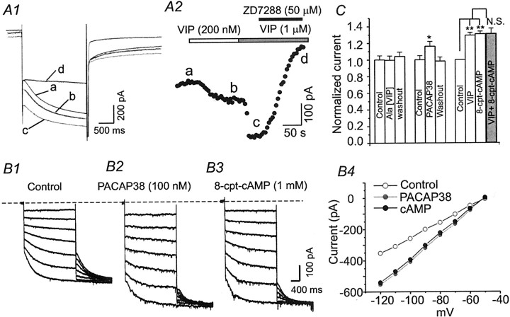
Ascending pathways mediated by monoamine neurotransmitters regulate the firing mode of thalamocortical neurons and modulate the state of brain activity. We hypothesized that specific neuropeptides might have similar actions. The effects of vasoactive intestinal peptide (VIP) and pituitary adenylate cyclase-activating polypeptide (PACAP) were tested on thalamocortical neurons using whole-cell patch-clamp techniques applied to visualized neurons in rat brain slices. VIP (2 microm) and PACAP (100 nm) reversibly depolarized thalamocortical neurons (7.8 +/- 0.6 mV; n = 16), reduced the membrane resistance by 33 +/- 3%, and could convert the firing mode from bursting to tonic. These effects on resting membrane potential and membrane resistance persisted in the presence of TTX. Morphologically diverse thalamocortical neurons located in widespread regions of thalamus were all depolarized by VIP and PACAP38. In voltage-clamp mode, we found that VIP and PACAP38 reversibly activated a hyperpolarization-activated cationic current (I(H)) in thalamocortical neurons and altered voltage- and time-dependent activation properties of the current. The effects of VIP on membrane conductance were abolished by the hyperpolarization-activated cyclic-nucleotide-gated channel (HCN)-specific antagonist ZD7288, showing that HCN channels are the major target of VIP modulation. The effects of VIP and PACAP38 on HCN channels were mediated by PAC(1) receptors and cAMP. The actions of PACAP-related peptides on thalamocortical neurons suggest an additional and novel endogenous neurophysiological pathway that may influence both normal and pathophysiological thalamocortical rhythm generation and have important behavioral effects on sensory processing and sleep-wake cycles.


Figures

Fig. 1.
Fig. 1.
VIP induces depolarizations of resting membrane potentials in thalamocortical neurons. A, Locally applied VIP (2 μm, 4 min) induced long-lasting depolarization (6 mV) of membrane potential. The effects of VIP recovered to baseline level after ∼20 min of washout. Thesolid black horizontal line indicates level of resting membrane potential in the control solution. B, Continuous current-clamp recording of another cell showing reversible effects of VIP (2 μm, 4 min, black bar) in the presence of TTX (1 μm). Vertical linesindicate responses to 500 msec current steps (−20 pA) applied at 0.1 Hz. The solid horizontal line indicates level of resting membrane potential in controls. The dashed horizontal line indicates control amplitude of membrane responses to hyperpolarizing current steps (−20 pA). C, Current-clamp recording from the same neuron depicted inA showing typical responses to a series of current steps ranging from −300 to +250 pA under control conditions (1) and during VIP application (2,3). C3, A steady hyperpolarizing current (−50 pA) was applied to the same neuron during VIP application to restore the resting membrane potential toward the control level, resulting in restoration of the directly evoked burst discharge.Black arrows in C1 and C3indicate bursts evoked by depolarizing current pulses. Note that the hyperpolarizing current evoked a rebound low-threshold spike during VIP application (C2, gray arrow) but not under control conditions or after the membrane was repolarized inC3. Traces in C were obtained at points 1–3 in A. The solid black horizontal line indicates level of resting membrane potential in control. The dashed horizontal line indicates amplitude of membrane responses to hyperpolarizing (−300 pA) current pulses. Theopen gray arrowhead in C3 shows the smaller voltage deflection obtained in the presence of VIP, indicating a conductance increase. D. C., Depolarizing current.
Fig. 2.
Fig. 2.
Morphologically distinct thalamocortical neurons are depolarized by VIP and PACAP. A, Resting membrane potential of a thalamocortical neuron during control, VIP application (2 μm, filled black bar), VIP washout, PACAP38 application (100 nm, filled gray bar), and PACAP38 washout. Locally applied VIP (2 μm, 3 min) induced long-lasting depolarization (6 mV) of membrane potential that was largely reversible on washout. PACAP38 (100 nm) mimicked the effects of VIP on resting membrane potential. B, Current-clamp responses evoked by current steps (100 pA, 0.5 sec) in the cell shown in A. Thedashed black line in A andB indicates control resting membrane potential. Traces 1–5 in B were obtained at points indicated by thenumbers in A. C, Photomicrograph of three biocytin-filled thalamocortical neurons in the ventral posterior nucleus. Scale bar, 50 μm. Arrowsshow a thalamocortical projecting axon, originating from cella and passing through the dendrites of cellsb and c, and branched collaterals in the reticular nucleus (RT). Inset, Biocytin-filled thalamocortical neuron (d) in the ventral lateral nucleus from a different slice. The membrane responses of these four cells and 12 others are shown in B. D, Resting membrane potentials in control solution (open circles) during VIP application (1 μm, black circles) and 20 min after VIP washout (gray circles) in 16 thalamocortical neurons.Rectangles indicate mean values for resting potentials of the population in control solution (open), at peak of the VIP-induced depolarization (black), and after ∼20 min of washout (gray). ***p< 0.001.
Fig. 3.
Fig. 3.
VIP-mediated effects on firing andIH in thalamocortical neurons.A1, Current-clamp recordings showing typical responses of a thalamocortical neuron to a current step (0.5 sec, 200 pA) in control solution (−68 mV, top, black trace) and during depolarization induced by VIP application (−54 mV, bottom, gray trace).A2, Raster plot of spikes evoked by current step (0.1 Hz) in the same experiment of A1. Thex-axis represents time within each response. They-axis represents time throughout the experiment (i.e., before drug, VIP, washout). Each point represents a single action potential. ‘burst’, Initial cluster of high-frequency spike firings (∼200 Hz) that occurred during burst discharge under control and washout conditions. Note that VIP application (gray bar) reversibly abolished burst firing.A3, Locally applied VIP (2 μm, 3 min) induced long-lasting depolarization (6 mV) of membrane potential in the same thalamocortical neuron as A1. The effects of VIP primarily recovered to baseline level after washout. D. C., Depolarizing current. Traces in A1 were obtained at points a and b inA2 and A3. The dashed lineindicates level of resting membrane potential in controls.B, Voltage-clamp recordings showing current traces elicited in a relay neuron by hyperpolarizing voltage steps (1 sec) from −40 to −130 mV in 10 mV increments,under control conditions (B1), and during VIP application (B2).B3, Superimposed traces from B1 andB2. Note that VIP increased the currents elicited by −120 mV steps but had very little effect on currents at −50 mV.Vhold = −50 mV inB1–B3.
Fig. 4.
Fig. 4.
Voltage-dependent modulation ofIH by VIP. A, Voltage-clamp recordings from a relay neuron showing currents elicited by hyperpolarizing voltage steps (1 sec) from −50 to −120 mV in 10 mV increments (above) under control conditions.Vhold = −50 mV. Note that voltage commands and current traces are shown on different time bases.Gray traces overlying black traces are single exponential fits of current traces. A2, The normalized conductance determined from tail currents (filled circles, measured at latency indicated byfilled gray bar in A1) was plotted versus voltage and fitted with a Boltzmann relationship,I/Imax = {1 + exp[(V+ V1/2)/K]}, under control conditions, where V1/2 = −86 mV and K = 10. The time constant of decay (τ), obtained from fitted curves in A1, was plotted versus voltage (open circles and gray line).B1, Normalized IHconductance, determined from mean tail current relative to that obtained at −140 mV, as a function of voltage fitted with a Boltzmann relationship in the absence (open circles andblack solid line; n = 11) and presence (filled circles and gray dashed line; n = 11) of VIP. B2, The half-activation voltages (V1/2) for IH in the absence (open circles) and presence (filled circles) of VIP for each neuron of B1. Open(controls) and filled (in VIP) squaresshow the averaged V1/2 values for each condition (p < 0.001; n= 11). B3, B4, Normalized mean peak current amplitude at −120 mV (B3) and −60 mV (B4) in control solution (open circles), during VIP application (black circles), and 20 min after VIP washout, measured from traces similar to those in A1 (n = 11).Open (controls) and filled (in presence of VIP) squares show averaged current values for each condition (***p < 0.001). Note that VIP caused an ∼60 ± 4% increase in currents elicited by steps to −60 mV but only a 30 ± 2% increase in currents elicited by steps to −120 mV.
Fig. 5.
Fig. 5.
Acceleration of IHactivation kinetics by VIP. A1, Currents elicited by hyperpolarizing steps to −130 mV from a holding potential of −50 mV under control conditions (a), during addition of VIP (b), and after VIP washout (c). Thin superimposed darker solid lines are single exponential fits to current traces.A2, The time constant of the exponential fit (τ) for the same experiment plotted versus time. Each circleshows the τ value for a single response evoked at 0.1 Hz.Black bar, VIP application. A3, Exponential time constants, obtained from a different relay neuron, plotted against test membrane potential under control conditions (open circles), during VIP application (black circles), and during washout (gray circles). B1, Mean exponential time constants for currents elicited by −100 mV steps in the control period, during VIP perfusion and after 20 min of washout. Columns show the average mean value for approximately eight responses evoked at 0.1 Hz in each of eight neurons; **p < 0.01.B2, Activation time constants for currents elicited by steps to −100 mV in the control period (open circles), during VIP perfusion (black circles), and after 20 min of washout (gray circles) for each neuron ofB1.
Fig. 6.
Fig. 6.
Effects of VIP on IHand membrane conductance are occluded by ZD7288. A1, Currents elicited by hyperpolarizing steps to −100 mV from a holding potential of −50 mV under control conditions (a,black trace), during the addition of 2 μmVIP (b, gray trace), during perfusion of 50 μm ZD7288 (c, black trace), and during application of both ZD7288 and 2 μm VIP (d, gray trace). Traces in A1 were obtained at points a–din A2. A2, Time series measurements in the cell of A1, showing that activation ofIH by VIP was blocked by ZD7288. ZD7288 perfusion eliminated VIP effects on both early (black circles, start) and late phases ofIH (open circles, end). During ZD7288 perfusion, a second application of VIP (gray bar on the right below d) had no effect on IH.Open andfilled circles in A1 andA2 indicate the time of measurements. B1, Current traces elicited by the voltage ramps (−50 to −130 mV over 2 sec, 0.2 Hz) shown in B2 after a 10 min initial application of ZD7288 (a), during the addition of 2 μm VIP (b), and after VIP washout (c). B2, Graph of currents measured at −100 mV under control conditions (black bar), after perfusion of ZD7288 (50 μm), after perfusion of ZD7288 plus 2 μm VIP, and ZD7288 alone after VIP washout (n = 5; NS vs ZD7288 alone).
Fig. 7.
Fig. 7.
Pharmacological profile of VIP-mediated effects onIH and involvement of cAMP.A1, Currents elicited by hyperpolarizing voltage steps to −100 mV from a holding potential of −50 mV under control conditions (a), during the addition of 200 nm (b, gray trace) or 1 μm (c) VIP, or during the addition of 1 μm VIP with ZD7288 (d). Traces are obtained at points a–d in A2.A2, Time series reflecting peak inward currents for the experiment in A1. Bars indicate time of drug applications. B, A family of current traces elicited by hyperpolarizing voltage steps (1 sec) from −60 to −120 mV in 10 mV increments from a different neuron under control conditions (B1), during perfusion of 100 nm PACAP38 (B2), and 20 min after PACAP38 washout during perfusion of 8-cpt-cAMP (1 mm; B3). B4,I–V plots of peak inward currents obtained from the neuron in B1–B3. C, Summary graph of normalized currents elicited by hyperpolarizing steps to −100 mV in different experimental conditions (n = 6 for each condition; *p < 0.05 and **p< 0.01 vs controls).

Similar articles

Cited by

References

    1. Ahnaou A, Laporte AM, Ballet S, Escourrou P, Hamon M, Adrien J, Bourgin P. Muscarinic and PACAP receptor interactions at pontine level in the rat: significance for REM sleep regulation. Eur J Neurosci. 2000;12:4496–4504. - PubMed
    1. Bayer L, Eggermann E, Saint-Mleux B, Machard D, Jones BE, Muhlethaler M, Serafin M. Selective action of orexin (hypocretin) on nonspecific thalamocortical projection neurons. J Neurosci. 2002;22:7835–7839. - PMC - PubMed
    1. Beaudet MM, Parsons RL, Braas KM, May V. Mechanisms mediating pituitary adenylate cyclase-activating polypeptide depolarization of rat sympathetic neurons. J Neurosci. 2000;20:7353–7361. - PMC - PubMed
    1. BoSmith RE, Briggs I, Sturgess NC. Inhibitory actions of ZENECA ZD7288 on whole-cell hyperpolarization activated inward current (If) in guinea-pig dissociated sinoatrial node cells. Br J Pharmacol. 1993;110:343–349. - PMC - PubMed
    1. Bourgin P, Lebrand C, Escourrou P, Gaultier C, Franc B, Hamon M, Adrien J. Vasoactive intestinal polypeptide microinjections into the oral pontine tegmentum enhance rapid eye movement sleep in the rat. Neuroscience. 1997;77:351–360. - PubMed

Publication types

MeSH terms

LinkOut - more resources