Comparative Study
. 2003 Apr 1;23(7):2789-96.
doi: 10.1523/JNEUROSCI.23-07-02789.2003.

Spinal cats on the treadmill: changes in load pathways

Affiliations
Comparative Study

Spinal cats on the treadmill: changes in load pathways

Marie-Pascale Côté et al. J Neurosci. .

Abstract

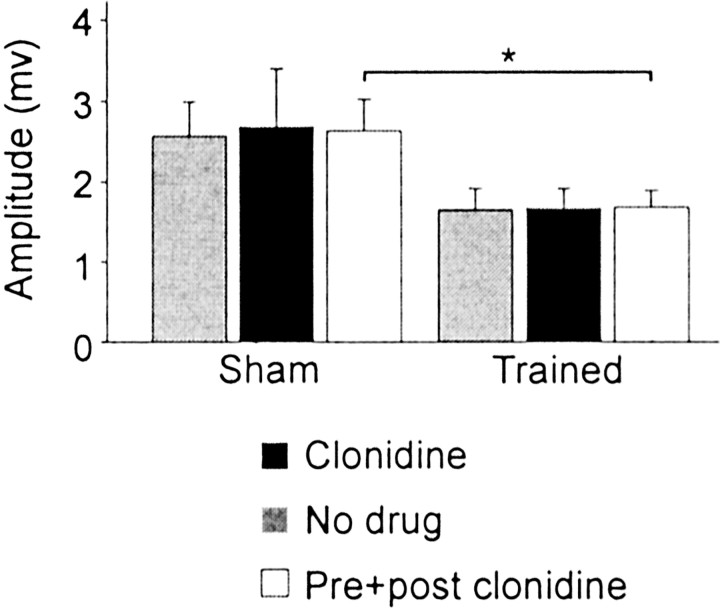
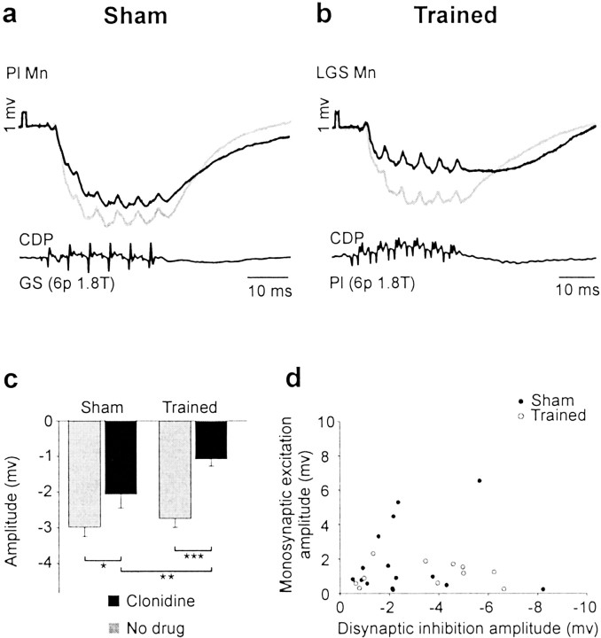
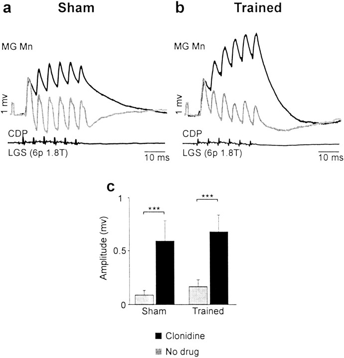
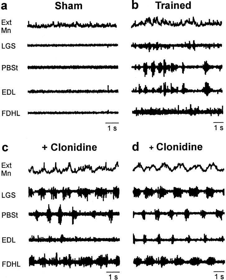
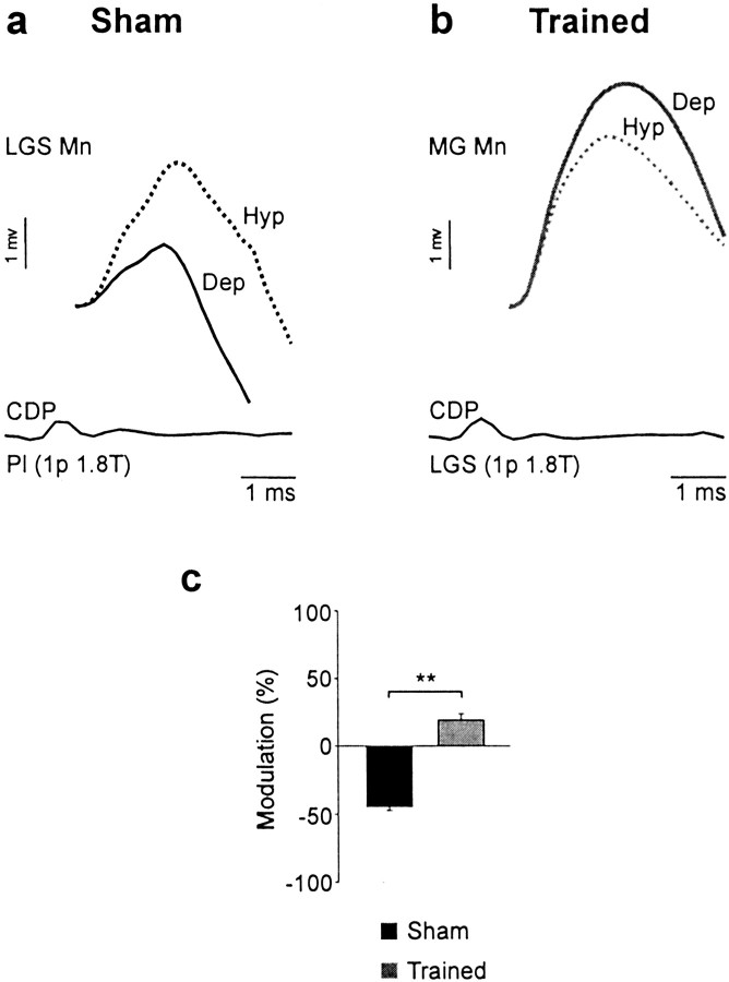
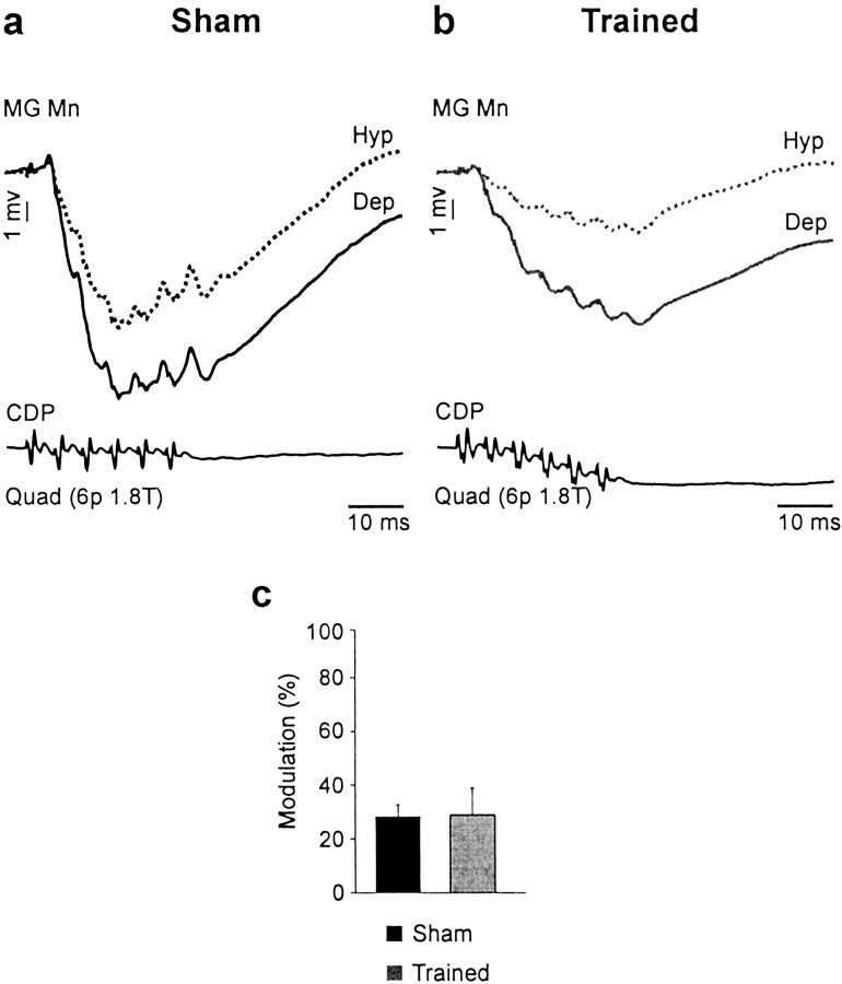
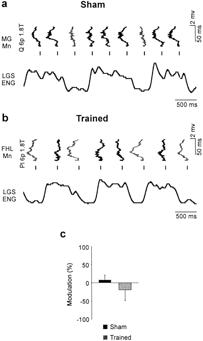
Treadmill training and clonidine, an alpha-2 noradrenergic agonist, have been shown to improve locomotion after spinal cord injury. We speculate that transmission in load pathways, which are involved in body support during stance, is specifically modified by training. This was evaluated by comparing two groups of spinal cats; one group (n = 11) was trained to walk until full-weight-bearing (3-4 weeks), and the other (shams; n = 7) was not. During an acute experiment, changes in group I pathways, monosynaptic excitation, disynaptic inhibition, and polysynaptic excitation were investigated by measuring the response amplitude in extensor motoneurons before and after clonidine injection. Monosynaptic excitation was not modified by clonidine but was decreased significantly by training. Disynaptic inhibition was significantly decreased by clonidine in both groups, but more significantly in trained cats, and significantly reduced by training after clonidine. Also, clonidine could reverse group IB inhibition into polysynaptic excitation in both groups but more frequently in trained cats. We also investigated whether fictive stepping revealed additional changes. In trained cats, the phase-dependent modulation of all three responses was similar to patterns reported previously, but in shams, modulation of monosynaptic and polysynaptic responses was not. Overall, training appears to decrease monosynaptic excitation and enhance the effects of clonidine in the reduction of disynaptic inhibition and reversal to polysynaptic excitation. Because it is believed that polysynaptic excitatory group I pathways transmit locomotor drive to extensor motoneurons, we suggest that the latter changes would facilitate the recruitment of extensor muscles for recovering weight-bearing during stepping.


Figures

Fig. 1.
Fig. 1.
Spinal proprioceptive pathways under study. A schematic representation of three sensory pathways transmitting inputs from muscle group I afferents to extensor motoneurons (Ext Mn) is shown to the left: the monosynaptic (stretch reflex) pathway (from group IA afferents originating in muscle spindles of extensors), the disynaptic inhibitory pathway (from group IB afferents of extensors originating in Golgi-tendon organs plus some group IA fibers), and the polysynaptic excitatory pathway (from group IB and IA afferents of extensors). In the acute spinal cat, this latter pathway shares interneurons with the network generating the excitatory locomotor drive in extensors (box E). Sample records of motoneuronal postsynaptic potentials used for measurements are on theright. a, The amplitude of monosynaptic EPSPs was measured at a latency of 1.4 msec (rising phase in this example; i.e., just before the onset of possible disynaptic components). b, The disynaptic IB inhibition was evoked by a short train of stimuli (6 pulses, 1.4–2.0 T, 200–300 Hz), and the IPSP amplitude was measured at the maximal negative deflection in the intracellular trace. Note that there were often monosynaptic EPSPs (six positive humps) overriding the inhibitory trough (dotted line). c, Polysynaptic excitation was evoked by a similar short train of stimuli, and the amplitude was measured at the maximal positive deflection (dotted line) underlying monosynaptic EPSPs.
Fig. 2.
Fig. 2.
Clonidine did not modify monosynaptic excitation. The mean amplitude of monosynaptic EPSPs (109 trials in 73 cells) evoked by the stimulation of knee and ankle extensor group I afferents (Quad, Pl, LGS, MG) was not changed significantly by clonidine injection. If we grouped all values together (preclonidine and postclonidine), there is a significant decrease in the amplitude monosynaptic EPSPs (*p < 0.05) caused by training.Filled bars, Clonidine; gray bars, no drug; open bars, preclonidine and postclonidine.
Fig. 3.
Fig. 3.
Training plus clonidine injection decreased disynaptic IB inhibition. a, b, IPSPs evoked by stimulation of GS group I afferents [6 p 1.8 T] in a Pl motoneuron in a sham (a) and Pl group I afferents (6 p 1.8 T) in an LGS motoneuron (similar AHP as the Pl cell) in a trained cat (b) before (gray trace) and after (black trace) clonidine. Clonidine decreased IB inhibition in both groups of cats.Mn, Motoneuron. c, Afferent volley was monitored by the CDP. Overall, disynaptic IPSPs (314 trials in 143 cells) evoked by stimulation of knee and ankle extensor group I afferents (Quad, Pl, LGS, MG, GS) were significantly decreased by clonidine in shams (30.5%; *p < 0.05) and even more significantly in trained cats (61.0%; ***p < 0.001). Training enhanced significantly the reduction of IB inhibition after clonidine (**p < 0.01).d, Plot of EPSP amplitude versus IPSP amplitude measured from the same cell in shams (filled circles) and trained cats (open circles).
Fig. 4.
Fig. 4.
Clonidine increased polysynaptic group I excitation in both groups of cats. a, b, EPSPs evoked by stimulation of LGS afferents [6 p 1.8 T] recorded in MG motoneurons (with similar AHPs) in a sham cat (a) and a trained cat (b) before and after clonidine. Here, clonidine reversed IB inhibition (gray trace) to polysynaptic excitation (black trace) both in sham and trained cats.Mn, Motoneuron. c, Overall, the amplitude of polysynaptic EPSPs (313 trials in 143 cells) evoked by stimulation of knee and ankle extensor group I afferents (Quad, Pl, LGS, MG, GS) was increased by clonidine in sham (535.6%; ***p < 0.001) and in trained (307.8%; ***p < 0.001) cats.
Fig. 5.
Fig. 5.
Fictive locomotion can be induced in shams and trained cats. a, b, Motoneuronal intracellular potential and ENG activity in flexor and extensor muscle nerves in a sham (a) and a trained (b) cat. Rhythmic bursts of activity evoked by perineal stimulation before clonidine injection were observed in trained cats (7 of 11) and in shams (2 of 7). c,d, After clonidine, perineal stimulation induced robust locomotor episodes in both groups of cats. Ext Mn, Extensor motoneuron.
Fig. 6.
Fig. 6.
Training could change the pattern of CPG-related modulation of monosynaptic excitation. a, The amplitude of monosynaptic EPSPs evoked by Pl stimulation [1 p 1.8 T] was larger during the hyperpolarized (Hyp) phase in an LGS motoneuron from a sham cat. b, The amplitude of monosynaptic EPSPs evoked by LGS stimulation (1 p, 1.8 T) was larger during the depolarized (Dep) phase in an MG motoneuron from a trained cat. Mn, Motoneuron. c, Training modified significantly the pattern of phase-dependent modulation of monosynaptic EPSPs (5 trials in 4 cells; **p < 0.01) evoked by group I afferents of ankle extensors (Pl, MG, LGS), with the maximum amplitude occurring during the hyperpolarized phase in sham cats and during the depolarized phase in trained cats.
Fig. 7.
Fig. 7.
Training did not change the pattern of CPG-related modulation of IB inhibition. a, b, IPSPs evoked by Quad (6 p, 1.8 T) in MG motoneurons during fictive locomotion in a sham (a) and a trained (b) cat. The amplitude of IPSPs (trough) was increased during the depolarized (dep) phase in the sham (black trace) and the trained (gray trace) cat.Mn, Motoneuron. c, The depth of modulation in IPSPs (40 trials in 33 cells) was not significantly changed by training. Hyp, hyperpolarized.
Fig. 8.
Fig. 8.
Different patterns of CPG-related modulation of polysynaptic excitation. a, EPSPs recorded in an MG motoneuron (tilted 90°) in a sham were evoked by Quad stimuli [6 p 1.8 T] at different moments in the step cycle illustrated by the rectified and filtered ENG activity of the LGS nerve. The amplitude of polysynaptic EPSPs was maximal (gray trace) when occurring during the active period of LGS (i.e., during the extension phase). b, The amplitude of polysynaptic EPSPs evoked by Pl stimulation and recorded in an FHL motoneuron (tilted 90°) from a trained cat (6 p, 1.8 T) was minimal (black trace) during the extension phase when LGS was maximally active.Mn, Motoneuron. c, Overall, the pattern of phase-dependent modulation of polysynaptic EPSPs (19 trials in 15 cells) tended to be opposite in shams and trained cats, but this difference was not statistically significant.

References

    1. Akazawa K, Aldridge JW, Steeves JD, Stein RB. Modulation of stretch reflexes during locomotion in the mesencephalic cat. J Physiol (Lond) 1982;329:553–567. - PMC - PubMed
    1. Baldissera F, Hultborn H, Illert M. Integration in spinal neuronal systems. In: Brookhart JM, Mountcastle VB, editors. Handbook of physiology. The nervous system, Vol II. American Physiological Society; Bethesda, MD: 1981. pp. 509–595.
    1. Barbeau H, Rossignol S. Recovery of locomotion after chronic spinalization in the adult cat. Brain Res. 1987;412:84–95. - PubMed
    1. Barbeau H, Rossignol S. Initiation and modulation of the locomotor pattern in the adult chronic spinal cat by noradrenergic, serotonergic and dopaminergic drugs. Brain Res. 1991;546:250–260. - PubMed
    1. Barbeau H, Julien C, Rossignol S. The effects of clonidine and yohimbine on locomotion and cutaneous reflexes in the adult chronic spinal cat. Brain Res. 1987;437:83–96. - PubMed

Publication types

MeSH terms

LinkOut - more resources