. 2003 Apr 1;23(7):2804-14.
doi: 10.1523/JNEUROSCI.23-07-02804.2003.

A chondroitin sulfate proteoglycan PTPzeta /RPTPbeta regulates the morphogenesis of Purkinje cell dendrites in the developing cerebellum

Affiliations

A chondroitin sulfate proteoglycan PTPzeta /RPTPbeta regulates the morphogenesis of Purkinje cell dendrites in the developing cerebellum

Masahiko Tanaka et al. J Neurosci. .

Abstract

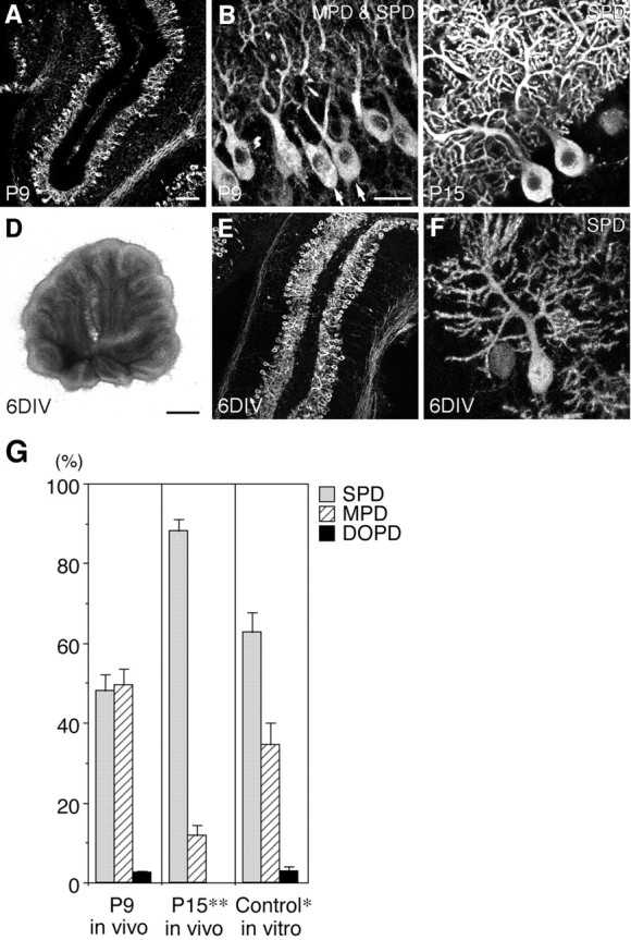
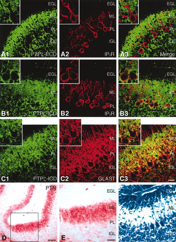
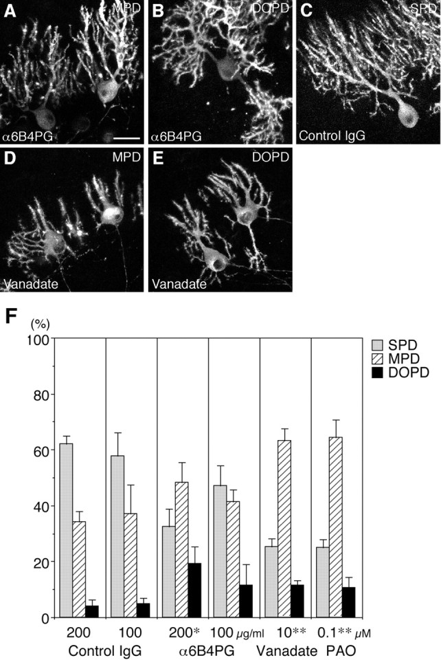
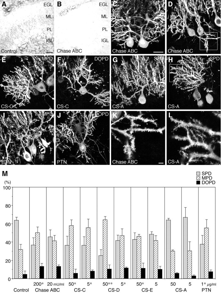
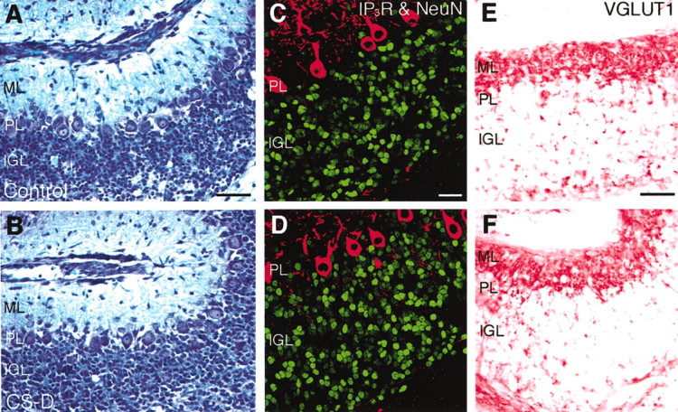
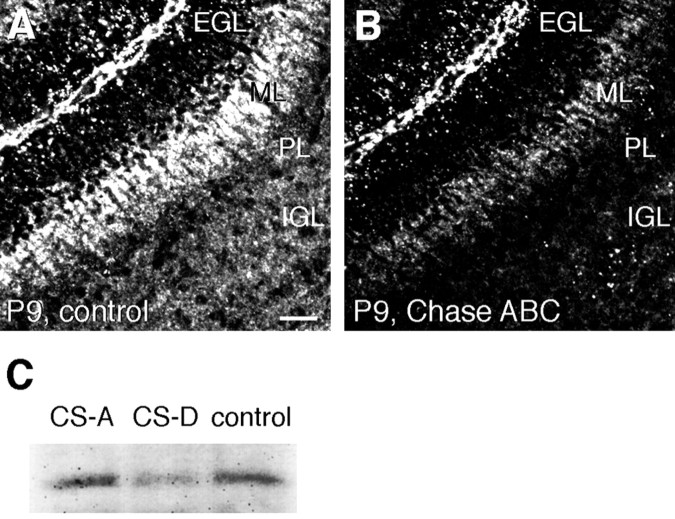
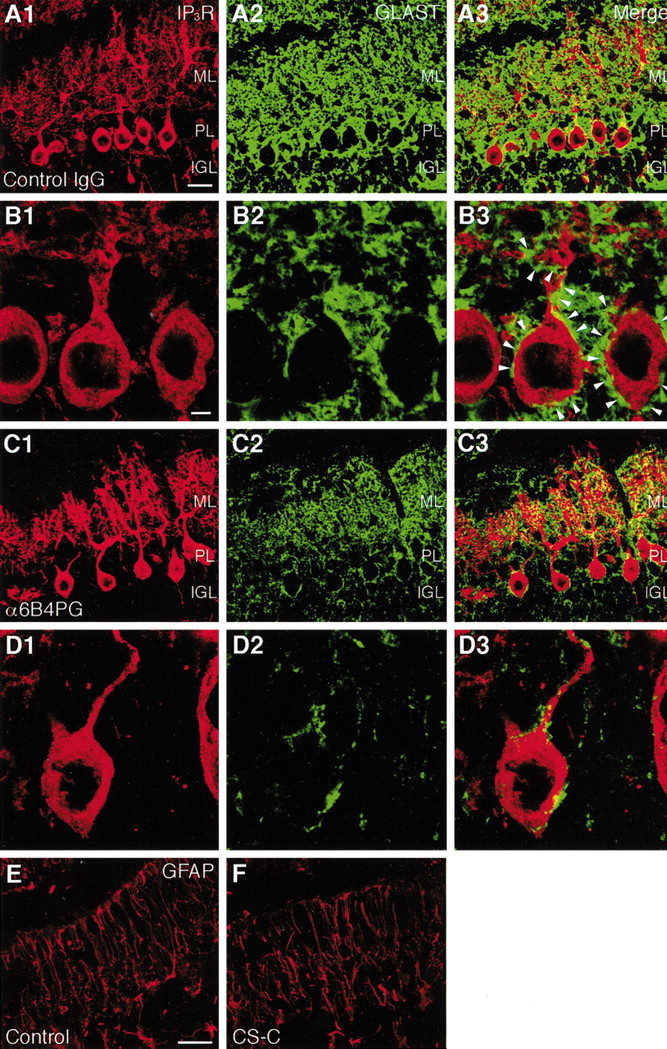
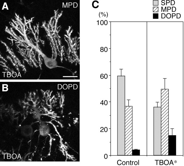
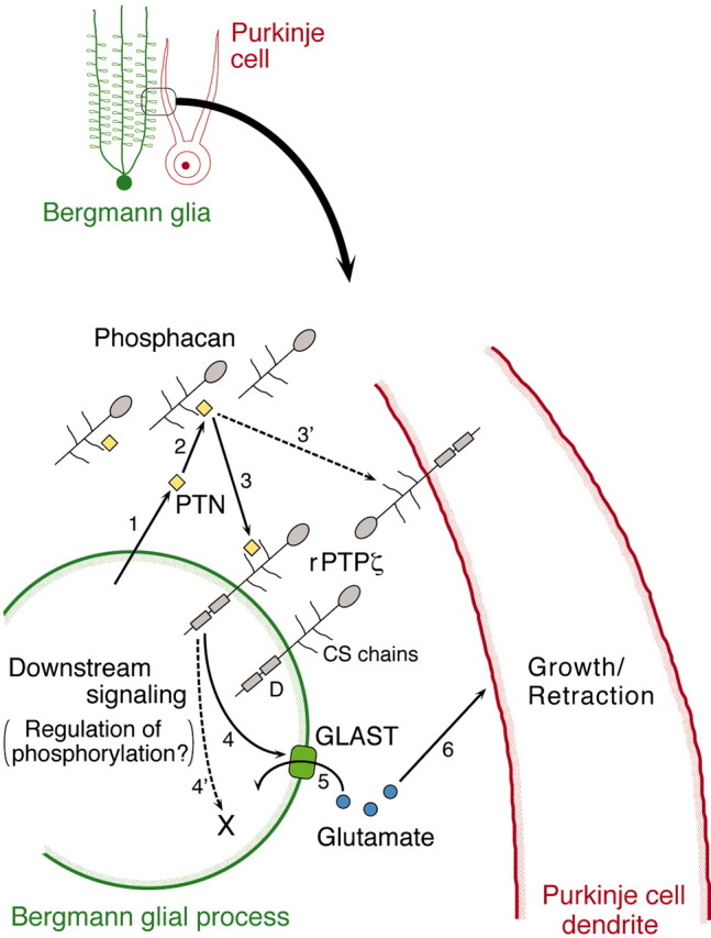
PTPzeta/RPTPbeta, a receptor-type protein tyrosine phosphatase synthesized as a chondroitin sulfate (CS) proteoglycan, uses a heparin-binding growth factor pleiotrophin (PTN) as a ligand, in which the CS portion plays an essential role in ligand binding. Using an organotypic slice culture system, we tested the hypothesis that PTN-PTPzeta signaling is involved in the morphogenesis of Purkinje cell dendrites. An aberrant morphology of Purkinje cell dendrites such as multiple and disoriented primary dendrites was induced in slice cultures by (1) addition of a polyclonal antibody against the extracellular domain of PTPzeta, (2) inhibition of protein tyrosine phosphatase activity, (3) enzymatic removal of the CS chains, (4) addition of exogenous CS chains, and (5) addition of exogenous PTN, all of which disturb PTN-PTPzeta signaling. These treatments also reduced the immunoreactivity to GLAST, a glial glutamate transporter, on Bergmann glial processes. Furthermore, a glutamate transporter inhibitor also induced the abnormal morphogenesis of Purkinje cell dendrites. Altogether, these findings suggest that PTN-PTPzeta signaling regulates the morphogenesis of Purkinje cell dendrites and that the mechanisms underlying that regulation involve the GLAST activity in Bergmann glial processes.


Figures

Fig. 1.
Fig. 1.
Morphogenesis of Purkinje cell dendrites in vivo and in slice cultures. A–C, Fluorescent immunohistochemistry using a monoclonal antibody against IP3R (4C11) showing the morphology of Purkinje cells in the cerebellum from P9 (A, B) and P15 (C) rats. A, Low-power view of an immunostained cryosection. B, C, Confocal microscopic images of immunostained slices. The multiple primary dendrite (MPD)-type (arrows) and single primary dendrite (SPD)-type Purkinje cells coexist on P9 (B), whereas most Purkinje cells are of the SPD type on P15 (C).DF, Overview (D) and fluorescent immunohistochemistry using 4C11 (E, F) of cerebellar slices derived from P9 rats and cultured for 6 d under control conditions. E, Low-power view of an immunostained cryosection. F, A confocal microscopic image of an immunostained slice culture. Scale bars: (in A) A, E, 100 μm; (inB) B, C, F, 25 μm;D, 1 mm. G, Quantitative representation of the ratios of the three types of Purkinje cells in vivo (P9 and P15) and in slice cultures at 6 DIV under control conditions. The ratio of SPD-type Purkinje cells significantly increased in slice cultures at 6 DIV compared with that on P9, although the increase was not as marked as in vivo. Cell counts were made in slices, not in cryosections, which enabled us to reliably distinguish the morphology of Purkinje cell dendrites.DOPD, Disoriented primary dendrite.n = 4. Error bars represent SEM. *p < 0.05, **p < 0.001 versus P9.
Fig. 2.
Fig. 2.
Expression patterns of PTPζ and PTN in postnatally developing cerebellum. A–C, Confocal microscopic images of cerebellar cryosections derived from P9 rats and double stained by fluorescent immunohistochemistry using antibodies against the extracellular domain (ECD) (A1) or the intracellular domain (ICD) (B1, C1) of PTPζ (Cy2) and IP3R (A2, B2) or GLAST (C2) (Cy3). A3, B3, andC3 are the merged images. The immunoreactivities to PTPζ-ICD and GLAST partially overlapped each other, especially around Purkinje cells. EGL, External granular layer;ML, molecular layer; PL, Purkinje cell layer; IGL, internal granular layer. Scale bar, 25 μm.D, E, A cerebellar cryosection derived from a P9 rat and stained by immunohistochemistry using an antibody against PTN. E is the high-magnification image of the enclosed area in D. PTN distributes abundantly in the ML. Scale bars: D, 50 μm; E, 25 μm.F, An adjacent section stained with toluidine blue. Scale bar, 25 μm.
Fig. 3.
Fig. 3.
Involvement of PTPζ in the morphogenesis of Purkinje cell dendrites. A–E, Effects of the polyclonal antibody against PTPζ-ECD (α6B4PG) (A, B), control IgG (C), and sodium vanadate (D, E) on the morphogenesis of Purkinje cell dendrites. Shown are confocal microscopic images of cerebellar slices derived from P9 rats, cultured for 6 d with culture medium containing α6B4PG (200 μg/ml) (A, B), control IgG (200 μg/ml) (C), and sodium vanadate (10 μm) (D, E) and stained by fluorescent immunohistochemistry using 4C11. On addition of α6B4PG and sodium vanadate, the MPD (A, D) and DOPD (B,E) types of Purkinje cells markedly increased. Control IgG did not influence the morphogenesis of Purkinje cell dendrites (C). Scale bar, 25 μm. F, Quantitative representation of the effects of α6B4PG, control IgG, sodium vanadate, and phenylarsine oxide (PAO) on the morphogenesis of Purkinje cell dendrites in slice cultures at 6 DIV.n = 3 (Vanadate, PAO) or 4 (α6B4PG, Control IgG). Error bars represent SEM. *p < 0.05 versus control IgG (200 μg/ml); **p < 0.005 versus control (data in Fig.1G).
Fig. 4.
Fig. 4.
Involvement of chondroitin sulfate (CS) and PTN in the morphogenesis of Purkinje cell dendrites. A, B, Distribution of CS in the control (A) and chondroitinase ABC (Chase ABC)-treated (B) slice cultures. Cryosections of cerebellar slices derived from P9 rats, cultured for 6 d without (A) or with (B) Chase ABC (200 mU/ml), and stained by immunohistochemistry using a monoclonal antibody against CS (CS-56). Scale bar, 25 μm. C–J, Effects of Chase ABC, CS, and PTN on the morphogenesis of Purkinje cell dendrites. Shown are confocal microscopic images of cerebellar slices cultured for 6 d with culture medium containing Chase ABC (200 mU/ml) (C,D), CS-C (50 μg/ml) (E,F), CS-A (50 μg/ml) (G,H), and PTN (1 μg/ml) (I,J), and stained by fluorescent immunohistochemistry using 4C11. On addition of Chase ABC, CS-C, and PTN, the MPD (C, E,I) and DOPD (D, F,J) types of Purkinje cells increased. In contrast, many Purkinje cells had an SPD under the CS-A-added conditions (G, H). Scale bar, 25 μm. K, L, High-power views of the enclosed areas in D, H. Spine formation on Purkinje cell dendrites did not differ between the Chase ABC (K)- and CS-A (L)-added conditions. Scale bar, 3 μm. M, Quantitative representation of the effects of Chase ABC, CS, and PTN on the morphogenesis of Purkinje cell dendrites in slice cultures at 6 DIV.n = 3 (CS chains) or 4 (Control, Chase ABC,PTN). Error bars represent SEM. *p < 0.05, **p < 0.005 versus control.
Fig. 5.
Fig. 5.
Development of granule cells in slice cultures under control and CS-D-added conditions. Cerebellar slices cultured for 6 d without (A,C,E) or with (B,D,F) CS-D were processed for several histological analyses.A, B, Nissl staining with toluidine blue. Migration of granule cells from the EGL to the IGL was nearly completed even in the CS-D-treated cultures. Scale bar, 50 μm.C, D, Confocal microscopic images of cryosections double stained by fluorescent immunohistochemistry using antibodies against NeuN (Cy2, green) and IP3R (Cy3, red). The density of granule cells in the IGL was not significantly different between these two conditions (Table 2). Scale bar, 25 μm. E,F, Cryosections stained by immunohistochemistry using an antibody against vesicular glutamate transporter 1 (VGLUT1). VGLUT1 was expressed in the ML under both conditions. Scale bar, 50 μm.
Fig. 6.
Fig. 6.
Effects of the Chase ABC and CS treatments on PTN contents in the cerebellum. A, B, Confocal microscopic images of adjacent cryosections derived from the cerebellum of a P9 rat and stained by fluorescent immunohistochemistry using an antibody against PTN without (A) or with (B) pretreatment by Chase ABC (20 mU/ml; 37°C; 40 min). The pretreatment with Chase ABC reduced PTN immunoreactivity. Scale bar, 25 μm. C, Western blotting analysis showing effects of CS-D and -A on PTN contents in cerebellar slice cultures. Addition of CS-D but not CS-A decreased the PTN contents in the slices.
Fig. 7.
Fig. 7.
Reduction in GLAST immunoreactivity on Bergmann glial processes by the reagents affecting PTN-PTPζ signaling. Shown are confocal microscopic images of cryosections of cerebellar slices cultured for 6 d with culture medium containing control IgG (A, B) and α6B4PG (C, D) (200 μg/ml) and double stained by fluorescent immunohistochemistry using antibodies against IP3R (A1, B1, C1, D1) and GLAST (A2, B2, C2, D2). A3, B3, C3, and D3 are the merged images. Although the cell bodies and dendrites of Purkinje cells were surrounded by the GLAST-positive lamellate processes of Bergmann glia under the control IgG-treated conditions (B, arrowheads), the GLAST immunoreactivity was markedly reduced under the α6B4PG-treated conditions (C, D). Scale bars: (inA) A, C, 25 μm; (inB) B, D, 5 μm.E, F, Distribution of GFAP in cerebellar slice cultures. Confocal microscopic images of cryosections of slices cultured for 6 d without (E) or with (F) CS-C (50 μg/ml) and stained by fluorescent immunohistochemistry using an antibody against GFAP. The GFAP-positive shaft processes of Bergmann glia did not differ between the two culture conditions. Scale bar, 25 μm.
Fig. 8.
Fig. 8.
Effects of a glutamate transporter inhibitor on the morphogenesis of Purkinje cell dendrites. A,B, Confocal microscopic images of cerebellar slices derived from P9 rats, cultured for 6 d with culture medium containing dl-threo-β-benzyloxyaspartate (TBOA) (10 μm), an inhibitor of glutamate transporters, and stained by fluorescent immunohistochemistry using 4C11. On addition of dl-TBOA, the MPD (A) and DOPD (B) types of Purkinje cells increased. Scale bar, 25 μm. C, Quantitative representation of the effects of dl-TBOA on the morphogenesis of Purkinje cell dendrites in slice cultures at 6 DIV. n = 4. Error bars represent SEM. *p < 0.05 versus control.
Fig. 9.
Fig. 9.
A model for regulation of the morphogenesis of Purkinje cell dendrites by PTN-PTPζ signaling. 1, PTN is produced by Bergmann glia. 2, Before binding to the transmembrane (receptor) form of PTPζ (rPTPζ), PTN may be pooled by binding to the secreted form of PTPζ (Phosphacan) in the extracellular matrix of the ML.3, PTN is suggested to bind with rPTPζ on Bergmann glia. 3′, The possibility that PTN directly binds with rPTPζ on Purkinje cells cannot be excluded. 4, PTPζ signaling controls the formation or maintenance, or both, of the GLAST-positive lamellate processes of Bergmann glia, the mechanism of which is not known at present. 4′, The existence of a GLAST-independent mechanism cannot be excluded. 5, 6, GLAST regulates the extracellular levels of glutamate, which can induce growth or retraction of Purkinje cell dendrites. D, Tyrosine phosphatase domain.

Similar articles

Cited by

References

    1. Altman J, Anderson WJ. Experimental reorganization of the cerebellar cortex. I. Morphological effects of eliminating all microneurons with prolonged X-irradiation started at birth. J Comp Neurol. 1972;146:355–406. - PubMed
    1. Amet LEA, Lauri SE, Hienola A, Croll SD, Lu Y, Levorse JM, Prabhakaran B, Taira T, Rauvala H, Vogt TF. Enhanced hippocampal long-term potentiation in mice lacking heparin-binding growth-associated molecule. Mol Cell Neurosci. 2001;17:1014–1024. - PubMed
    1. Armengol J-A, Sotelo C. Early dendritic development of Purkinje cells in the rat cerebellum. A light and electron microscopic study using axonal tracing in “in vitro” slices. Dev Brain Res. 1991;64:95–114. - PubMed
    1. Bandtlow CE, Zimmermann DR. Proteoglycans in the developing brain: new conceptual insights for old proteins. Physiol Rev. 2000;80:1267–1290. - PubMed
    1. Baptista CA, Hatten ME, Blazeski R, Mason CA. Cell-cell interactions influence survival and differentiation of purified Purkinje cells in vitro. Neuron. 1994;12:243–260. - PubMed

Publication types

MeSH terms

LinkOut - more resources