. 2003 Apr 1;23(7):2815-23.
doi: 10.1523/JNEUROSCI.23-07-02815.2003.

TUC-4b, a novel TUC family variant, regulates neurite outgrowth and associates with vesicles in the growth cone

Affiliations

TUC-4b, a novel TUC family variant, regulates neurite outgrowth and associates with vesicles in the growth cone

Christopher C Quinn et al. J Neurosci. .

Abstract

The TUC (TOAD-64/Ulip/CRMP) proteins are homologs of UNC-33, a protein that is required for axon extension and guidance in Caenorhabditis elegans. The TUC proteins are expressed in newly born neurons in the developing nervous system and have been implicated in semaphorin signaling and neuronal polarity. Here, we identify several new variants of the TUC family, each of which is expressed during distinct periods of neural development. We cloned and characterized TUC-4b, a variant of TUC-4a that includes a unique N-terminal extension. The functional relevance of this N-terminal domain is demonstrated by the finding that overexpression of TUC-4b, but not TUC-4a, results in increased neurite length and branching. Furthermore, whereas TUC-4a is expressed throughout life, TUC-4b is expressed exclusively during embryonic development. TUC-4b is localized to SV2 (synaptic vesicle protein 2)-positive vesicles in the central domain of the growth cone, suggesting a potential role in growth cone vesicle transport. Furthermore, TUC-4b interacts with the SH3A (Src homology 3A) domain of intersectin, an endocytic-exocytic adaptor protein. Together, these data suggest that TUC-4b can regulate neurite extension and branching through a mechanism that may involve membrane transport in the growth cone.


Figures
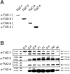
Fig. 1.
Fig. 1.
Isoform-specific TUC antibodies recognize 64 and 75 kDa proteins. A, Each TUC isoform-specific antibody was tested by immunoblotting lysates from HEK293 cells transfected with cDNA encoding each of the TUC proteins. Each antibody recognized its target isoform and did not cross react with the other isoforms.B, Brain homogenates from rats from E12 to adult (Ad) were immunoblotted with each TUC isoform-specific antibody. Whereas the antibody to TUC-3 recognized only a single 64 kDa protein, each of the other antibodies recognized proteins at 64 and 75 kDa. The antibody to TUC-2 also recognized two proteins at ∼68 kDa.
Fig. 2.
Fig. 2.
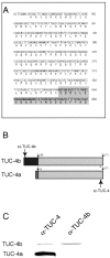
Identification of TUC-4b as a variant of TUC-4. A, Nucleotide sequence encoding the unique N-terminal extension of TUC-4b. The predicted amino acid sequence of the TUC-4b N-terminal extension is shown below the nucleotide sequence. The shaded area represents the beginning of the region of sequence identity with TUC-4a. The full nucleotide sequence of TUC-4b can be found in GenBank (accession number AF398465).B, In TUC-4b, the first 13 amino acids of TUC-4a, which are derived from exon 1, are replaced by a unique 126 amino acid sequence. The TUC-4 antibody (αTUC-4) recognizes an epitope that is present at the C-terminal end of TUC-4a and TUC-4b. The TUC-4b antibody (αTUC-4b) recognizes an epitope that is unique to TUC-4b.C, Western blot of E18 rat brain lysate with the TUC-4 and TUC-4b antibodies. The TUC-4 antibody recognizes proteins at 64 and 75 kDa, whereas the TUC-4b antibody only recognizes a 75 kDa protein.
Fig. 3.
Fig. 3.
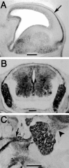
TUC-4b is expressed in the CNS and PNS of E17 rat embryos. A, Sagittal section through the telencephlon stained with the antibody to TUC-4b. TUC-4b was expressed at low levels in the ventricular zone and at higher levels in the cortical plate (arrow). Scale bar, 500 μm. B, Coronal section of E17 spinal cord stained with an antibody to TUC-4b. TUC-4b was expressed in the dorsal root ganglia and in the spinal cord. Scale bar, 200 μm. C, Sagittal section through the trigeminal ganglia. TUC-4b was detected in the trigeminal ganglia and in its peripheral (arrow) and central (arrowhead) processes. Scale bar, 200 μm.
Fig. 4.
Fig. 4.
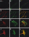
TUC-4b is enriched in the central domain of growth cones from DRG neurons. A–F, DRG explant cultures were stained with antibodies to TUC-4b (A), TUC-4 (D), or tubulin (B,E). The merged images are shown inC and F. Scale bar, 100 μm. TUC-4b immunoreactivity was weak in neurites and strong in the growth cones (see arrows in A). TUC-4 immunoreactivity was strong throughout the neurite and growth cones.G–L, High-power images of growth cones stained with antibodies to TUC-4b (G), TUC-4 (J), or phalloidin (H,K). The merged images are shown inI and L. Scale bar, 10 μm. TUC-4b immunoreactivity was localized to punctate structures in the central domain of the growth cone. TUC-4 immunoreactivity was distributed throughout the growth cone, extending into the filopodia.
Fig. 5.
Fig. 5.
TUC-4b is localized to vesicles in the growth cone. A–C, DRG explant cultures were colabeled with antibodies to TUC-4b (A) and SV2 (B), and the merged image is seen inC. D–F DRG explant cultures were colabeled with antibodies to TUC-4b (D) and Rab5 (E), and the merged image is seen inF. Scale bar, 10 μm.
Fig. 6.
Fig. 6.
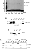
The TUC family interacts with the SH3A domain of intersectin. A, GST fusion proteins encoding the individual SH3 domains of intersectin (SH3A–SH3E) were precoupled to glutathione-Sepharose and incubated with Triton X-100-soluble proteins from E18 rat brains (+ extract) or with buffer alone (− extract). The beads were washed, and specifically bound material was eluted and processed for SDS-PAGE along with an aliquot of the soluble extract [starting material (SM)] equivalent to 120 of the protein added to the beads. The gels were stained with Coomassie blue to reveal the full protein profile. An unknown protein (TUCs) bound to the SH3A and SH3E domains and was later determined to belong to the TUC family. The strong bands below the 45 kDa marker are the GST fusion proteins that elute from the beads. The band at 110 kDa represents dynamin as indicated and as described previously (Yamabhai et al., 1998). The band at 97 kDa is an Escherichia coli-derived protein, as evidenced by its presence in the no-extract lanes. B, Binding assays were conducted with each of the five SH3 domains of intersectin, and bound proteins were immunoblotted with the pan-TUC antibody. Ten percent of the starting material (SM) was included on each gel for reference. The TUC family bound to the SH3A domain and to a lesser degree the SH3E domain but not to the SH3B, SH3C, or SH3D domains.C, Proteins that bound to the SH3A domain were immunoblotted with the TUC-4 antibody, which recognizes both TUC-4a and TUC-4b. Whereas TUC-4a bound at levels <10% of the starting material (SM), TUC-4b bound at levels substantially >10% of the starting material. D, Binding assays were conducted with the SH3A domain and lysates from cells that were transfected with either TUC-4a or TUC-4b, or TUC-4aΔ12. Bound fractions were immunoblotted with an antibody that recognizes a region in common between TUC-4a and TUC-4b. To assess possible nonspecific binding, the bound fractions were also immunoblotted with an antibody against actin. Whereas TUC-4a was slightly enriched relative to 10% of the starting material (SM), TUC-4b was highly enriched relative to 10% of the starting material. Neither TUC-4aΔ12 nor actin bound to the SH3A domain.
Fig. 7.
Fig. 7.
Overexpression of TUC-4b increases neurite outgrowth. A–C, Digital camera lucida drawings illustrating representative examples of E18 cortical neurons cotransfected with GFP and PRK-5 control vector (A), TUC-4a (B), or TUC-4b (C). Scale bar, 100 μm.D, Graph representing the average length of the longest neurite for neurons transfected with PRK5 (n = 88), TUC-4a (n = 90), or TUC-4b (n = 101). Transfection of neurons with TUC-4b increased neurite length by 27% relative to neurons transfected with PRK5, a statistically significant result (p < 0.05; unpairedt test). E, Graph representing the average number of branch points for each neuron transfected with PRK5 (n = 91), TUC-4a (n = 83), or TUC-4b (n = 103). Transfection of neurons with TUC-4b increased neurite branching by 71% relative to neurons transfected with PRK5, a statistically significant result (p < 0.05; unpaired ttest).
Fig. 8.
Fig. 8.
The TUC family is based on a TUC-4 core region that is 33.5% identical to the C. elegans 55 kDa isoform of UNC-33. TUC-4a consists of the core region plus a 12 amino acid N-terminal extension. TUC-4b consists of the core region plus a 126 amino acid extension. The TUC-4a and TUC-4b N-terminal extensions are unique and do not have any homology to UNC-33 or any other known proteins. The 55 kDa isoform of UNC-33 represents a core region that is 33.5% identical to the TUC-4 core region. The 72 kDa isoform of UNC-33 consists of the UNC-33 core region plus a 156 amino acid N-terminal extension. The 90 kDa UNC-33 isoform consists of the entire sequence of the 72 kDa isoform plus an additional 175 amino acid N-terminal extension.

Similar articles

Cited by

References

    1. Bouchard JF, Roux PP, Shekarbi M, Barker PA, Kennedy TE. cAMP elevation rapidly translocates DCC to the plasma membrane and promotes netrin-1-dependent axon extension. Soc Neurosci Abstr. 2002;28:729.14.
    1. Buckley K, Kelly RB. Identification of a transmembrane glycoprotein specific for secretory vesicles of neural and endocrine cells. J Cell Biol. 1985;100:1284–1294. - PMC - PubMed
    1. Bunge MB. Initial endocytosis of peroxidase or ferritin by growth cones of cultured nerve cells. J Neurocytol. 1977;6:407–439. - PubMed
    1. Byk T, Dobransky T, Cifuentes-Diaz C, Sobel A. Identification and molecular characterization of Unc-33-like phosphoprotein (Ulip), a putative mammalian homolog of the axonal guidance-associated unc-33 gene product. J Neurosci. 1996;16:688–701. - PMC - PubMed
    1. Byk T, Ozon S, Sobel A. The Ulip family phosphoproteins—common and specific properties. Eur J Biochem. 1998;254:14–24. - PubMed

Publication types

MeSH terms

Associated data