. 2003 Apr 1;23(7):2824-32.
doi: 10.1523/JNEUROSCI.23-07-02824.2003.

The predominant neural stem cell isolated from postnatal and adult forebrain but not early embryonic forebrain expresses GFAP

Affiliations

The predominant neural stem cell isolated from postnatal and adult forebrain but not early embryonic forebrain expresses GFAP

Tetsuya Imura et al. J Neurosci. .

Abstract

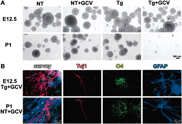
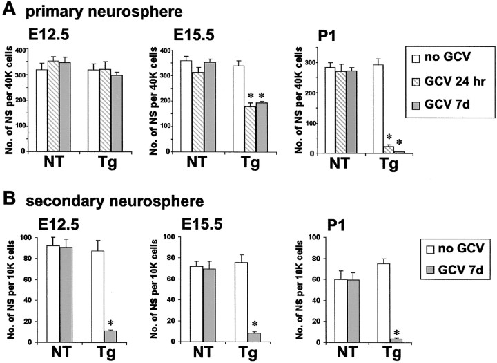
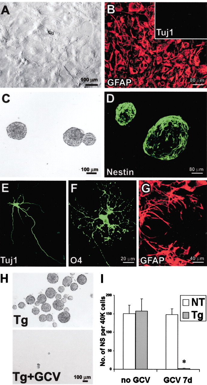
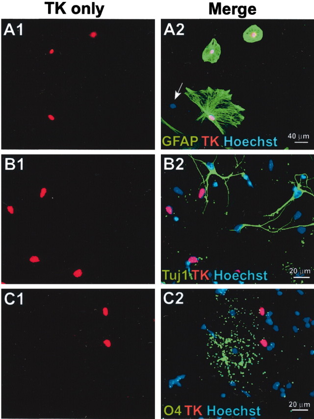
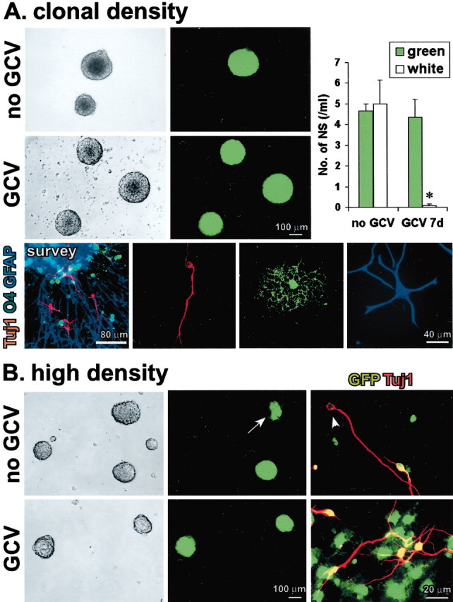
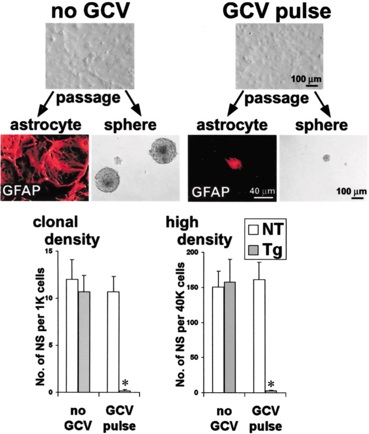
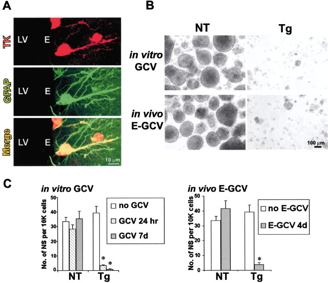
Periventricular germinal zones (GZs) of developing and adult brain contain neural stem cells (NSCs), the cellular identities and origins of which are not defined completely. We used tissue culture techniques and transgenic mice expressing herpes simplex virus thymidine kinase (HSV-TK) from the mouse glial fibrillary acid protein (GFAP) promoter to test the hypothesis that certain NSCs express GFAP. To do so, we determined the relative proportions of multipotent neurospheres that are formed by GFAP-expressing cells derived from GZs at different stages of development. In this transgenic model, dividing GFAP-expressing cells are ablated selectively by treatment with the antiviral agent ganciclovir (GCV). Single-cell analysis showed that transgene-derived HSV-TK was present only in GFAP-expressing cells. GCV applied in vitro eliminated growth of multipotent neurospheres from GZs of postnatal and adult transgenic mice but not early embryonic (embryonic day 12.5) transgenic mice. GCV prevented growth of secondary multipotent neurospheres prepared after passage of primary transgenic neurospheres derived from all three of these developmental stages. In addition, GCV prevented growth of multipotent neurospheres from transgenic astrocyte-enriched cell cultures derived from postnatal GZ, and elaidic acid GCV given for 4 d to adult transgenic mice in vivo abolished the ability to grow multipotent neurospheres from GZ. Extensive control experiments, including clonal analysis, demonstrated that failure of neurosphere growth was not merely secondary to loss of GFAP-expressing support cells or the result of a nonspecific toxic effect. Our findings demonstrate that the predominant multipotent NSCs isolated from postnatal and adult but not early embryonic GZs express GFAP, and that NSCs exhibit heterogeneous expression of intermediate filaments during developmental maturation.


Figures

Fig. 1.
Fig. 1.
GFAP-expressing cells are required to form multipotent neurospheres from early postnatal but not early embryonic GZs. A, Phase-contrast images of live, floating neurospheres prepared from E12.5 or P1 tissue from nontransgenic (NT) or GFAP-TK transgenic (Tg) mice in the presence or absence of GCV. GCV completely prevented sphere growth from P1 transgenic mice but did not reduce growth from transgenic E12.5 or nontransgenic mice. B, Tricolored immunofluorescence of markers for neurons (Tuj1, red), oligodendrocytes (O4, green) and astrocytes (GFAP,blue) after differentiation of neurospheres that were grown from E12.5 transgenic mice or P1 nontransgenic mice in the presence of GCV. Differentiation was induced in the absence of GCV. Photomicrographs show low-magnification surveys tripled labeled for all three markers, as well as details of each individual cell type.
Fig. 2.
Fig. 2.
Quantitative analysis of multipotent neurosphere formation at different stages of development, with and without ablation of GFAP-expressing cells. Graphs show mean ± SEM number of neurospheres (NS) prepared from E12.5, E15.5, or P1 tissue from nontransgenic (NT) or GFAP-TK transgenic (Tg) mice in the presence or absence of either 24 hr or 7 d of GCV. A, Effects of GCV on the number of primary spheres formed per 40,000 cells derived directly from GZs. GCV treatment had no significant effect on the number of primary spheres derived directly from nontransgenic GZs at any age. GCV also had no significant effect on the number of primary spheres from E12.5 GZs but significantly reduced sphere formation by one-half at E15.5 and abolished sphere formation at P1. B, Effects of GCV on the number of secondary spheres formed per 10,000 cells derived after passage of primary spheres. GCV essentially abolished (>90% reduction) the formation of secondary spheres derived after passage of primary spheres from GZ at all three developmental stages.n = 3–8 separate cultures prepared from different mice. *Significantly different from nontransgenic or non-GCV-treated mice (p < 0.001; ANOVA plus post hoc pairwise analysis).
Fig. 3.
Fig. 3.
GFAP-expressing cells are required to form multipotent neurospheres from primary astrocyte cultures prepared from P1 GZs. A, Phase-contrast image of primary astrocytesin vitro showing high density of cells that have reached confluence after 21 DIV. B, Immunofluorescence of primary astrocyte culture at cell confluence after 21 DIV. Most cells stain for GFAP (red). No cells stain for Tuj1 (inset). C, Live floating neurospheres prepared from primary astrocyte culture. D, Fixed floating neurospheres prepared from primary astrocyte culture and stained for nestin (green). E–G, Immunofluorescence of chemical markers for neurons (Tuj1,E), oligodendrocytes (O4,F), and astrocytes (GFAP, G) shows cells of all three types derived by differentiation of neurospheres prepared from primary astrocyte cultures. H, Phase-contrast images of live floating neurospheres prepared from primary astrocyte cultures from GFAP-TK transgenic (Tg) mice in the absence or presence of GCV. I, Graph shows mean ± SEM number of neurospheres (NS) formed per 40,000 cells prepared after passage of primary astrocyte cultures from nontransgenic (NT) or transgenic mice in the presence or absence of GCV. GCV completely prevented the growth of spheres from transgenic mice. n = 3–5 separate cultures prepared from different mice. *Significantly different from nontransgenic or non-GCV-treated mice (p < 0.001; ANOVA plus post hoc pairwise analysis).
Fig. 4.
Fig. 4.
Transgene-derived HSV-TK is expressed only in GFAP-expressing cells in vitro. A–C, Primary astrocyte cultures derived from P1 GZs of GFAP-TK transgenic mice were double stained by immunofluorescence for TK (red) and either GFAP (A), Tuj1 (B), or O4 (C) (green); Hoechst blue was used as a general cytological counterstain. Pairs of images show the same microscopic fields with single-channel visualization of TK only (red) and merged visualization of all three fluorescent channels (red, green,blue). In merged images, red TK staining overlaps completely with blue stain, so that nuclei appear purple. A1, A2, All TK cells (red) are GFAP-positive (green) and vice versa. A cell negative for both GFAP and TK is indicated by the blue nucleus (arrow). B1, B2, No TK-positive cells (red) are positive for the neuronal marker Tuj1 (green). C1, C2, No TK-positive cells (red) are positive for the oligodendrocyte marker O4 (green).
Fig. 5.
Fig. 5.
GFAP-expressing cells are not merely support cells, and their ablation is not nonspecifically toxic to neurosphere formation. A, Phase-contrast and fluorescent images of the same live floating neurospheres prepared at clonal cell density (1000 cells/ml; 500 green cells plus 500 white cells) from primary P1 astrocyte cultures with mixed single-cell suspensions of GFP and GFAP-TK cells. Without GCV, both GFP-only (green) and GFAP-TK-only (white) spheres are present. There are no mixed GFP plus GFAP-TK (green and white) spheres. In the presence of GCV, only GFP (green) spheres are present. Graph shows mean ± SEM number of green or white neurospheres (NS) formed per 1000 cells in the presence or absence of GCV. n = 3 separate cultures prepared from different mice for each value. *Significantly different from non-GCV-treated or non-TK-expressing mice (p< 0.01; ANOVA plus post hoc pairwise analysis). Tricolored immunofluorescence of markers for neurons (Tuj1,red), oligodendrocytes (O4,green), and astrocytes (GFAP, blue) shows that on differentiation, clonal spheres gave rise to different cells expressing markers of all three neural cell types, as depicted in a triple-labeled survey and details of individual cells of each type.B, Phase-contrast and fluorescent images of the same live floating neurospheres prepared at high cell density (40,000 cells/ml) from primary P1 astrocyte cultures with mixed suspensions of GFP and GFAP-TK cells. In the absence of GCV, three types of spheres formed: GFP only (green), GFAP-TK only (white), and mixed GFP and GFAP-TK (green and white;arrow). In the presence of GCV, only GFP-only (green) spheres formed. Merged double-labeled images of immunocytochemistry for Tuj1 (red) plus GFP (green) show that differentiation of spheres grown without GCV gave rise to both GFP-positive (red plus green =yellow) and GFP-negative (red,arrowhead), Tuj1-positive neurons, whereas after differentiation of spheres grown with GCV, all Tuj1-positive neurons were also GFP positive (red plusgreen = yellow).
Fig. 6.
Fig. 6.
Ablation of GFAP-expressing cells prevents the onset of neurosphere formation. Confluent, nondividing primary P1 astrocyte cultures prepared from GFAP-TK transgenic mice were exposed to GCV for 24 hr before passage (GCV pulse), with no detectable cell loss. Cultures not exposed to GCV could be passaged readily to form either more primary astrocytes or multipotent neurospheres. Cultures exposed to GCV failed to form either primary astrocytes or neurospheres after passage and the induction of cell division. Graphs show mean ± SEM number of neurospheres (NS) prepared after passage of primary astrocyte cultures from nontransgenic (NT) or GFAP-TK transgenic (Tg) mice in the presence or absence of GCV pulse under clonal (1000 cells/ml) or high-density (40,000 cells/ml) conditions. The GCV pulse had no significant effect on the number of spheres formed by nontransgenic cells at either clonal or high density but abolished sphere formation by transgenic cells. n = 3 separate cultures prepared from different mice. *Significantly different from non-GCV-treated or nontransgenic mice (p < 0.01; ANOVA plus post hoc pairwise analysis).
Fig. 7.
Fig. 7.
Ablation of GFAP-expressing cells by delivery of GCV in vitro or in vivo abolishes the ability to derive NSCs from adult GZ. A, Tissue section of GZ adjacent to lateral ventricle (LV) from an adult GFAP-TK transgenic mouse, double stained by immunofluorescence for TK (red) and GFAP (green). Confocal scanning laser microscopic images show separate visualization of red (TK),green (GFAP), and merged channels. Several cells are double labeled for both TK and GFAP. TK immunoreactivity is intense in cell nuclei and somata and in some cases is detectable in cell processes. GFAP immunoreactivity is intense in intracellular filaments located in cell processes and that sometimes traverse the cell body but is weak in somatic cytoplasm. All TK-positive cells (red) are GFAP-positive (green) and vice versa. Ependymal cells (E) are not visible, and GFAP/TK-positive cells are in the subependymal zone. B, C, Phase-contrast images (B) and quantitative analysis (C) of live floating multipotent neurospheres prepared from GZ of adult nontransgenic (NT) or GFAP-TK transgenic (Tg) mice in the presence or absence of GCV in vitro or after 4 d of E-GCV treatmentin vivo. Graphs show mean ± SEM number of neurospheres (NS) formed per 10,000 cells derived directly from adult GZs of nontransgenic or transgenic mice. Both GCVin vitro and E-GCV in vivo prevented sphere growth from adult GZ of transgenic but not nontransgenic mice.n = 3–5 separate cultures prepared from different mice. *Significantly different from nontransgenic or non-GCV-treated mice (p < 0.001; ANOVA plus post hoc pairwise analysis).

References

    1. Alvarez-Buylla A, Garcia-Verdugo JM, Tramontin AD. A unified hypothesis on the lineage of neural stem cells. Nat Rev Neurosci. 2001;2:287–293. - PubMed
    1. Anderson DJ. Stem cells and pattern formation in the nervous system: the possible versus the actual. Neuron. 2001;30:19–35. - PubMed
    1. Andrae J, Bongcam-Rudloff E, Hansson I, Lendahl U, Westermark B, Nister M. A 1.8 kb GFAP-promoter fragment is active in specific regions of the embryonic CNS. Mech Dev. 2001;107:181–185. - PubMed
    1. Balzarini J, Degreve B, Andrei G, Neyts J, Sandvold M, Myhren F, de Clercq E. Superior cytostatic activity of the ganciclovir elaidic acid ester due to the prolonged intracellular retention of ganciclovir anabolites in herpes simplex virus type 1 thymidine kinase gene-transfected tumor cells. Gene Ther. 1998;5:419–426. - PubMed
    1. Bush TG, Savidge TC, Freeman TC, Cox HJ, Campbell EA, Mucke L, Johnson MH, Sofroniew MV. Fulminant jejuno-ileitis following ablation of enteric glia in adult transgenic mice. Cell. 1998;93:189–201. - PubMed

Publication types