Premotor neurons encode torsional eye velocity during smooth-pursuit eye movements
- PMID: 12684484
- PMCID: PMC6742083
- DOI: 10.1523/JNEUROSCI.23-07-02971.2003
Premotor neurons encode torsional eye velocity during smooth-pursuit eye movements
Abstract
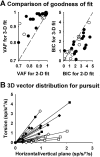
Responses to horizontal and vertical ocular pursuit and head and body rotation in multiple planes were recorded in eye movement-sensitive neurons in the rostral vestibular nuclei (VN) of two rhesus monkeys. When tested during pursuit through primary eye position, the majority of the cells preferred either horizontal or vertical target motion. During pursuit of targets that moved horizontally at different vertical eccentricities or vertically at different horizontal eccentricities, eye angular velocity has been shown to include a torsional component the amplitude of which is proportional to half the gaze angle ("half-angle rule" of Listing's law). Approximately half of the neurons, the majority of which were characterized as "vertical" during pursuit through primary position, exhibited significant changes in their response gain and/or phase as a function of gaze eccentricity during pursuit, as if they were also sensitive to torsional eye velocity. Multiple linear regression analysis revealed a significant contribution of torsional eye movement sensitivity to the responsiveness of the cells. These findings suggest that many VN neurons encode three-dimensional angular velocity, rather than the two-dimensional derivative of eye position, during smooth-pursuit eye movements. Although no clear clustering of pursuit preferred-direction vectors along the semicircular canal axes was observed, the sensitivity of VN neurons to torsional eye movements might reflect a preservation of similar premotor coding of visual and vestibular-driven slow eye movements for both lateral-eyed and foveate species.
Figures


Similar articles
-
Activity of smooth pursuit-related neurons in the monkey periarcuate cortex during pursuit and passive whole-body rotation.J Neurophysiol. 2000 Jan;83(1):563-87. doi: 10.1152/jn.2000.83.1.563. J Neurophysiol. 2000. PMID: 10634896
-
Validity of Listing's law during fixations, saccades, smooth pursuit eye movements, and blinks.Exp Brain Res. 1996 Nov;112(1):135-46. doi: 10.1007/BF00227187. Exp Brain Res. 1996. PMID: 8951416
-
Brain stem pursuit pathways: dissociating visual, vestibular, and proprioceptive inputs during combined eye-head gaze tracking.J Neurophysiol. 2003 Jul;90(1):271-90. doi: 10.1152/jn.01074.2002. J Neurophysiol. 2003. PMID: 12843311
-
Three-dimensional organization of otolith-ocular reflexes in rhesus monkeys. II. Inertial detection of angular velocity.J Neurophysiol. 1996 Jun;75(6):2425-40. doi: 10.1152/jn.1996.75.6.2425. J Neurophysiol. 1996. PMID: 8793754
-
Stopping smooth pursuit.Philos Trans R Soc Lond B Biol Sci. 2017 Apr 19;372(1718):20160200. doi: 10.1098/rstb.2016.0200. Philos Trans R Soc Lond B Biol Sci. 2017. PMID: 28242734 Free PMC article. Review.
Cited by
-
Mechanics of the orbita.Dev Ophthalmol. 2007;40:132-57. doi: 10.1159/000100353. Dev Ophthalmol. 2007. PMID: 17314483 Free PMC article. Review.
-
Canal-otolith interactions and detection thresholds of linear and angular components during curved-path self-motion.J Neurophysiol. 2010 Aug;104(2):765-73. doi: 10.1152/jn.01067.2009. Epub 2010 Jun 16. J Neurophysiol. 2010. PMID: 20554843 Free PMC article.
-
Current concepts of mechanical and neural factors in ocular motility.Curr Opin Neurol. 2006 Feb;19(1):4-13. doi: 10.1097/01.wco.0000198100.87670.37. Curr Opin Neurol. 2006. PMID: 16415671 Free PMC article. Review.
-
Differential lateral rectus compartmental contraction during ocular counter-rolling.Invest Ophthalmol Vis Sci. 2012 May 14;53(6):2887-96. doi: 10.1167/iovs.11-7929. Invest Ophthalmol Vis Sci. 2012. PMID: 22427572 Free PMC article.
-
Dynamic visuomotor synchronization: quantification of predictive timing.Behav Res Methods. 2013 Mar;45(1):289-300. doi: 10.3758/s13428-012-0248-3. Behav Res Methods. 2013. PMID: 22956395 Free PMC article.
References
-
- Angelaki DE. Dynamic polarization vector of spatially tuned neurons. IEEE Trans Biomed Eng. 1991;38:1053–1060. - PubMed
-
- Angelaki DE, Dickman JD. Spatiotemporal processing of linear acceleration: primary afferent and central vestibular neuron responses. J Neurophysiol. 2000;84:2113–2132. - PubMed
-
- Caines PE. Linear stochastic systems. Wiley; Toronto: 1988.
-
- Cannon SC, Robinson DA. Loss of the neural integrator of the oculomotor system from brain stem lesions in monkey. J Neurophysiol. 1987;57:1383–1409. - PubMed
Publication types
MeSH terms
Grants and funding
LinkOut - more resources
Full Text Sources
