. 2003 Apr 1;23(7):2980-92.
doi: 10.1523/JNEUROSCI.23-07-02980.2003.

Transmission security for single kinesthetic afferent fibers of joint origin and their target cuneate neurons in the cat

Affiliations

Transmission security for single kinesthetic afferent fibers of joint origin and their target cuneate neurons in the cat

Gordon T Coleman et al. J Neurosci. .

Abstract

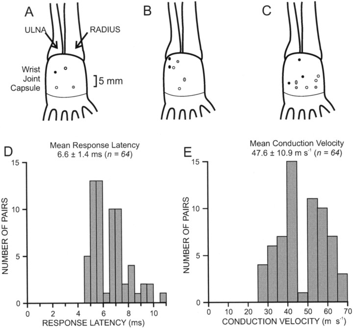
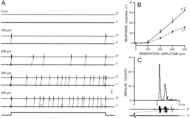
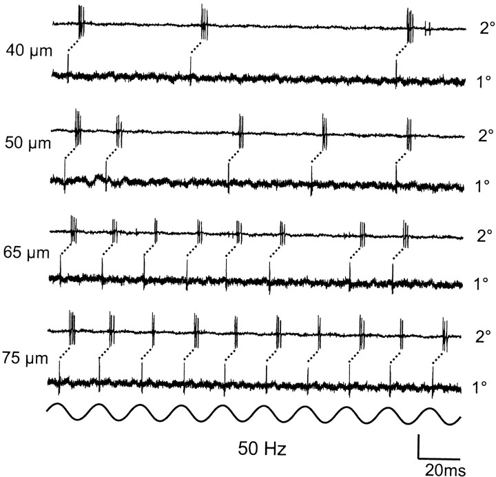
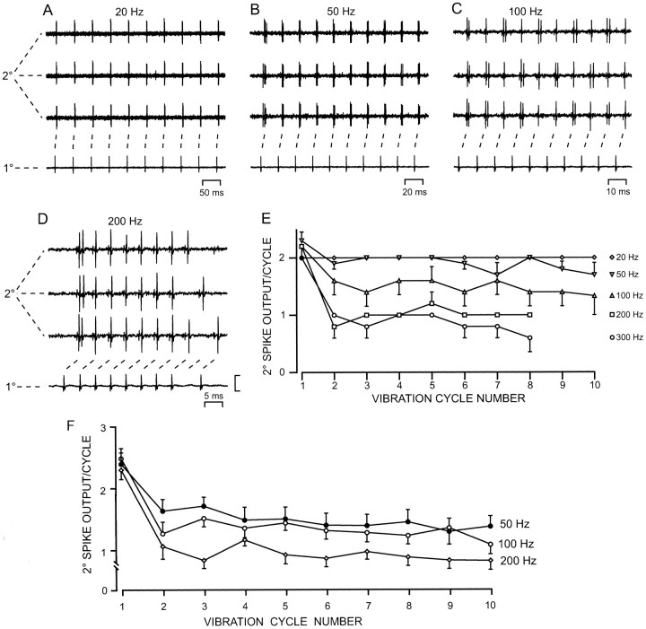
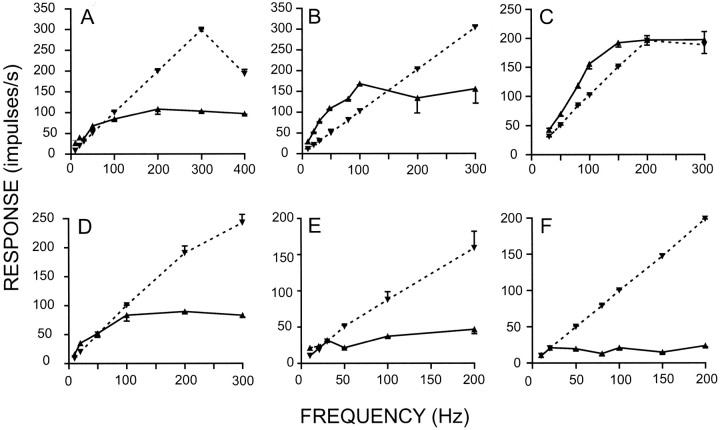
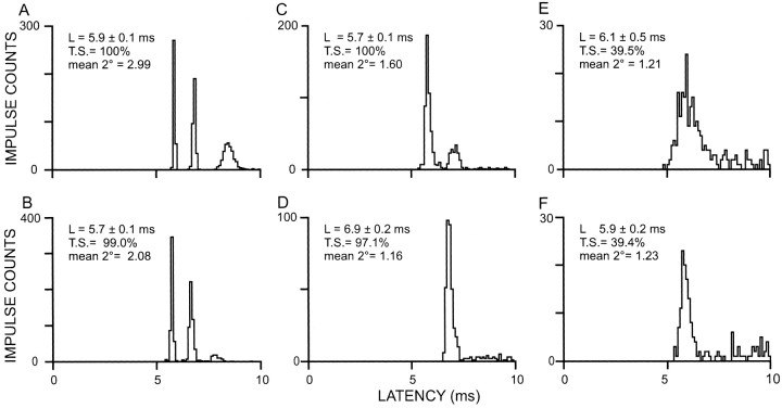
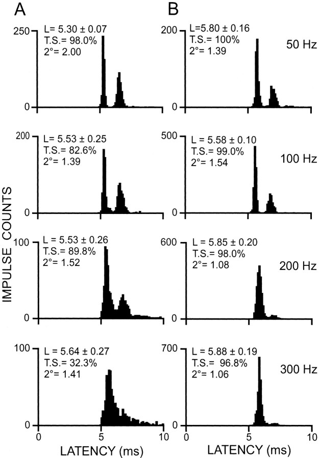
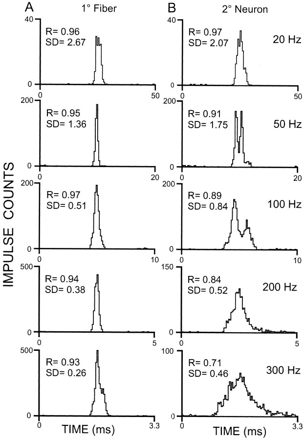
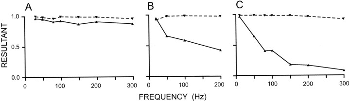
Transmission between single identified, kinesthetic afferent fibers of joint origin and their central target neurons of the cuneate nucleus was examined in anesthetized cats by means of paired electrophysiological recording. Fifty-three wrist joint afferent-cuneate neuron pairs were isolated in which the single joint afferent fiber exerted suprathreshold excitatory actions on the target cuneate neuron. For each pair, the minimum kinesthetic input, a single spike, was sufficient to generate cuneate spike output, often amplified as a pair or burst of spikes, particularly at input rates up to 50-100 impulses per second. The high security was confirmed quantitatively by construction of stimulus-response relationships and calculation of transmission security measures in response to both static and dynamic vibrokinesthetic disturbances applied to the joint capsule. Graded stimulus-response relationships demonstrated that the output for this synaptic connection between single joint afferents and cuneate neurons could provide a sensitive indicator of the strength of joint capsule stimuli. The transmission security measures, calculated as the proportion of joint afferent spikes that generated cuneate spike output, were high (>85-90%) even at afferent fiber discharge rates up to 100-200 impulses per second. Furthermore, tight phase locking in the cuneate responses to vibratory stimulation of the joint capsule demonstrated that the synaptic linkage preserved, with a high level of fidelity, the temporal information about dynamic kinesthetic perturbations that affected the joint. The present study establishes that single kinesthetic afferents of joint origin display a capacity similar to that of tactile afferent fibers for exerting potent synaptic actions on central target neurons of the major ascending kinesthetic sensory pathway.


Figures

Fig. 1.
Fig. 1.
Receptive field, latency, and conduction velocity characteristics for wrist joint afferent–cuneate neuron pairs. Receptive fields are shown in A for four neurons activated by probing a single point on the capsule, in Bfor three neurons each activated at a suprathreshold level by two identified afferent fibers from two discrete points on the capsule (filled, gray, and open symbolsfor the three pairs of foci), and in C for three neurons each activated by three afferent fibers from three discrete foci (filled, gray, and open symbolsfor the fields of the three neurons). D, The histogram shows the distribution of latencies for cuneate neuron responses to 64 single identified wrist joint afferent fibers measured from the occurrence time of the peripheral spike recorded from the wrist joint nerve in the mid forearm. For each of these latency measurements, the afferent fiber was activated at low rates (∼1 Hz). E, Distribution of approximate conduction velocities for the wrist joint afferent fibers on the basis of cuneate latency values inD, with an allowance of 1 msec for central delay time across the synaptic linkage.
Fig. 2.
Fig. 2.
The capacity of single spikes in a wrist joint afferent fiber (1°) to generate spike output from a cuneate target neuron (2°) in response to static indentation of the joint capsule. The paired simultaneous recordings in A show the responses to focal static indentation (1 sec duration) of the joint capsule at amplitudes up to 400 μm. The asterisk in the peripheral response trace at 400 μm marks the occasional recruitment of a second fiber that fails to elicit any response from this cuneate neuron. Vertical calibration, 0.28 mV for the 1° traces and 0.50 mV for the 2° traces. The stimulus–response relationships in B show the graded input–output functions for peripheral (1°) and central (2°) elements (dashedand solid lines, respectively) and reveal the amplification of output across this synaptic connection. InC, the RCH and superimposed impulse traces for 1° and 2° elements illustrate the pairs or bursts of spikes elicited in the cuneate neuron in response to the single input spike. The transient onset segment (∼15 msec) of the indentation period was excluded from the impulse counts plotted in B and from the RCH inC because additional fibers were sometimes recruited by the abrupt onset of the step indentation. In A andC, the connecting dots emphasize the correlation between peripheral and central spike activity.
Fig. 3.
Fig. 3.
High transmission security and amplification across the connection between a single joint afferent fiber (1°) and a cuneate target neuron (2°) at high rates of afferent drive generated by focal vibration (50 Hz) of the joint capsule at a range of amplitudes (40–75 μm). Where the peripheral fiber failed to respond during a vibration cycle, there was a correlated failure in the response of the cuneate neuron. This close correlation between peripheral and central activity confirmed that the fiber for which activity is shown in the peripheral trace was uniquely responsible for these responses of the cuneate neuron. Vertical calibration, 0.10 mV for the 1° traces and 0.34 mV for the 2° traces.
Fig. 4.
Fig. 4.
Response traces and quantified output measures for the linkage in wrist joint afferent–cuneate neuron pairs as a function of the frequency and cycle number for focal vibration delivered to the joint capsule. A–D, For one pair, one representative trace is shown of the response of the peripheral fiber (1°) and three representative traces are shown for the central neuron (2°) over the first 10 cycles at each frequency from 20 to 200 Hz. At each frequency, the vibration amplitude applied was sufficient to produce a one-to-one following in the fiber over the first 10 cycles, except at 200 Hz, where one-to-one following could only be sustained over the first seven to eight cycles. E, Graph of the spike output of this cuneate neuron (2° spike output per cycle) as a function of vibration cycle number when the fiber was driven at a one-to-one level over the first 10 cycles. Each data point represents the mean ± SE response level of the cuneate neuron for five repetitions of the stimulus. At 200 and 300 Hz, a one-to-one response level could not be sustained in the input fiber beyond seven to eight vibration cycles. The vertical calibration in D represents 0.10 mV for the afferent fiber traces and 0.20 mV for the cuneate neuron traces.F, Security of the linkage for a sample of wrist joint afferent–cuneate neuron pairs quantified as the averaged spike output of the neuron on successive cycles of the focal vibratory stimulus. This measure of cuneate neuron output was obtained at vibratory stimulus frequencies of 50, 100, and 200 Hz for each pair when the wrist joint afferent fiber was responding to the vibration with a regular one-to-one pattern of activity. The mean spike output on each cycle was calculated, up to cycle number 10, and plotted along with SE (error bars). The averaged responses from 14 pairs are shown for the 50 Hz plot and for 15 pairs for the 100 Hz plot. At 200 Hz, the number of pairs that contributed to the plot fell from 11 in each of the first eight cycles to 10 over the last two cycles.
Fig. 5.
Fig. 5.
Bandwidth of vibrokinesthetic responsiveness for wrist joint afferent fibers (dashed lines) and their target cuneate neurons (solid lines). Frequency–response graphs are shown for six wrist joint afferent–cuneate neuron pairs. The response levels plotted at each vibration frequency were for the lowest amplitude that produced a one-to-one (or near one-to-one) impulse rate in the peripheral fiber over the entire 1 sec period of the vibration stimulus. For the six joint afferent fibers in A–F, the average vibration amplitudes required for one-to-one firing in the frequency range of 20–200 Hz (where most observations were made and where one-to-one discharge was attained) were as follows: 78 μm at 20 Hz (n = 6), 88 μm at 30 Hz (n = 4), 77 μm at 50 Hz (n = 6), 68 μm at 80 Hz (n = 3), 87 μm at 100 Hz (n = 6), 70 μm at 150 Hz (n = 2), and 77 μm at 200 Hz (n = 6). These 33 values ranged from 20 to 140 μm and showed little systematic change as a function of vibration frequency.
Fig. 6.
Fig. 6.
RCHs constructed for six wrist joint afferent–cuneate neuron pairs when the peripheral fiber was driven at a one-to-one or near one-to-one level by a 50 Hz vibration train that lasted 1 sec and that was applied focally to the joint capsule. The security of the linkages for the representative pairs illustrated ranged from transmission security (T.S.) values of >95% (A–D) to linkages, for which values were ∼40% (E, F). The mean cuneate response (mean 2°) and its latency (L, ± SD) are indicated on each histogram (see Results).
Fig. 7.
Fig. 7.
Changes in transmission characteristics for two representative wrist joint afferent–cuneate neuron pairs (A, B) as a function of increased rates of peripheral drive. RCHs were constructed for each pair in response to focal stimulation of the joint capsule at vibration frequencies that ranged from 50 to 300 Hz. Transmission security (T.S.), latency (L, ± SD), and mean central response (mean 2°) are indicated on each RCH.
Fig. 8.
Fig. 8.
Comparison of the precision of temporal patterning observed in the responses of a wrist joint afferent fiber and its target cuneate neuron to vibration, applied to the receptive field focus on the joint capsule, at several frequencies (20–300 Hz). CHs were constructed from the responses of the fiber (1° fiber) on the left side and the central target neuron on the right (2° neuron). The width of the horizontal axis for each CH corresponds to the duration of one vibration cycle period. Resultant vectors (R) and SDs were calculated from the CH distributions of the fiber and its target neuron as measures of phase locking and absolute temporal dispersion, respectively, in the responses of the wrist joint afferent and its target neuron.
Fig. 9.
Fig. 9.
Quantitative evaluation of changes in the tightness of phase locking in responses of three representative wrist joint afferent fiber–cuneate neuron pairs (A–C) as a function of the frequency parameter of focal vibratory stimuli applied to the joint capsule. Thedashed lines plot the vector strength (R) derived from the afferent fiber responses, whereas the solid lines plot these values for the synaptically linked cuneate neurons. The pair that retained precise temporal patterning in transmission across this synaptic linkage (A) also had a high transmission security (e.g., 100% at 50 Hz), whereas those in B and Cthat had less faithful retention of temporal patterning had poorer measures of transmission security (40 and 36%, respectively, at 50 Hz).

Similar articles

Cited by

References

    1. Andersen P, Eccles JC, Schmidt RF, Yokota T. Identification of relay cells and interneurons in the cuneate nucleus. J Neurophysiol. 1964;27:1085–1095. - PubMed
    1. Andersen P, Etholm B, Gordon G. Presynaptic and postsynaptic inhibition elicited in the cat's dorsal column nuclei by mechanical stimulation of the skin. J Physiol (Lond) 1970;210:435–455. - PMC - PubMed
    1. Asanuma H, Zarzecki P, Jankowska E, Hongo T, Marcus S. Projection of individual pyramidal tract neurons to lumbar motor nuclei of the monkey. Exp Brain Res. 1979;34:73–89. - PubMed
    1. Baxendale RH, Ferrell WR, Wood L. Responses of quadriceps motor units to mechanical stimulation of knee joint receptors in the decerebrate cat. Brain Res. 1988;453:150–156. - PubMed
    1. Berkley KJ, Budell RJ, Blomqvist A, Bull M. Output systems of the dorsal column nuclei in the cat. Brain Res. 1986;396:199–225. - PubMed

Publication types

LinkOut - more resources