. 2003 Apr 1;23(7):3039-51.
doi: 10.1523/JNEUROSCI.23-07-03039.2003.

Directional avoidance turns encoded by single interneurons and sustained by multifunctional serotonergic cells

Affiliations

Directional avoidance turns encoded by single interneurons and sustained by multifunctional serotonergic cells

Jian Jing et al. J Neurosci. .

Abstract

Avoidance turns in the sea slug Pleurobranchaea are responses to noxious stimuli and replace orienting turns to food stimuli after avoidance conditioning or satiation. Avoidance turns proved to be centrally patterned behaviors, the fictive expression of which could be elicited in reduced preparations and the isolated CNS. Activity in one of a bilateral interneuron pair, the A4 cells, was necessary and sufficient to drive the avoidance turn toward the contralateral side. Single A4 cells appeared to encode both turn direction and angle, in contrast to directional behaviors of other animals in which displacement angle is usually encoded by multiple units. The As1-4 cells, bilateral serotonergic cell clusters, excited the prolonged A4 burst during the turn through electrical and chemical coupling. However, during the escape swim, As1-4 became integral elements of the swim motor network, and A4 activity was entrained to the swim rhythm by alternating excitatory-inhibitory inputs, with only weak spiking. This provides a likely mechanism for the previously observed suppression of the avoidance turn by escape swimming. These observations add significant new aspects to the multiplying known functions of As1-4 and their homologs in other molluscs and point to a pivotal role of these neurons in the organization of gastropod behavior. Simple functional models predict (1) the essential actions of inhibitor neurons in the directionality of the turning network motor output and (2) an integrating role for As1-4 in the behavioral switch between turning avoidance and swimming escape, on the basis of their response to increasing stimulus intensity.


Figures
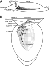
Fig. 1.
Fig. 1.
The lateral body wall muscles (LBWM). A, Schematic drawing of the position of the LBWM viewed from the left side of an intact animal.B, Distribution of muscle bands and nerve innervation in dorsal view in a dissected preparation. Lateral body wall muscles are shown on the left. The LBWM of the right side is generally similar, with slight asymmetry caused by the presence of reproductive organs, anus, and heart (data not shown). The muscle bandsA, B, and C of LBWM are shown, as are the large fascicles of the LMF and the circular muscles of the foot. The major innervation of LBWM is from the posterior lateral body wall nerve (pLBWN) of the pedal ganglion. Other innervation comes from the anterior lateral body wall nerve (aLBWN) of the pedal ganglion and the body wall nerve (BWN) of the cerebropleural ganglion. Foot muscles and pedal cilia are innervated by the medial pedal nerve (mPN), posterior pedal nerve (pPN), and anterior pedal nerve (aPN) [which goes below the branch A1 of band A of LBWM (Table 1) to innervate the foot there; data not shown] of the pedal ganglion.
Fig. 2.
Fig. 2.
The progression of the avoidance turn of a living animal, viewed dorsally. Drawings were from video frames of the avoidance turn to the right side induced by electric shock (Probe) to the left oral veil. Initial responses were head withdrawal, body shortening, and turn initiation. At ∼30 sec, the animal extended its body and locomoted away from the stimulus. The tail, which remained attached to substrate, did not change position during the actual turning phase. Arrows show direction faced by the animal, based on extension of a line joined by midpoints of oral veil and neck. Inset, Plot of turn angle versus time relative to the initial direction of the animal.
Fig. 3.
Fig. 3.
The posterior lateral body wall nerve shows directionally specific motor activity after unilateral shock (bars) to the head region in a reduced preparation. Initial activity (<5 sec), presumed to represent head withdrawal, is present in both nerves. Subsequent activity is greatest in the nerve contralateral to the shock application, consistent with fictive avoidance turning. A, Shock to left side.B, Shock to right side. Mean spike frequency is plotted below the nerve recordings (bin size 1 sec; see Materials and Methods);dotted lines are baseline frequency. Vertical rectangles indicate stimulation interval.LpLBWN, Left pLBWN; RpLBWN, right pLBWN.
Fig. 4.
Fig. 4.
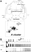
Relative location and morphology of A4 and its activity during a fictive swim. A, The axon of A4 courses laterally to the ipsilateral aCPC, branching in the cerebropleural neuropile before entering the connective (n = 9). The axon enters the pedal ganglion to branch there in the neuropil. Inset, Locations of the interneurons in the A cluster (dorsal surface). Somata are shown only unilaterally for convenience. Cerebropleural ganglion:BWN, body wall nerve; sBWN, small body wall nerve; CBC, cerebrobuccal connective;aCPC, anterior cerebropedal connective;pCPC, posterior cerebropedal connective;CVC, cerebrovisceral connective; LOVN, large oral veil nerve; MN, mouth nerve;RN, rhinophore nerve; SCC, subcerebral commissure; SOVN, small oral veil nerve;TN, tentacle nerve. Pedal ganglion:aLBWN, anterior lateral body wall nerve;pLBWN, posterior lateral body wall nerve;PC, pedal commissure; pPC, parapedal commissure; aPN, anterior pedal nerve;mPN, medial pedal nerve; pPN, posterior pedal nerve. B, A4 (bottom) was weakly active during escape swimming elicited by BWN stimulation (bar). The membrane potential of A4 oscillated in phase with As2/3 (middle), the activity of which led slightly that of the A1 swim interneuron (top).
Fig. 5.
Fig. 5.
Asymmetric spiking activity of A4 interneurons during avoidance turns in isolated CNS and whole animals.A, In isolated CNS, shocks to left (A1) or right (A2) LOVN (bars) elicited fictive avoidance turns represented in strong motor activity in right pLBWN (RpLBWN, A1) and left pLBWN (LpLBWN, A2), respectively. The spike activity of left and right A4 cells was directionally specific: the A4 ipsilateral to the stimulus was active whereas the contralateral A4 was inactive. After the fictive turn, the A4 showed slow bursting activity in anti-phase to each other, corresponding to similar slow oscillation in the bilateral pLBWN (A2). Records ofA1 and A2 are separated by ∼10 min. Records in A2 are continuous. B, In a whole-animal preparation, an A4 fired a prolonged burst during an avoidance turn induced by mechanical stimulation of the ipsilateral oral veil (bar).
Fig. 6.
Fig. 6.
Directional sensitivity of As2/3 response to LOVN shock. A, Ipsilateral oral veil nerve stimulation (bar) induced short-latency firing (0.6 sec) at moderate spike rate (6–8 Hz). B, Contralateral stimulation (bar) induced a response with longer latency (>3 sec) and lower frequency (3–5 Hz). As1 also showed directional sensitivity to oral veil stimulation (see Results).
Fig. 7.
Fig. 7.
Synaptic connectivity of A4. A, Synaptic connections between A4 and the ipsilateral As1–3.A1, A4 was electrically coupled to As2 and As3 because A4 and As3 were hyperpolarized when As2 was injected with a hyperpolarizing current (bar). As2 and As3 are indistinguishable, so name assignment for these two cells is arbitrary.A2, Depolarization of As1 (bar) to spike elicited slow EPSPs in A4 and As2/3. A3, Depolarization of A4 (bar) to spike elicited slow EPSPs in As1. All recordings were made in high divalent saline. B, Synaptic connections between A4 and the ipsilateral A1. Depolarization of A1 (bar) to spike induced biphasic effects in A4, an early excitation followed by inhibition. Spikes in A4 were clipped. Recordings were made in normal saline.
Fig. 8.
Fig. 8.
As1 strongly inhibits the contralateral A4. Driving an As1 with depolarizing current injection (bar) elicited depolarization in both ipsilateral and contralateral A4 (i-A4, c-A4), which was subsequently overridden by inhibition in the c-A4. Inhibition was slow, with occasional phasic IPSPs. Inhibitory potentials induced in i-A4 were less effectual. Recordings were made in high divalent saline.
Fig. 9.
Fig. 9.
Hyperpolarization of A4 blocked fictive turning.A, Control. Fictive avoidance turning was elicited by brief electrical stimulation (bar) of the right LOVN, ipsilateral to the right A4 (R-A4). Fictive turning was evident in the prominent, long-lasting (>30 sec) activity in left posterior lateral body wall nerve (LpLBWN; contralateral to R-A4). Less activity was present in the right (ipsilateral) pLBWN (RpLBWN).B, Hyperpolarization of A4 (betweenarrowheads) after right LOVN stimulation suppressed the long-lasting activity in LpLBWN. The initial activity burst (<10 sec) immediately after LOVN stimulation in both LpLBWN and RpLBWN, presumed to represent head and anterior body withdrawal, was unaffected.Traces below nerve recordings are mean spike frequency plots (bin size, 1 sec). See Figure 3 legend for details.
Fig. 10.
Fig. 10.
Role of As2/3 during avoidance turning.A, Stimulation of right LOVN (bar) activated both right As2/3 (R-As2/3) and right A4 (R-A4) and strong output in left pLBWN (LpLBWN), indicative of an avoidance turn to the left. B, Hyperpolarization of As2/3 (arrowheads) suppressed fictive avoidance turning output in LpLBWN, until R-A4 escaped inhibition and fired a characteristic bursting that lasted ∼25 sec, when the turning activity was restored, at least partially, in LpLBWN. Traces below nerve recordings are mean spike frequency plots (bin size, 1 sec). See Figure3 legend for details.
Fig. 11.
Fig. 11.
A4 drives fictive avoidance turning. Activation (bar) of the left A4 (L-A4) induced strong activity in right pLBWN (A, RpLBWN), suggesting a contralateral turn. Likewise, activation (bar) of the right A4 (R-A4) induced strong activity in left pLBWN (B, LpLBWN).Traces below nerve recordings are mean spike frequency plots (bin size, 1 sec). See Figure 3 legend for details.
Fig. 12.
Fig. 12.
As2/3 activates A4 and concomitant activity in pLBWN. A, Driving a right As2/3 (R-As2/3,bar) induced strong activity in the right, but not left, pLBWN. Note that the left As2/3 (L-As2/3) was not activated. Traces below nerve recordings are mean spike frequency plots (see details in Fig. 3 legend). B, In a different preparation, driving a right As2/3 (bar) induced a prolonged burst in the left A4, with corresponding spike activity in right pLBWN.
Fig. 13.
Fig. 13.
A network model for avoidance turning. In this model, appropriate sensory input is presumed to act on A4 and As1–4 interneurons unilaterally. The prolonged A4 burst of the turn is initiated and sustained by reverberating excitation among As1–4 and A4 neurons. Inhibition of the contralateral A4 is mediated by a hypothetical inhibitory neuron (I) primarily excited by As1 (Fig. 8). A4 would also excite the “I” neuron through its excitation to As1–4, if not directly as well. Potentially, the I neuron might also be activated by unilateral sensory inputs (not shown). Finally, A4 excites the contralateral turning motor neurons to cause a contralateral turn.
Fig. 14.
Fig. 14.
A network model for behavioral choice between avoidance turning and escape swimming, based on state transition in neural networks by cooption of motive elements. As1–4 provide the driving excitation in both the avoidance turning (the avoidance turning interneuron A4) and the escape swimming CPG (Jing and Gillette, 1999) (the A1/A10 ensemble). Noxious stimulation of the head primarily excites As1–4. At moderate levels of As1–4 activity, the avoidance network formed by A4/As1–4 is expressed. At higher levels of excitation and stronger activation of As1–4, the A1/A10 ensemble is recruited to form the operational swim network, which then coopts As1–4 into rhythmic burst activity phase-locked to the swim cycle. Cyclic excitation and inhibition of A4 by swim interneurons during the swim prevents the prolonged A4 burst and thereby suppresses avoidance turning. As1-driven inhibition from the hypothetical inhibitory neurons of the avoidance turning network (Fig. 8) may also contribute.IVS, Ventral swim interneuron.

Similar articles

Cited by

References

    1. Bablanian GM, Weiss KR, Kupfermann I. Motor control of the appetitive phase of feeding behavior in Aplysia. Behav Neural Biol. 1987;48:394–407. - PubMed
    1. Brace RC. Shell attachment and associated musculature in the Notaspidea and Anaspidea (Gastropoda: Opisthobranchia). Trans Zool Soc Lond. 1977a;34:31–42.
    1. Brace RC. The functional anatomy of the mantle complex and columellar muscle of tectibranch molluscs (Gastropoda: Opisthobranchia), and its bearing on the evolution of opisthobranch organization. Philos Trans R Soc Lond B Biol Sci. 1977b;277:1–54. - PubMed
    1. Comer CM, Robertson RM. Identified nerve cells and insect behavior. Prog Neurobiol. 2001;63:409–439. - PubMed
    1. Cook DG, Carew TJ. Operant conditioning of head-waving in Aplysia. I. Identified muscles involved in the operant response. J Neurosci. 1989;9:3097–3106. - PMC - PubMed

Publication types

MeSH terms