. 2003 Apr 29;100(9):5319-23.
doi: 10.1073/pnas.0730719100. Epub 2003 Apr 8.

Impaired colonization of the gonads by primordial germ cells in mice lacking a chemokine, stromal cell-derived factor-1 (SDF-1)

Affiliations

Impaired colonization of the gonads by primordial germ cells in mice lacking a chemokine, stromal cell-derived factor-1 (SDF-1)

Toshiaki Ara et al. Proc Natl Acad Sci U S A. .

Abstract

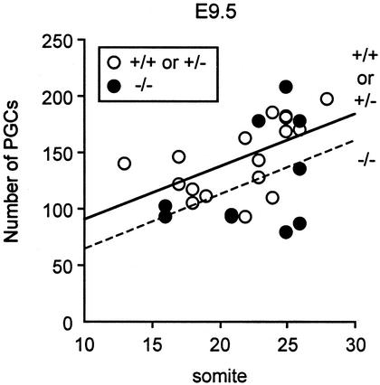
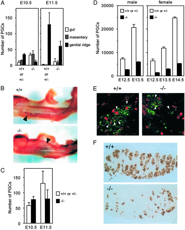
Primordial germ cells (PGCs) are the founders of sperm or oocytes. PGCs migrate through the tissues of the embryos and colonize the gonads during development. However, the cytokines essential for colonization of the gonads by PGCs in mammals remain unclear. Stromal cell-derived factor-1 (SDF-1, also called PBSF and CXCL12) is a member of chemokines, a family of structurally related chemoattractive cytokines. SDF-1 and its primary physiologic receptor CXCR4 have multiple essential functions in development including colonization of bone marrow by hematopoietic cells and neuron localization within cerebellum during embryogenesis as well as B lymphopoiesis and cardiovasculogenesis. Here, we have shown that PGCs have cell-surface expression of CXCR4 and that, in SDF-1(-/-) mice, PGCs undergo directed migration through tissues of embryos, but the numbers of PGCs in the gonads are significantly reduced. The proliferation of PGCs within the gonads seems normal in the mutant mice. These findings reveal the essential role for SDF-1 in murine PGC development likely by controlling colonization of the gonads by PGCs.


Figures

Figure 1
Figure 1
Relatively normal numbers of PGCs in the route of migration in SDF-1−/− embryos at E9.5. Linear regression analysis of PGC numbers (counted in whole-mount) vs. somite numbers in control and SDF-1−/− embryos at E9.5.
Figure 2
Figure 2
SDF-1−/− embryos have defective colonization of the gonads by PGCs. (A) The location of PGCs in control and SDF-1−/− embryos at E10.5 and E11.5. The numbers of PGCs in hindgut endoderm, hindgut mesentery, and the genital ridges in control and SDF-1−/− embryos. (B) Whole-mount alkaline phosphatase staining in wild-type (Upper) and SDF-1−/− (Lower) embryos at E10.5. PGCs (arrowheads) are found predominantly in the genital ridges in wild type, but PGCs (arrowheads) that remain in hindgut endoderm or mesentery are easily detectable in the mutants. (C) Total numbers of PGCs within the migration route and genital ridges in control and SDF-1−/− embryos at E10.5 and E11.5. (D) The numbers of PGCs in the gonads in control and SDF-1−/− embryos at E12.5, E13.5, and E14.5. (E) Proliferating PGCs in the control and SDF-1−/− gonads at E12.5. Fluorescence and immunostaining microscopic analysis with antibodies to BrdUrd (red) and a PGC-specific marker 4C9 (green). Arrowheads point at BrdUrd-labeled PGCs. (F) Immunohistochemistry with antibodies to PGC-specific marker 4C9 on paraffin sections of the male E13.5 gonads from control and SDF-1−/− embryos.
Figure 3
Figure 3
Expression of CXCR4 and SDF-1 in murine PGCs or the developing genital ridge. (A) Flow cytometric analysis of cell-surface expression of CXCR4 on PGCs at E11.5 and E13.5, using transgenic mice expressing GFP in PGCs. GFP+ PGCs have significant cell-surface expression of CXCR4. Solid lines represent staining with CXCR4. Dashed lines represent background staining with isotype-matched controls. (B) RT-PCR analysis of CXCR4 gene expression with E11.5 or E13.5 PGCs. M, molecular marker; lane 1, E11.5 GFP+ PGCs; lane 2, E13.5 male GFP+ PGCs; lane 3, E11.5 GFP cells; lane 4, pre-B cell clone, DW34 (10). The CXCR4 RT-PCR products of 76 bp are identified. (C) The expression pattern of SDF-1 in the E12.5 gonad (go) and subjacent mesonephric tissue (me) from the embryo in which GFP gene is knocked into the SDF-1 locus.

Similar articles

Cited by

References

    1. Wylie C. Cell. 1999;96:165–174. - PubMed
    1. Lawson K A, Dunn N R, Roelen B A, Zeinstra L M, Davis A M, Wright C V, Korving J P, Hogan B L. Genes Dev. 1999;13:424–436. - PMC - PubMed
    1. Ying Y, Liu X M, Marble A, Lawson K A, Zhao G Q. Mol Endocrinol. 2000;14:1053–1063. - PubMed
    1. Mintz B, Russell E S. J Exp Zool. 1957;134:207–237. - PubMed
    1. Godin I, Deed R, Cooke J, Zsebo K, Dexter M, Wylie C C. Nature. 1991;352:807–809. - PubMed

Publication types