Incidental pancreatic cysts: clinicopathologic characteristics and comparison with symptomatic patients
- PMID: 12686529
- PMCID: PMC4403874
- DOI: 10.1001/archsurg.138.4.427
Incidental pancreatic cysts: clinicopathologic characteristics and comparison with symptomatic patients
Abstract
Hypothesis: Widespread use of computed tomography and ultrasound has led to the identification of increasing numbers of patients with asymptomatic cystic lesions of the pancreas.
Design: Retrospective case series of patients with pancreatic cystic lesions.
Setting: University-affiliated tertiary care referral center.
Patients: Two hundred twelve patients with pancreatic cystic lesions seen in our surgical practice during 5 years (April 1997-March 2002).
Main outcome measures: Presence or absence of symptoms, cyst size and location, cytologic or pathologic diagnosis, surgical treatment, and outcome.
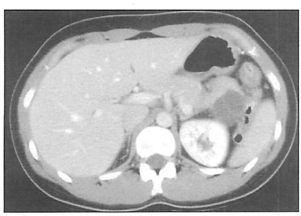
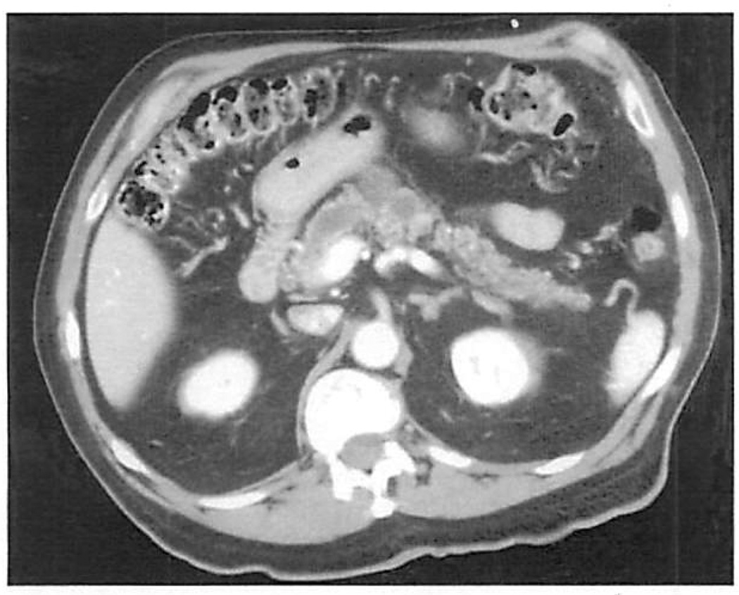
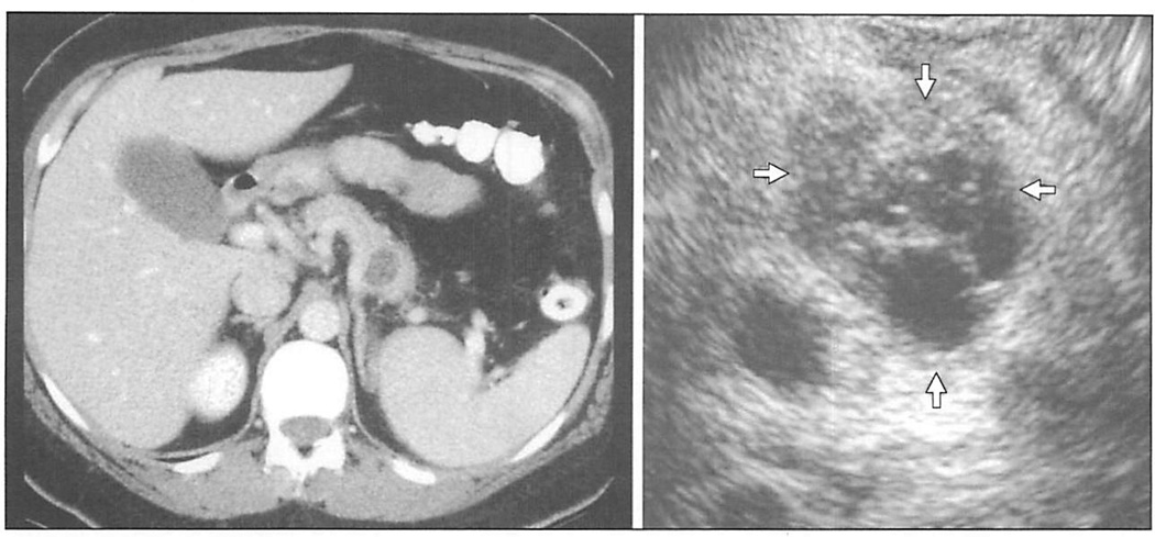
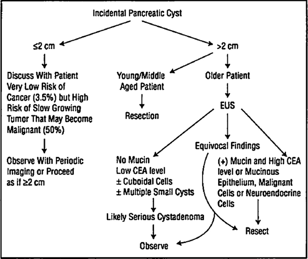
Results: Seventy-eight (36.7%) of 212 patients were asymptomatic. Incidental cysts were smaller (3.3 +/- 1.9 vs 4.6 +/- 2.7 cm; P<.001) and were found in older patients (65 +/- 13 vs 56 +/- 15 years; P<.001). Seventy-eight percent of the asymptomatic patients and 87% of those with symptoms underwent surgery, with a single operative death in the entire group (0.5%). Seventeen percent of asymptomatic cysts were serous cystadenomas; 28%, mucinous cystic neoplasms; 27%, intraductal papillary mucinous neoplasms; and 2.5%, ductal adenocarcinomas. The respective numbers for symptomatic cysts were 7%, 16%, 40%, and 9%. Ten percent of asymptomatic patients had a variety of other cystic lesions, and in 12%, no definitive cytologic or pathologic diagnosis was obtained. Overall, 17% of asymptomatic patients had in situ or invasive cancer, and 42% had a premalignant lesion. When evaluated as a function of size, only 1 (3.5%) of 28 asymptomatic cysts smaller than 2 cm had cancer compared with 13 (26%) of 50 cysts larger than 2 cm (P =.04). The proportion of premalignant lesions, however, remained high in both groups (46% and 38%, respectively). Pseudocysts comprised only 3.8% of asymptomatic cysts compared with 19.4% of symptomatic cysts (P =.003).
Conclusions: Incidental pancreatic cysts are common, occur in older patients, are smaller than symptomatic cysts, and are unlikely to be pseudocysts. More than half of them are either malignant or premalignant lesions and therefore cannot be dismissed.
Figures
References
-
- Gaines PA, Sampson MA. The prevalence and characterization of simple hepatic cysts by ultrasound examination. Br J Radiol. 1989;62:335–337. - PubMed
-
- Terada N, Ichioka K, Matsuta Y, Okubo K, Yoshimura K, Arai Y. The natural history of simple renal cysts. J Urol. 2002;167:21–23. - PubMed
-
- Thompson LDR, Becker RC, Prygodzki RM, Adair CF, Heffess CS. Mucinous cystic neoplasm (mucinous cystadenocarcinoma of low-grade malignant potential) of the pancreas: a clinicopathological study of 130 cases. Am J Surg Pathol. 1999;23:1–16. - PubMed
-
- Loftus EV, Olivares-Pakzad BA, Batts KP, et al. Intraductal papillary-mucinous tumors of the pancreas: clinicopathologic features, outcome, and nomenclature. Gastroentemlogy. 1996;110:1909–1918. - PubMed
Publication types
MeSH terms
Grants and funding
LinkOut - more resources
Full Text Sources
Medical
Miscellaneous