. 2003 Apr;14(4):1295-307.
doi: 10.1091/mbc.e02-07-0406.

ADP-ribosylation factor 6 and a functional PIX/p95-APP1 complex are required for Rac1B-mediated neurite outgrowth

Affiliations

ADP-ribosylation factor 6 and a functional PIX/p95-APP1 complex are required for Rac1B-mediated neurite outgrowth

Chiara Albertinazzi et al. Mol Biol Cell. 2003 Apr.

Abstract

The mechanisms coordinating adhesion, actin organization, and membrane traffic during growth cone migration are poorly understood. Neuritogenesis and branching from retinal neurons are regulated by the Rac1B/Rac3 GTPase. We have identified a functional connection between ADP-ribosylation factor (Arf) 6 and p95-APP1 during the regulation of Rac1B-mediated neuritogenesis. P95-APP1 is an ADP-ribosylation factor GTPase-activating protein (ArfGAP) of the GIT family expressed in the developing nervous system. We show that Arf6 has a predominant role in neurite extension compared with Arf1 and Arf5. Cotransfection experiments indicate a specific and cooperative potentiation of neurite extension by Arf6 and the carboxy-terminal portion of p95-APP1. Localization studies in neurons expressing different p95-derived constructs show a codistribution of p95-APP1 with Arf6, but not Arf1. Moreover, p95-APP1-derived proteins with a mutated or deleted ArfGAP domain prevent Rac1B-induced neuritogenesis, leading to PIX-mediated accumulation at large Rab11-positive endocytic vesicles. Our data support a role of p95-APP1 as a specific regulator of Arf6 in the control of membrane trafficking during neuritogenesis.


Figures
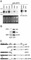
Figure 1
Figure 1
Expression of p95-APP1 in neural cells. (A) Northern blot analysis (top) on total RNA (bottom) prepared from different chick E10 tissues, from E6 retina, and from CEFs. The filter was incubated with a cDNA probe specific for p95-APP1. Then 20 μg of total RNA was loaded in each lane. (B) Characterization of the endogenous p95-APP1 complex in E6 neural retina. A lysate from E6 neural retinas was incubated with agarose beads bound to GST-Rac1B loaded with GDP-β-S (left lane) or GTPγS (right lane). Eluates were immunoblotted with anti-p95-APP1 (top), anti-PIX (middle), and anti-paxillin antibodies (bottom). (C) P95-APP1–derived constructs used in this study. ANK, three ankyrin repeats; LZ, coiled coil region with leucine zipper; PBS, paxillin binding subdomain.
Figure 2
Figure 2
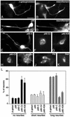
Effect of p95-APP1 ArfGAP mutants on neuritogenesis and localization at large vesicles. (A–K) Localization of p95-derived constructs expressed in E6 retinal neurons. Expression of full-length p95 (C) and p95-C (E) showed a diffuse distribution of the proteins along the neurites. Expression of either the truncated p95-C2 (F and H–K) or the p95-K39 mutant (G) inhibited neuritogenesis and induced the accumulation of the transfected proteins at large cytopalsmic vesicles. Endogenous paxillin (I), overexpressed PIX (J), and overexpressed PAK (K) colocalized with p95-C2 at the large endocytic vesicles. Bar, 10 μm (A–E); 5 μm (F and G); and 7 μm (H–K). (L) Effects of the expression of different p95-APP1 mutants on neuritogenesis. Data are expressed as percentage of neurofilament-positive neurons with no, short, or long neurites, as detailed in MATERIALS AND METHODS. Control neurons were transfected with β-galactosidase. Values are means ± SD from two experiments.
Figure 3
Figure 3
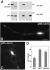
Expression of the p95-ΔSHD mutant in retinal neurons. (A) COS-7 cells were cotransfected to express wild-type βPIX together with either full-length FLAG-p95-APP1 (A) or with FLAG-p95-ΔSHD (B). Lysates (LT-A and LT-B) were first immunoprecipitated with preimmune serum (c-A and c-B); the unbound material was then immunoprecipitated with anti-PIX immune serum (IP-A and IP-B). Samples were prepared in duplicate. One set was used for immunoblotting with anti-FLAG mAb (top filters); the second set was used for immunoblotting with the anti-PIX pAb (bottom filters). (B and C) Retinal neurons were transfected with pFLAG-p95-ΔSHD and treated for immunofluorescence with the anti-FLAG mAb. (D) Quantitation of the total neurite length/neuron in retinal neurons transfected with β-galactosidase, p95-C, or pFLAG-p95-ΔSHD. Quantitation was done on 30 neurons for each type of transfectant. Bars, SEM.
Figure 4
Figure 4
Specific localization of p95-C2 at large Rab11-positive vesicles. Neurons expressing p95-C2 were costained with antibodies against markers for distinct intracellular compartments. P95-C2 colocalized with the recycling endosomal marker Rab11 (A and B), whereas no colocalization was evident with the early endosomal marker EEA1 (D and E), the lysosomal marker LEP100 (F and G), and the Golgi marker βCOP (H–I). In C is shown the normal appearance of the Rab11 compartment in untransfected retinal neurons. Bar, 5 μm.
Figure 5
Figure 5
Wild-type Arf6 is required for neuritogenesis from retinal neurons. E6 retinal neurons were transfected with wild-type (A), GTP-binding–defective (B), or GTP-hydrolysis–defective (C) Arf1, Arf5, or Arf6 GTPases. Data are expressed as percentage of transfected, neurofilament-positive neurons with no, short, or long neurites, as detailed in MATERIALS AND METHODS. Control neurons were transfected with β-galactosidase. Values are means ± SD from two experiments. (D) Effects on neuritogenesis of the coexpression of p95-C with wild-type or mutant Arf proteins. Quantitation was performed by evaluating the total neurite length/cell in 30 neurons for each experimental condition. Bars, SEM.
Figure 6
Figure 6
Arf6 and Arf5 colocalize with p95-N. Neurons were cotransfected with the amino-terminal p95-N construct together with N27Arf6 (A and B), N31Arf1 (C and D), or N31Arf5 (E and F). Analysis by confocal fluorescence microscopy of cells fixed one day after transfection shows colocalization of 95-N with N27Arf6 and N31Arf5 (arrows), but not with N31Arf1. (G) Distribution of N31Arf5 in neurons transfected with N31Arf5 only. Bar, 5 μm.
Figure 7
Figure 7
Arf6 colocalizes with p95-C2. (A–C) Neurons were transfected with wild-type Arf6, or Arf1, and analyzed by immunofluorescence 1 d after transfection. In contrast to the diffuse distribution of Arf6 (A), Arf1 (B) colocalized with the endogenous Golgi marker βCOP (C). In retinal cells cotransfected with wild-type Arf proteins and p95-C2, a clear colocalization at large vesicles of wild-type Arf6 with p95-C2 could be observed (D and E). In contrast, neither wild-type Arf1 (F and G) nor wild-type Arf5 (H and I) colocalized with p95-C2–positive large vesicles. (J and K) distribution of overexpressed L67Arf6 in retinal neurons. Bars, 5 μm.
Figure 8
Figure 8
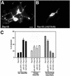
N27Arf6- and ArfGAP-deficient p95-C2 inhibit Rac1B-enhanced neurite branching. Enhanced neuritogenesis in E6 retinal neurons overexpressing wild-type Rac1B alone (A) was inhibited by coexpression of the GTP-binding–defective N27Arf6 mutant (B). Bar, 5 μm. (C) Quantitation ± SD of the effects of the expression of the indicated constructs on neurite branching; for each experimental condition, branching was evaluated in a total of 100 neurons from two independent experiments.

Similar articles

Cited by

  • CRL5-dependent regulation of the small GTPases ARL4C and ARF6 controls hippocampal morphogenesis.
    Han JS, Hino K, Li W, Reyes RV, Canales CP, Miltner AM, Haddadi Y, Sun J, Chen CY, La Torre A, Simó S. Han JS, et al. Proc Natl Acad Sci U S A. 2020 Sep 15;117(37):23073-23084. doi: 10.1073/pnas.2002749117. Epub 2020 Sep 1. Proc Natl Acad Sci U S A. 2020. PMID: 32873638 Free PMC article.
  • G-protein-coupled receptor kinase-interacting proteins inhibit apoptosis by inositol 1,4,5-triphosphate receptor-mediated Ca2+ signal regulation.
    Zhang S, Hisatsune C, Matsu-Ura T, Mikoshiba K. Zhang S, et al. J Biol Chem. 2009 Oct 16;284(42):29158-69. doi: 10.1074/jbc.M109.041509. Epub 2009 Aug 25. J Biol Chem. 2009. PMID: 19706611 Free PMC article.
  • Arf6 as a therapeutic target: Structure, mechanism, and inhibitors.
    Sun D, Guo Y, Tang P, Li H, Chen L. Sun D, et al. Acta Pharm Sin B. 2023 Oct;13(10):4089-4104. doi: 10.1016/j.apsb.2023.06.008. Epub 2023 Jun 14. Acta Pharm Sin B. 2023. PMID: 37799386 Free PMC article. Review.
  • WAVE regulatory complex activation by cooperating GTPases Arf and Rac1.
    Koronakis V, Hume PJ, Humphreys D, Liu T, Hørning O, Jensen ON, McGhie EJ. Koronakis V, et al. Proc Natl Acad Sci U S A. 2011 Aug 30;108(35):14449-54. doi: 10.1073/pnas.1107666108. Epub 2011 Aug 15. Proc Natl Acad Sci U S A. 2011. PMID: 21844371 Free PMC article.
  • Genome-wide haplotype association study identifies the FRMD4A gene as a risk locus for Alzheimer's disease.
    Lambert JC, Grenier-Boley B, Harold D, Zelenika D, Chouraki V, Kamatani Y, Sleegers K, Ikram MA, Hiltunen M, Reitz C, Mateo I, Feulner T, Bullido M, Galimberti D, Concari L, Alvarez V, Sims R, Gerrish A, Chapman J, Deniz-Naranjo C, Solfrizzi V, Sorbi S, Arosio B, Spalletta G, Siciliano G, Epelbaum J, Hannequin D, Dartigues JF, Tzourio C, Berr C, Schrijvers EM, Rogers R, Tosto G, Pasquier F, Bettens K, Van Cauwenberghe C, Fratiglioni L, Graff C, Delepine M, Ferri R, Reynolds CA, Lannfelt L, Ingelsson M, Prince JA, Chillotti C, Pilotto A, Seripa D, Boland A, Mancuso M, Bossù P, Annoni G, Nacmias B, Bosco P, Panza F, Sanchez-Garcia F, Del Zompo M, Coto E, Owen M, O'Donovan M, Valdivieso F, Caffarra P, Scarpini E, Combarros O, Buée L, Campion D, Soininen H, Breteler M, Riemenschneider M, Van Broeckhoven C, Alpérovitch A, Lathrop M, Trégouët DA, Williams J, Amouyel P. Lambert JC, et al. Mol Psychiatry. 2013 Apr;18(4):461-70. doi: 10.1038/mp.2012.14. Epub 2012 Mar 20. Mol Psychiatry. 2013. PMID: 22430674 Free PMC article.

References

    1. Adler R, Jerdan J, Hewitt AT. Responses of cultured neural retinal cells to substratum-bound laminin and other extracellular matrix molecules. Dev Biol. 1985;112:100–114. - PubMed
    1. Albertinazzi C, Gilardelli D, Paris S, Longhi R, de Curtis I. Overexpression of a neural-specific Rho family GTPase, cRac1B, selectively induces enhanced neuritogenesis and neurite branching in primary neurons. J Cell Biol. 1998;142:815–825. - PMC - PubMed
    1. Bernard V, Pohl BP, Bokoch GM. Characterization of Rac and Cdc42 activation in chemoattractant-stimulated human neutrophils using a novel assay for active GTPases. J Biol Chem. 1999;274:13198–13204. - PubMed
    1. Chomczynski P, Sacchi N. Single-step method of RNA isolation by acid guanidinium thiocyanate-phenol-chloroform extraction. Anal Biochem. 1987;162:156–159. - PubMed
    1. Craig AM, Wyborski RJ, Banker G. Preferential addition of newly synthesized membrane protein at axonal growth cones. Nature. 1995;375:592–594. - PubMed

Publication types

MeSH terms