. 2003 Apr;14(4):1418-32.
doi: 10.1091/mbc.e02-04-0235.

Extracellular signal-regulated kinase mediates phosphorylation of tropomyosin-1 to promote cytoskeleton remodeling in response to oxidative stress: impact on membrane blebbing

Affiliations

Extracellular signal-regulated kinase mediates phosphorylation of tropomyosin-1 to promote cytoskeleton remodeling in response to oxidative stress: impact on membrane blebbing

François Houle et al. Mol Biol Cell. 2003 Apr.

Abstract

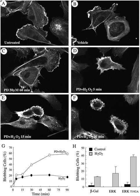
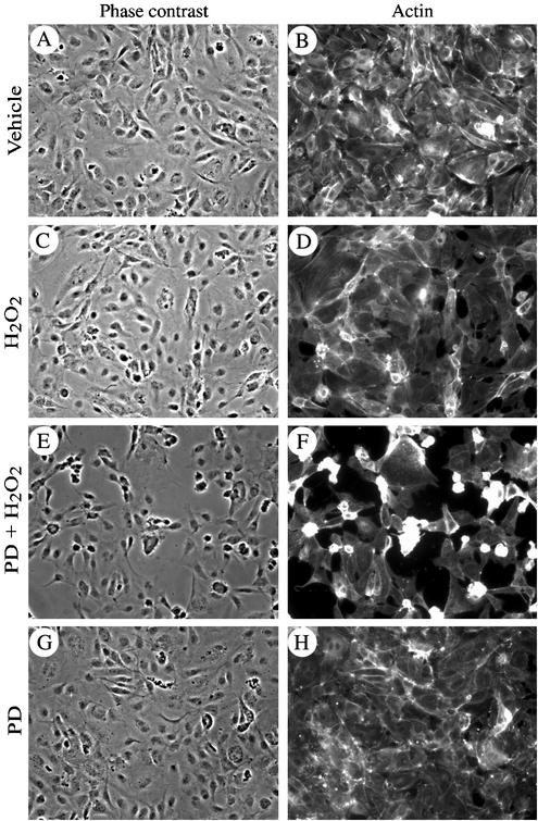
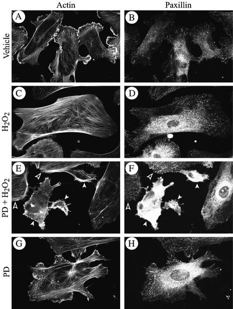
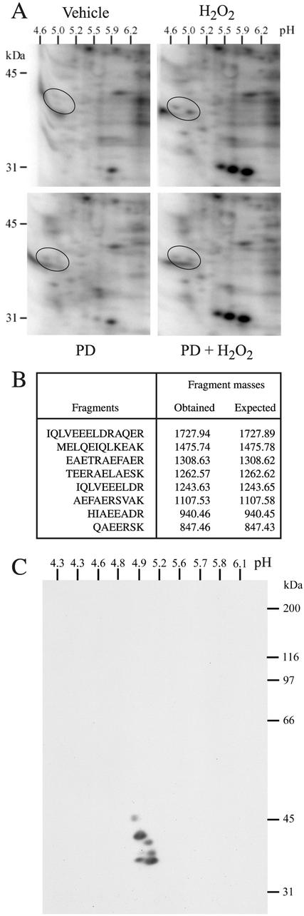
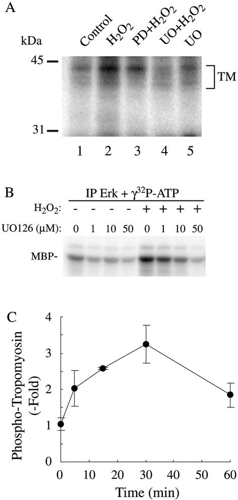
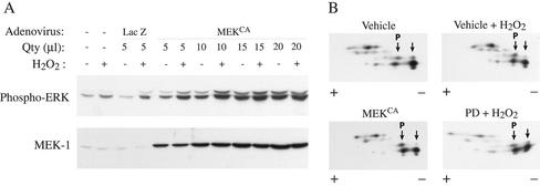
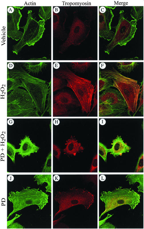
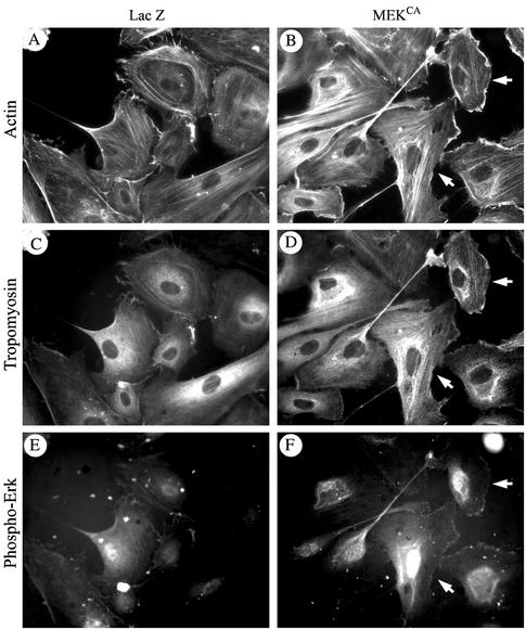
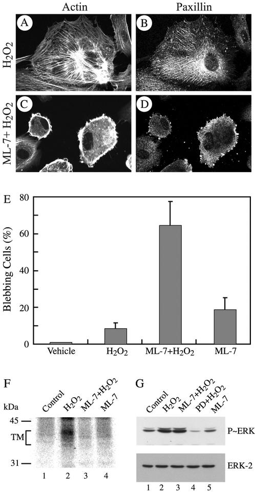
Oxidative stress induces in endothelial cells a quick and transient coactivation of both stress-activated protein kinase-2/p38 and extracellular signal-regulated kinase (ERK) mitogen-activated protein kinases. We found that inhibiting the ERK pathway resulted, within 5 min of oxidative stress, in a misassembly of focal adhesions characterized by mislocalization of key proteins such as paxillin. The focal adhesion misassembly that followed ERK inhibition with the mitogen-activated protein kinase kinase (MEK) inhibitor PD098059 (2'-amino-3'-methoxyflavone) or with a kinase negative mutant of ERK in the presence of H(2)O(2) resulted in a quick and intense membrane blebbing that was associated with important damage to the endothelium. We isolated by two-dimensional gel electrophoresis a PD098059-sensitive phosphoprotein of 38 kDa that we identified, by mass spectrometry, as tropomyosin-1. In fact, H(2)O(2) induced a time-dependent phosphorylation of tropomyosin that was sensitive to inhibition by PD098059 and UO126 (1,4-diamino-2,3-dicyano-1,4-bis[2-aminophenylthio]butanediane). Tropomyosin phosphorylation was also induced by expression of a constitutively activated form of MEK1 (MEK(CA)), which confirms that its phosphorylation resulted from the activation of ERK. In unstimulated cells, tropomyosin-1 was found diffuse in the cells, whereas it quickly colocalized with actin and stress fibers upon stimulation of ERK by H(2)O(2) or by expression of MEK(CA). We propose that phosphorylation of tropomyosin-1 downstream of ERK by contributing to formation of actin filaments increases cellular contractility and promotes the formation of focal adhesions. Incidentally, ML-7 (1-[5iodonaphthalene-1-sulfonyl]homopiperazine, HCl), an inhibitor of cell contractility, inhibited phosphorylation of tropomyosin and blocked the formation of stress fibers and focal adhesions, which also led to membrane blebbing in the presence of oxidative stress. Our finding that tropomyosin-1 is phosphorylated downstream of ERK, an event that modulates its interaction with actin, may lead to further understanding of the role of this protein in regulating cellular functions associated with cytoskeletal remodeling.


Figures

Figure 1
Figure 1
Inhibition of ERK MAP kinase in the presence of H2O2 leads to early membrane blebbing. Exponentially growing HUVECs were either untreated (A) or treated with vehicle alone (0.25% DMSO, 60 min; B) or they were pretreated with the MEK inhibitor PD098059 (50 μM) for 60 min and treated or not (C) with 250 μM H2O2 for 5 min (D), 15 min (E), or 30 min (F). In G, exponentially growing HUVECs were pretreated with vehicle (0.25% DMSO, 60 min; filled circles) or with PD098059 (50 μM, 60 min; unfilled circles) and then were treated with 250 μM H2O2 for increasing periods of times. Blebbing cells were visualized by fluorescence microscopy, counted, and means from two separate experiments were calculated. In H, PAECs were transfected with vector expressing either β-galactosidase, wild-type ERK1 MAP kinase fused to HA tag, or kinase dead ERK1T192A MAP kinase fused to HA tag. To make sure that MAP kinase kinase upstream of ERK was not limiting, transfection with ERK1 was supplemented with vector-expressing wild-type MEK1. Cells were treated (gray bars) or not (black bars) with 500 μM H2O2 for 60 min. Blebbing HA-stained cells were visualized by fluorescence microscopy, counted, and means from two separate experiments were calculated. Membrane blebbing has been quantitated by counting the number of blebbing cells over a total of 500 cells counted in different representative fields. In each experiment, cells were fixed, permeabilized, and stained for F-actin by using FITC-phalloidin and for HA tag by using 12Ca5 antibody (H only) as described in MATERIALS AND METHODS.
Figure 2
Figure 2
Inhibition of ERK MAP kinase in the presence of H2O2 is associated with disruption of the endothelial layer. HUVECs (4 × 10⁴) were plated in Lab-Tek chambers and grown to confluence (72 h). They were then pretreated with vehicle (0.25% DMSO, 60 min; A–D) or with PD098059 (50 μM, 60 min; E–H) and thereafter were treated or not (A, B, G, and H) with 250 μM H2O2 for 30 min (C–F). After treatments, cells were fixed, permeabilized, and stained for F-actin by using FITC-phalloidin as described in MATERIALS AND METHODS. Endothelial layers were visualized at 20× magnification by phase contrast (A, C, E, and G) and by fluorescence microscopy after staining for F-actin (B, D, F, and H).
Figure 3
Figure 3
Early membrane blebbing is associated with focal adhesion misassembly. Exponentially growing HUVECs were pretreated with vehicle (0.25% DMSO, 60 min; A–D) or with PD059098 (50 μM, 60 min; E–H), and then they were treated or not (A, B, G, and H) with 250 μM H2O2 for 5 min; (C–F). After treatments, cells were fixed, permeabilized, and stained for F-actin by using FITC-phalloidin and for paxillin by using anti-paxillin monoclonal antibody as described in MATERIALS AND METHODS. Open arrowheads show intact cells and white arrow heads show cells that begin to bleb.
Figure 4
Figure 4
MALDI-TOF identification of tropomyosin-1 as a downstream target of ERK. (A) Exponentially growing HUVECs were labeled with H3[32P]O4 and were pretreated with vehicle (0.25% DMSO, 60 min; top) or with PD098059 (50 μM, 60 min; bottom). They were then treated or not (left) with 250 μM H2O2 for 15 min (right). Proteins were extracted and run into IEF (pH 4–6) gels and in a second dimension into 8.5% SDS-PAGE. Representative autoradiograms from four separate experiments are shown. (B) In a separate experiment, spots circled in A were excised and processed for MALDI-TOF analysis. Database searching identified the protein as tropomyosin-1 with eight matching peptides that represent 23% sequence coverage. In C, exponentially growing HUVECs were extracted and processed for 2D electrophoresis as in A. The gel was transferred on nitrocellulose membrane and processed for immunodetection by using a monoclonal anti-tropomyosin antibody that cross-reacted with the different isoforms of tropomyosin.
Figure 5
Figure 5
H2O2 induces a time-dependent phosphorylation of tropomyosin that is inhibited by inhibitors of the ERK pathway. (A) Exponentially growing HUVECs were labeled with H3[32P]O4 and were pretreated simultaneously with the phosphatase inhibitor NaF (1 mM) and with vehicle (0.25% DMSO, 60 min; lanes 1 and 2), or with PD098059 (50 μM, 60 min; lane 3) or with UO126 (50 μM, 60 min; lanes 4 and 5) and treated for 30 min with 250 μM H2O2 (lanes 2–4). Proteins were extracted, immunoprecipitated using anti-tropomyosin antibody, and antigen–antibody complexes were run into 8.5% SDS-PAGE. Representative autoradiogram from two separate experiments is shown. (B) Exponentially growing HUVECs were pretreated with vehicle (0.25% DMSO, 60 min) or for 60 min with increasing concentration of MEK inhibitor UO126 and treated or not with 500 μM H2O2 for 30 min. After treatment, cells were extracted and processed for ERK kinase assay using myelin basic protein as substrate as described in MATERIALS AND METHODS. (C) Exponentially growing HUVECs were labeled with H3[32P]O4 and were pretreated with vehicle (0.25% DMSO, 60 min) along with phosphatase inhibitor NaF (1 mM) and were treated for increasing time with 250 μM H2O2. Proteins were extracted and immunoprecipitated using mouse monoclonal anti-tropomyosin antibody. Antigen–antibody complexes were run into 8.5% SDS-PAGE, and tropomyosin bands were quantified using PhosphorImager. Means with error bars from three separate experiments are shown.
Figure 6
Figure 6
Tropomyosin is phosphorylated in cells in which ERK is activated by expression of a constitutive activated form of MEK1. (A) Exponentially growing HUVECs were infected or not with varying quantity of adenoviral vector carrying β-galactosidase or MEKCA. Twenty-four hours later, cells were treated with 250 μM H2O2. Proteins were extracted, run into 8.5% SDS-PAGE, and transferred on nitrocellulose membrane. Immunodectections with monoclonal mouse anti-phospho-ERK antibody and rabbit polyclonal anti-MEK1 #9 antibody are shown. (B) Exponentially growing HUVECs were pretreated or not with vehicle (0.25% DMSO, 60 min) or with PD098059 (50 μM, 60 min; PD) and were treated or not for 30 min with 250 μM H2O2. Bottom left, exponentially growing HUVECs were infected with 15 μl of adenoviral vector carrying MEKCA. Proteins were extracted in IPG buffer (pH 4.5–5.5) and separated on IPGPhor as described in MATERIALS AND METHODS. IEF gels were run into 8.5% SDS-PAGE for the second dimension. The gel was transferred on nitrocellulose membrane and processed for immunodetection with monoclonal anti-tropomyosin antibody that cross-reacted with the different isoforms of tropomyosin. Shift of the relevant tropomyosin-1 spots toward the acidic forms (P, phosphorylated) is indicated by arrows. Representative autoradiogram from two separate experiments is shown.
Figure 7
Figure 7
Phosphorylation of tropomyosin-1 induces its colocalization with F-actin. Exponentially growing HUVECs were pretreated with vehicle (0.25% DMSO, 60 min; A–F) or with PD098059 (50 μM, 60 min; G–L), and then they were treated or not (A–C and J–L) with 250 μM H2O2 for 5 min (D–I). After treatments, cells were fixed, permeabilized, and stained for F-actin by using FITC-phalloidin and for tropomyosin by using anti-tropomyosin monoclonal antibody as described in MATERIALS AND METHODS. F-actin staining (A, D, G, and J), tropomyosin staining (B, E, H, and K and merged picture (C, F, I, and L) are shown. In merged picture, the yellow areas represent colocalization of both proteins. Representative fields from two separate experiments are shown.
Figure 8
Figure 8
Phosphorylation of tropomyosin-1 by expression of MEKCA induces stress fiber formation and colocalization with F-actin. Exponentially growing HUVECs were infected with adenoviral vector carrying β-galactosisdase or MEKCA. Forty-eight hours later, cells were fixed, permeabilized, and stained for F-actin by using rhodamine-phalloidin, for tropomyosin by using anti-tropomyosin monoclonal antibody, and for phospho-ERK (to monitor MEKCA-expressing cells) by using anti-phospho-ERK polyclonal antibody. F-actin staining (A and B), tropomyosin staining (C and D), and phospho-ERK staining (E and F) are shown. Arrows indicate that cells that stain more strongly for phospho-ERK also stain more strongly for both fibrillar actin and tropomyosin. Representative fields from two separate experiments are shown.
Figure 9
Figure 9
Inhibition of cellular contractility with ML-7 induces early membrane blebbing. Exponentially growing HUVECs were pretreated with vehicle (0.25% DMSO, 60 min; A and B) or with MLCK inhibitor ML-7 (25 μM, 60 min; C and D) and treated with 250 μM H2O2 for 5 min (A–D). After treatments, cells were fixed, permeabilized and stained for F-actin and paxillin as in Figure 3. In E, exponentially growing HUVECs were pretreated with vehicle (0.25% DMSO, 60 min) or with MLCK inhibitor ML-7 (25 μM, 60 min) and treated or not with 250 μM H2O2 for 30 min. After treatments, cells were processed as in A–D. Blebbing cells were counted, as in Figure 1, and mean from three separate experiments was calculated. (F) Exponentially growing HUVECs were labeled with H3[32P]O4 and were pretreated simultaneously with the phosphatase inhibitor NaF (1 mM) and with vehicle (0.25% DMSO, 60 min; lanes 1 and 2), or with ML-7 (25 μM, 60 min; lanes 3 and 4) before being treated or not (lanes 1 and 4) for 30 min with 250 μM H2O2 (lanes 2 and 3). Proteins were extracted, immunoprecipitated using anti-tropomyosin antibody, and antigen–antibody complexes were run into 8.5% SDS-PAGE. Representative autoradiogram from two separate experiments is shown. (G) Exponentially growing HUVECs were pretreated with vehicle (0.25% DMSO, 60 min; lanes 1 and 2), or with ML-7 (25 μM, 60 min; lanes 3 and 5), or with PD098059 (50 μM, 60 min; lane 4) before being treated or not (lanes 1 and 5) for 5 min with 250 μM H2O2 (lanes 2, 3, and 4). Proteins were extracted, were run into 10% SDS-PAGE, and transferred on nitrocellulose membrane. Immunodectections with mouse monoclonal anti-phospho-ERK antibody (top) and rabbit polyclonal anti-ERK antibody (bottom) are shown.

Similar articles

Cited by

References

    1. Amano M, Ito M, Kimura K, Fukata Y, Chihara K, Nakano T, Matsuura Y, Kaibuchi K. Phosphorylation and activation of myosin by Rho-associated kinase (Rho-kinase) J Biol Chem. 1996;271:20246–20249. - PubMed
    1. Becker LC, Ambrosio G. Myocardial consequences of reperfusion. Prog Cardiovasc Dis. 1987;30:23–44. - PubMed
    1. Chrzanowska-Wodnicka M, Burridge K. Rho-stimulated contractility drives the formation of stress fibers and focal adhesions. J Cell Biol. 1996;133:1403–1415. - PMC - PubMed
    1. Coleman ML, Sahai EA, Yeo M, Bosch M, Dewar A, Olson MF. Membrane blebbing during apoptosis results from caspase-mediated activation of ROCK I. Nat Cell Biol. 2001;3:339–345. - PubMed
    1. Cooper JA. Actin dynamics: tropomyosin provides stability. Curr Biol. 2002;12:R523–R525. - PubMed

Publication types

LinkOut - more resources