. 2003 Apr;14(4):1448-59.
doi: 10.1091/mbc.e02-06-0342.

Autoinhibition of Jak2 tyrosine kinase is dependent on specific regions in its pseudokinase domain

Affiliations

Autoinhibition of Jak2 tyrosine kinase is dependent on specific regions in its pseudokinase domain

Pipsa Saharinen et al. Mol Biol Cell. 2003 Apr.

Abstract

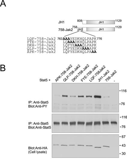
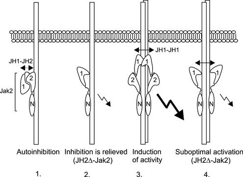
Jak tyrosine kinases have a unique domain structure containing a kinase domain (JH1) adjacent to a catalytically inactive pseudokinase domain (JH2). JH2 is crucial for inhibition of basal Jak activity, but the mechanism of this regulation has remained elusive. We show that JH2 negatively regulated Jak2 in bacterial cells, indicating that regulation is an intrinsic property of Jak2. JH2 suppressed basal Jak2 activity by lowering the V(max) of Jak2, whereas JH2 did not affect the K(m) of Jak2 for a peptide substrate. Three inhibitory regions (IR1-3) within JH2 were identified. IR3 (residues 758-807), at the C terminus of JH2, directly inhibited JH1, suggesting an inhibitory interaction between IR3 and JH1. Molecular modeling of JH2 showed that IR3 could form a stable alpha-helical fold, supporting that IR3 could independently inhibit JH1. IR2 (725-757) in the C-terminal lobe of JH2, and IR1 (619-670), extending from the N-terminal to the C-terminal lobe, enhanced IR3-mediated inhibition of JH1. Disruption of IR3 either by mutations or a small deletion increased basal Jak2 activity, but abolished interferon-gamma-inducible signaling. Together, the results provide evidence for autoinhibition of a Jak family kinase and identify JH2 regions important for autoregulation of Jak2.


Figures

Figure 1
Figure 1
Autoinhibition of Jak2 in E. coli. Gst-fusion proteins for JH1 (JH1-Gst) and JH1–2 (JH1–2-Gst) domains of Jak2 were expressed in bacterial cells as described in MATERIALS AND METHODS. The cells were lysed by boiling in reducing Laemmli sample buffer. Lysates were separated in 7.5% SDS-PAGE, and analyzed in anti-HA (right) and anti-phosphotyrosine (left) immunoblots. Arrows indicate the migration of JH1-Gst and JH1–2-Gst. The mobilities of the molecular mass markers (in kilodaltons) are shown on the right.
Figure 2
Figure 2
Kinetic analysis of the catalytic activity of Jak2. (A) Expression plasmids for JH1-HA and JH1–2-HA were transfected into 293T cells, and cell lysates were immunoprecipitated using anti-HA antibody. Aliquots of the immunoprecipitates (left) and cell lysates (right) were separated in 4–15% SDS-PAGE and analyzed in an anti-HA immunoblot. The mobilities of the molecular mass markers (in kilodaltons) are shown on the right. (B) Immunoprecipitated JH1-HA and JH1–2-HA were subjected to in vitro kinase assay by using Jak2-derived peptide (1.2 mM) as a substrate. The reactions were stopped at various time points and the peptides were separated in 20% SDS-PAGE followed by quantification using PhosphorImager. (C) Immunoprecipitated JH1-HA and JH1–2-HA were subjected to in vitro kinase assay by using as substrate a range of different Jak2 peptide concentrations, as indicated. The reaction time was 30 min. The peptides were separated in 20% SDS-PAGE followed by quantification with PhosphorImager. (D) Data from C are shown as percentages of maximal JH1 or JH1–2 activities (the values for JH1 are divided by the highest JH1 value, and the values for JH1–2 are divided by the highest JH1–2 value). In B and C, [γ-33P]ATP was used, and the total concentration of ATP was 250 μM.
Figure 3
Figure 3
Molecular model of the three-dimensional structure of the JH2 domain of human Jak2. Different colors represent sequential deletions of 584-Jak2 (magenta), 619-Jak2 (blue), 671-Jak2 (green), 725-Jak2 (red), and 758-Jak2 (yellow) constructs. Ball and stick representation of indicated amino acids. IR1 (green), IR2 (yellow), and IR3 (gray).
Figure 4
Figure 4
Comparison of the effect of two different JH2 deletions on the activity of Jak2. (A) Schematic presentation of Jak2 constructs. (B) Expression plasmids for Jak2-HA, JH2Δ-HA, and BglIIΔ-HA were transfected into 293T cells and cell lysates were immunoprecipitated using anti-HA antibody. Aliquots of the immunoprecipitates were separated in 7.5% SDS-PAGE and analyzed in anti-phosphotyrosine (top) and anti-HA immunoblots (middle). Aliquots of cell lysates were separated in 7.5% SDS-PAGE and analyzed in anti-HA immunoblot (bottom). (C) Expression plasmids for Jak2-HA, JH2Δ-HA, and BglIIΔ-HA were transfected into 293T cells, and cell lysates were immunoprecipitated using anti-HA antibody. Aliquots of the immunoprecipitates were subjected to in vitro kinase assay by using [γ-32P]ATP and Stat5-derived peptide as a substrate. The peptides were separated in 20% SDS-PAGE followed by quantification using PhosphorImager. Aliquots of the immunoprecipitates (top) and cell lysates (bottom) were also separated in 7.5% SDS-PAGE and analyzed in anti-HA immunoblot. (D) Stat5 expression plasmid was transfected either alone or together with expression plasmids for Jak2-HA, JH2Δ-HA, and BglIIΔ-HA into 293T cells, and cell lysates were immunoprecipitated using anti-Stat5 antibody. Aliquots of the immunoprecipitates were separated in 7.5% SDS-PAGE and analyzed in anti-phosphotyrosine (top) and anti-Stat5 (middle) immunoblots. Cell lysates were separated in 7.5% SDS-PAGE and analyzed by immunoblotting with anti-HA antibody (bottom). The mobilities of the molecular mass markers (in kilodaltons) are shown on the right.
Figure 5
Figure 5
Mapping of the inhibitory region in JH2. (A) Schematic presentation of Jak2 constructs. (B) Stat5 expression plasmid was transfected into 293T cells either alone or together with HA-tagged Jak2, 584-Jak2, 619-Jak2, 671-Jak2, 725-Jak2, or 758-Jak2, as indicated. Cell lysates were immunoprecipitated using anti-Stat5 antibody, and immunoprecipitates were separated in 7.5% SDS-PAGE followed by immunoblotting with anti-phosphotyrosine (top) and anti-Stat5 antibodies (bottom). Cell lysates were separated in 4–15% SDS-PAGE and analyzed by immunoblotting with anti-HA antibody (right). (C) Stat5 expression plasmid was transfected into 293T cells either alone or together with HA-tagged JH1-Jak2 or 758-Jak2. Cell lysates were immunoprecipitated using anti-Stat5 antibody, and immunoprecipitates were separated in 7.5% SDS-PAGE followed by immunoblotting with anti-phosphotyrosine (top) and anti-Stat5 (bottom) antibodies. The mobilities of the molecular mass markers (in kilodaltons) are shown on the right.
Figure 6
Figure 6
Mutations in IR3 increase the activity of Jak2. (A) Schematic presentation of Jak2 constructs. (B) Stat5 expression plasmid was transfected into 293T cells either alone or together with HA-tagged JH1-Jak2, 758-Jak2, LQF-758-Jak2, FYE-758-Jak2, DKH-758-Jak2, QLP-758-Jak2, or APK-758-Jak2, as indicated. Cell lysates were immunoprecipitated using anti-Stat5 antibody, and immunoprecipitates were separated in 7.5% SDS-PAGE followed by immunoblotting with anti-phosphotyrosine (top) and anti-Stat5 antibodies (middle). Cell lysates were separated in 4–15% SDS-PAGE and analyzed by immunoblotting with anti-HA antibody (bottom). The mobilities of the molecular mass markers (in kilodaltons) are shown on the right.
Figure 7
Figure 7
IR3-deletion results in increased basal Jak2 activity and lack of cytokine-inducible signaling. (A) Schematic presentation of Jak2 constructs. (B) Expression plasmids for Jak2-HA, JH2Δ-HA, and 761Δ-HA were transfected into 293T cells, and cell lysates were immunoprecipitated using anti-HA antibody. Aliquots of the immunoprecipitates were separated in 7.5% SDS-PAGE and analyzed in anti-phosphotyrosine (top) and anti-HA immunoblots (middle). Aliquots of cell lysates were separated in 7.5% SDS-PAGE and analyzed in anti-HA immunoblot (bottom). (C) γ2A cells were transfected with Stat1-dependent luciferase reporter vector, pRLTK control vector, and Jak2, JH2Δ-Jak2, AflIIΔ-Jak2, or 761Δ-Jak2 constructs or with empty vector as a control. Five hours after transfection, the cells were changed into serum-free medium and starved for 15 h. The cells were stimulated with IFN-γ (1000 U/ml) for 5 h or left unstimulated. Luciferase activity was measured as described in MATERIALS AND METHODS. Shown is the mean from three independent experiments and the SEs of the mean.
Figure 8
Figure 8
A model for the function of the JH2 domain in activation of Jak2 by cytokine receptors. In the absence of cytokine, Jak2 is autoinhibited through a possibly intramolecular JH2–JH1 interaction (1). Cytokine binding results in receptor aggregation and displacement of the inhibitory JH1–JH1 interaction (2), possibly through engagement of JH1 in a homotypic JH1–JH1 interaction, leading to increased Jak activity. Induction of maximal activity of Jak2 requires a functional JH2 domain, via a still unknown mechanism (3). In the absence of JH2, maximal Jak activity is not achieved by cytokine stimulation (4).

References

    1. Abola EE, Sussman JL, Prilusky J, Manning NO. Protein Data Bank archives of three-dimensional macromolecular structures. Methods Enzymol. 1997;277:556–571. - PubMed
    1. Al-Lazikani B, Sheinerman FB, Honig B. Combining multiple structure and sequence alignments to improve sequence detection and alignment: application to the SH2 domains of Janus kinases. Proc Natl Acad Sci USA. 2001;98:14796–14801. - PMC - PubMed
    1. Andreotti AH, Bunnell SC, Feng S, Berg LJ, Schreiber SL. Regulatory intramolecular association in a tyrosine kinase of the Tec family. Nature. 1997;385:93–97. - PubMed
    1. Candotti F, et al. Structural and functional basis for JAK3-deficient severe combined immunodeficiency. Blood. 1997;90:3996–4003. - PubMed
    1. Chen M, Cheng A, Candotti F, Zhou YJ, Hymel A, Fasth A, Notarangelo LD, O'Shea JJ. Complex effects of naturally occurring mutations in the JAK3 pseudokinase domain: evidence for interactions between the kinase and pseudokinase domains. Mol Cell Biol. 2000;20:947–956. - PMC - PubMed

Publication types

LinkOut - more resources