. 2003 Apr;14(4):1468-78.
doi: 10.1091/mbc.e02-08-0545.

Nestin promotes the phosphorylation-dependent disassembly of vimentin intermediate filaments during mitosis

Affiliations

Nestin promotes the phosphorylation-dependent disassembly of vimentin intermediate filaments during mitosis

Ying-Hao Chou et al. Mol Biol Cell. 2003 Apr.

Abstract

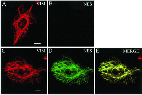
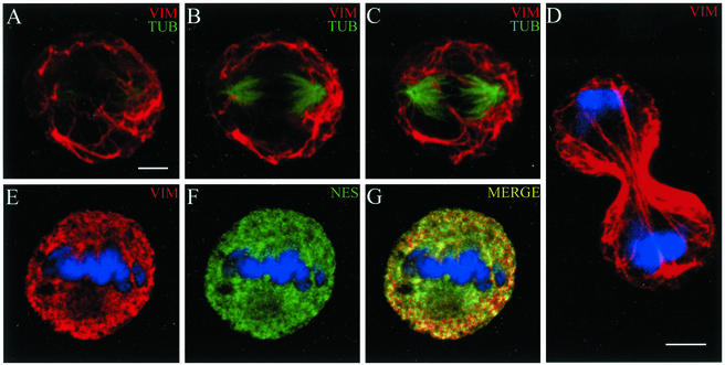
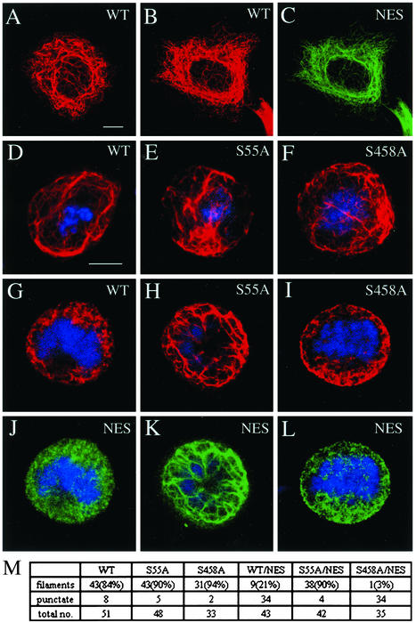
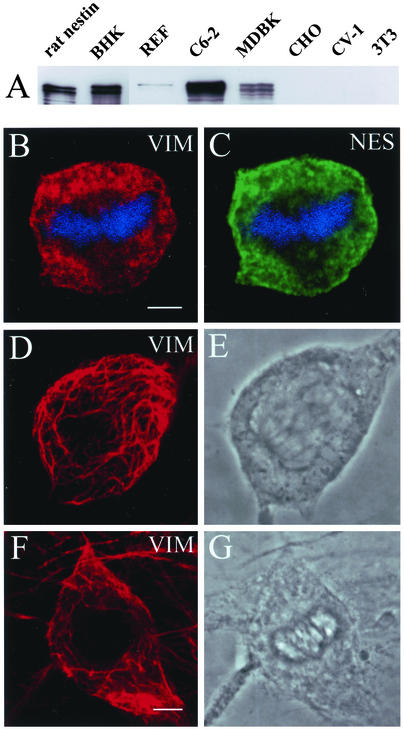
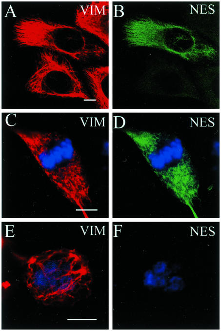
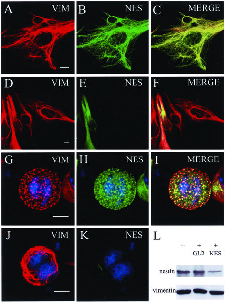
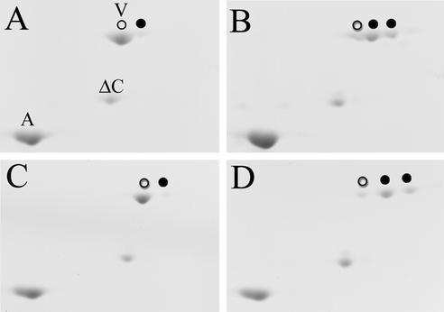
The expression of the intermediate filament (IF) protein nestin is closely associated with rapidly proliferating progenitor cells during neurogenesis and myogenesis, but little is known about its function. In this study, we examine the effects of nestin expression on the assembly state of vimentin IFs in nestin-free cells. Nestin is introduced by transient transfection and is positively correlated with the disassembly of vimentin IFs into nonfilamentous aggregates or particles in mitotic but not interphase cells. This nestin-mediated disassembly of IFs is dependent on the phosphorylation of vimentin by the maturation/M-phase-promoting factor at ser-55 in the amino-terminal head domain. In addition, the disassembly of vimentin IFs during mitosis appears to be a unique feature of nestin-expressing cell types. Furthermore, when the expression of nestin is downregulated by the nestin-specific small interfering RNA in nestin-expressing cells, vimentin IFs remain assembled throughout all stages of mitosis. Previous studies suggest that nonfilamentous vimentin particles are IF precursors and can be transported rapidly between different cytoplasmic compartments along microtubule tracks. On the basis of these observations, we speculate that nestin may play a role in the trafficking and distribution of IF proteins and potentially other cellular factors to daughter cells during progenitor cell division.


Figures

Figure 1
Figure 1
Nestin expression in interphase CHO cells. In untransfected interphase cells, vimentin (VIM, red) is seen in a typical filamentous pattern (A). These cells express no detectable nestin (NES, green) after staining with nestin antibody (B). When nestin is expressed in CHO cells, it is incorporated into the endogenous vimentin IF network with no apparent impact on its assembly state or organization (C–E). Bar, 10 μm.
Figure 2
Figure 2
Nestin expression in mitotic CHO cells. During mitosis, vimentin IFs remain intact and form a filamentous network surrounding the mitotic spindle. The relationship between vimentin IFs (red) and spindle tubulin microtubules (green) in an untransfected mitotic cell is shown in three consecutive optical sections (A–C). The vimentin IFs persist into mid to late cytokinesis (D). In mitotic cells expressing nestin, the endogenous vimentin IFs are disassembled and vimentin (red) and nestin (green) are extensively colocalized in punctate and diffuse structures (E–G). Mitotic cells were identified by the presence of condensed chromosomes (blue). Bar, 5 μm.
Figure 3
Figure 3
Phosphorylation of vimentin at ser-55 is required for the nestin-mediated disassembly of IFs during mitosis. Wild-type vimentin (WT) or either the ser-55:ala (S55A) or thr-457:ala/ser-458:ala (S458A) mutant vimentins were transfected individually or together with nestin in MCF-7 cells. Subsequently, the assembly state of vimentin was examined by indirect immunofluorescence with a rabbit antibody directed against vimentin (red) and a monoclonal antibody directed against rat nestin (NES, green). In interphase cells, typical vimentin IF networks assembled from WT vimentin alone (A) or from WT vimentin and nestin as demonstrated by double-label immunofluorescence (B and C). During mitosis, all three forms of vimentin, when expressed individually, retained their filamentous networks (D–F). However, when vimentin was coexpressed with nestin, IFs formed with WT vimentin/nestin (G and J) or with thr-457:ala/ser-458:ala-vimentin/nestin (I and L) were disassembled into punctate and diffuse structures, whereas IFs assembled with ser-55:ala-vimentin/nestin remained intact (H and K). Images G and J, H and K, and I and L are pairs of the same cells using double immunofluorescence. Mitotic cells were identified by the presence of condensed chromosomes (blue). (M) Summary table of the quantitative results of singly and doubly transfected mitotic MCF-7 cells. Bar in interphase (A–C) and mitotic (D–L) cells, 10 μm.
Figure 4
Figure 4
Nestin expression in cultured cell types. (A) Immunoblotting analysis of IF-enriched cytoskeletal samples from seven cultured cell lines with an affinity-purified polyclonal nestin antibody (see Materials and Methods). In cell types such as C6–2, vimentin (VIM, red) and nestin (NES, green) are present in a nonfilamentous punctate pattern during mitosis (B and C). The mitotic state (metaphase) of this cell is indicated by the presence of condensed chromosomes (blue). In cell types such as 3T3 (D and E) and CV-1 (F and G) that do not express nestin, the vimentin IFs remain filamentous, as shown in an anaphase 3T3 cell (D and E) and a metaphase/early anaphase CV-1 cell (F and G). Bar for B–E, F, and G, 5 μm.
Figure 5
Figure 5
Nestin expression and the assembly state of vimentin in MDBK cells. (A and B) All cells display a filamentous vimentin pattern (VIM, red), but fewer are positive for nestin (NES, green), as determined by double indirect immunofluorescence. During mitosis, cells exhibiting brighter nestin fluorescence contain a rather punctate vimentin and nestin pattern (C and D), whereas cells expressing no or low levels of nestin have a filamentous vimentin network (E and F). Mitotic chromosomes are stained with toto-3 (blue). Bar, 10 μm.
Figure 6
Figure 6
Nestin-specific siRNA prevents the disassembly of vimentin IFs in mitotic BHK-21 cells. Interphase cells display filamentous vimentin (VIM, red) and nestin (NES, green) networks that are very similar to each other (A–C, double-label and merged images). After cells were treated with nestin siRNA, nestin fluorescence was greatly reduced or nondetectable in many cells. Reduction of nestin expression had no detectable effect on the expression or the organization of endogenous vimentin (D–F, double-label and merged images). Untreated mitotic cells had both vimentin and nestin in a punctate pattern (G–I). In mitotic cells, in which the nestin signal was greatly reduced by nestin siRNA, the vimentin remained in a filamentous pattern (J and K). The mitotic state of the cells was identified by condensed chromosomes in blue. (L) Immunoblotting analysis of vimentin and nestin in cell lysates derived from cells untreated (−) or treated (+) with luciferase GL2 siRNA (GL2) or nestin siRNA (NES). Bar, 10 μm.
Figure 7
Figure 7
The expression of nestin does not affect the phosphorylation state of endogenous vimentin. IF-enriched preparations from untransfected (A and B) and nestin-transfected (38% transfection rate; C and D) CHO cells were analyzed by two-dimensional gel electrophoresis, and the phosphorylation states of vimentin were determined by the relative abundance of unphosphorylated and phosphorylated (more acidic) electrophoretic variants. In interphase samples (A and C), vimentin is largely unphosphorylated (marked as V, open circle), and only a small fraction of it exits as a more acidic variant (closed circle). However, when cells enter mitosis, the phosphorylation state of vimentin changes significantly (B and D), with the majority of the vimentin present as two more acidic variants (closed circles), and only a small fraction of it remains unphosphorylated (open circle). The positions of the endogenous actin (A), which is frequently associated with crude IF preparations, and the exogenously added ΔC-vimentin (ΔC) were used as mobility reference points.

Similar articles

Cited by

References

    1. Aubin JE, Osborn M, Franke WW, Weber K. Intermediate filaments of the vimentin-type and the cytokeratin-type are distributed differently during mitosis. Exp Cell Res. 1980;129:149–165. - PubMed
    1. Bellin RM, Sernett SW, Becker B, Ip W, Huiatt TW, Robson RM. Molecular characteristics and interactions of the intermediate filament protein synemin: interactions with alpha-actinin may anchor synemin-containing heterofilaments. J Biol Chem. 1999;274:29493–29499. - PubMed
    1. Celis JE, Larsen PM, Fey SJ, Celis A. Phosphorylation of keratin and vimentin polypeptides in normal and transformed mitotic human epithelial amnion cells: behavior of keratin and vimentin filaments during mitosis. J Cell Biol. 1983;97:1429–1434. - PMC - PubMed
    1. Chou YH, Bischoff JR, Beach D, Goldman RD. Intermediate filament reorganization during mitosis is mediated by p34cdc2 phosphorylation of vimentin. Cell. 1990;62:1063–1071. - PubMed
    1. Chou YH, Opal P, Quinlan RA, Goldman RD. The relative roles of specific N- and C-terminal phosphorylation sites in the disassembly of intermediate filament in mitotic BHK-21 cells. J Cell Sci. 1996;109:817–826. - PubMed

Publication types

MeSH terms