. 2003 Apr;14(4):1529-44.
doi: 10.1091/mbc.e02-04-0240.

etramps, a new Plasmodium falciparum gene family coding for developmentally regulated and highly charged membrane proteins located at the parasite-host cell interface

Affiliations

etramps, a new Plasmodium falciparum gene family coding for developmentally regulated and highly charged membrane proteins located at the parasite-host cell interface

Tobias Spielmann et al. Mol Biol Cell. 2003 Apr.

Abstract

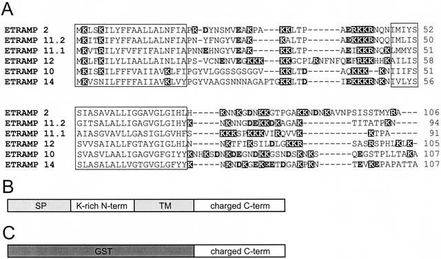
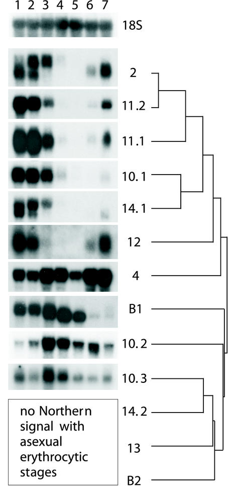
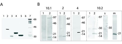
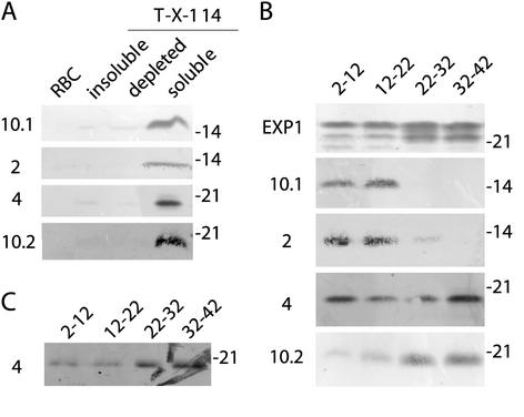
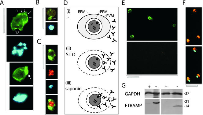
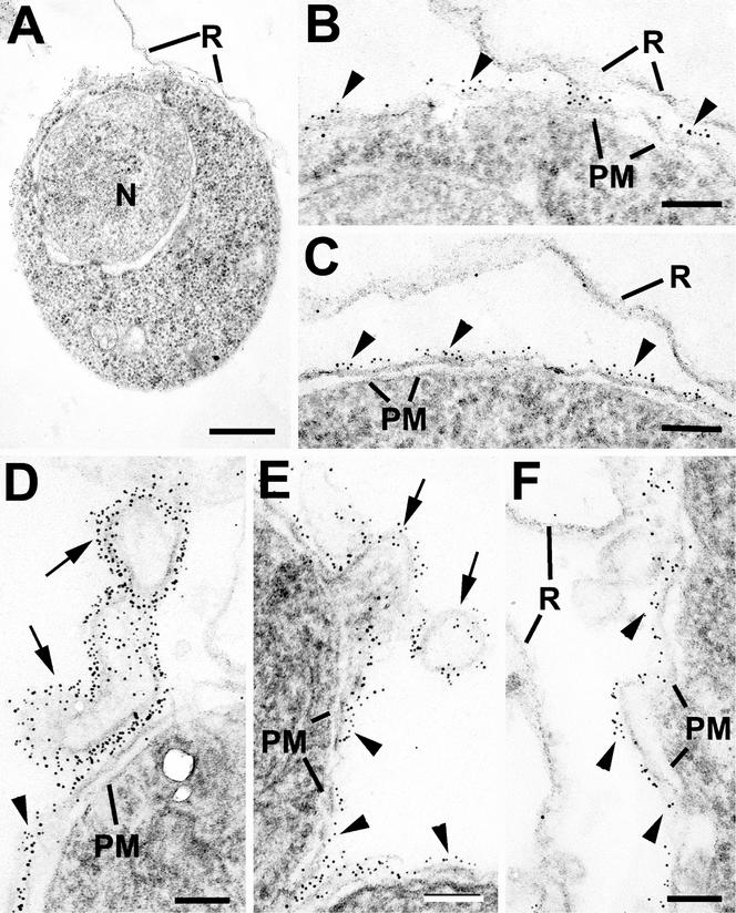
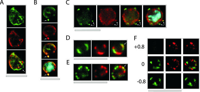
After invasion of erythrocytes, the human malaria parasite Plasmodium falciparum resides within a parasitophorous vacuole and develops from morphologically and metabolically distinct ring to trophozoite stages. During these developmental phases, major structural changes occur within the erythrocyte, but neither the molecular events governing this development nor the molecular composition of the parasitophorous vacuole membrane (PVM) is well known. Herein, we describe a new family of highly cationic proteins from P. falciparum termed early transcribed membrane proteins (ETRAMPs). Thirteen members were identified sharing a conserved structure, of which six were found only during ring stages as judged from Northern and Western analysis. Other members showed different stage-specific expression patterns. Furthermore, ETRAMPs were associated with the membrane fractions in Western blots, and colocalization and selective permeabilization studies demonstrated that ETRAMPs were located in the PVM. This was confirmed by immunoelectron microscopy where the PVM and tubovesicular extensions of the PVM were labeled. Early expressed ETRAMPs clearly defined separate PVM domains compared with the negatively charged integral PVM protein EXP-1, suggesting functionally different domains in the PVM with an oppositely charged surface coat. We also show that the dynamic change of ETRAMP composition in the PVM coincides with the morphological changes during development. The P. falciparum PVM is an important structure for parasite survival, and its analysis might provide better understanding of the requirements of intracellular parasites.


Figures

Figure 1
Figure 1
Conserved structure of ETRAMPs. (A) Alignment of the predicted aa sequence of the six most closely related ETRAMPs. The two hydrophobic regions containing the predicted signal peptide and TM are shown in boxes. Amino acids with positively charged side chains are shadowed in black; aa with negatively charged side chains are boldface and shaded in gray. The high number of charged residues is evident. (B) General structure of ETRAMPs. (C) Schematic view of GST-ETRAMP fusion used for recombinant expression. SP, signal peptide.
Figure 2
Figure 2
Developmental regulation of etramp transcription. Northern analysis was performed with filters containing total RNA of highly synchronized asexual blood stage cultures harvested at 0–6, 6–12, 12–18, 18–24, 24–30, 33–39, and 40–4 hpi (lanes 1–7). etramps used as probes are shown on the right side, including a dendrogram to show the degree of relative aa sequence similarities between different ETRAMPs. The six ring-transcribed etramps show the highest degree of similarity. A second group contains only one actively transcribed family member (etramp10.3). Two identical filters that were stripped and reprobed were used for all experiments. A 18s rRNA gene probe was used to demonstrate equal loading and transfer.
Figure 3
Figure 3
Characterization of anti-ETRAMP sera. (A) ETRAMPs are recognized by natural antibodies. Western blot containing recombinant GST (lane 1), MSP-2 (lane 2), ETRAMP2-GST (lane 3), ETRAMP4-GST (lane 4), ETRAMP10.1-GST (lane 5), ETRAMP13-GST (lane 6), and ETRAMP10.2-GST (lane 7) were probed with a serum pool of individuals living in a high endemicity malaria region. ETRAMP10.2 shows some degradation in E. coli. MSP-2 served as a positive control, and GST alone as negative control. (B) Western blots of total protein extracts generated from uninfected RBCs (lanes 1) or parasites released by saponin lysis (lanes 2) were probed with mouse antisera against ETRAMP10.1, 2, 4, and 10.2 (shown on top of corresponding boxes). The faint signal in the RBC lane is due to red staining originating from hemoglobin. Single bands for ETRAMP10.1, 2, and 4 show specificity of the corresponding antisera. ETRAMP10.2 seems to be processed into a soluble (lane s) and a membrane portion (lane m). Molecular mass markers (in kilodaltons) are shown on the right of each filter.
Figure 4
Figure 4
ETRAMPs are detected in the membrane fraction only and are developmentally expressed. (A) Western analysis with filters containing insoluble, soluble Triton X-114–depleted and Triton X-114–soluble parasite protein. All ETRAMPs were found in the membrane (Triton X-114–soluble) fraction only except for a 28-kDa ETRAMP10.2 signal in the detergent-depleted fraction (our unpublished data). (B) Western analysis with filters containing membrane protein harvested at four time points of synchronized P. falciparum in vitro cultures. Lanes 1 (2–12 hpi) and lane 2 (12–22 hpi) correspond to early and late ring stage, respectively; lane 3 (22–32 hpi) trophozoite stage; lane 4 (32–42 hpi) schizont stage. ETRAMP10.1 and 2 are only expressed in ring stages, whereas ETRAMP4 is found throughout the cycle. ETRAMP10.2 shows the highest expression after the ring stage. An antiserum against EXP-1 was used to confirm equal loading. (C) Formaldehyde (1%) cross-linking before extraction of membrane protein greatly diminished ETRAMP4 signal in rings. Antisera used are shown on the left; molecular mass is indicated in kilodaltons on the right of each filter.
Figure 5
Figure 5
ETRAMPs are located in the PVM with their C termini facing the RBC cytosol. (A–C) Distribution of ETRAMP4 around the parasite periphery in fixed cells. Top two panels of A depict ETRAMP4 staining (FITC) and nuclear staining (DAPI) of a schizont stage parasite; spots of more intense staining are indicated by small arrows. Bottom two panels of A show ETRAMP4 (FITC) and nuclear staining (DAPI) of a late trophozoite stage parasite. The single patch of strong staining is indicated by a large arrow. (B) Extensions from the parasite body are labeled by anti-ETRAMP4 serum, indicating localization in blebbing vesicles or the TVM. The top panel shows ETRAMP4 (FITC) detected in a young trophozoite; below the nucleus is shown (DAPI). The extension is indicated by an arrow. (C) Costaining with a rabbit anti-aldolase serum verifies that the periphery of the parasite is decorated by ETRAMP4 antiserum; the four panels represent the same section depicting a young trophozoite. From top to bottom, ETRAMP4 (FITC), aldolase (Cy3), nucleus (DAPI), and aldolase and ETRAMP4 merged. Bars, 5 μm. (D) Representation of the selective permeabilization scheme: 1) an untreated IRBC (P, parasite; EPM, erythrocyte plasma membrane; PPM, parasite plasma membrane); 2) a cell permeabilized with SLO, enabling access of antibodies through the perforated RBC membrane, resulting in a fluorescence signal if the corresponding antigen is located in the PVM facing the RBC cytosol; 3) a cell permeabilized with saponin, which allows to detect antigens facing the outer side of the PPM or both sides of the PVM. (E) Formaldehyde (1%)-fixed and saponin-permeabilized ring stage parasites showed a signal for ETRAMP10.1 (green, FITC, top), but not for aldolase (red, Cy3, bottom). Both panels show the same section. (F) Aldolase is strongly detected under similar fixation conditions and treatment with Triton X-100, indicating that aldolase is well detectable in ring stages. (G) Western analysis with extracts of saponin-released parasites that were either treated with (+) or without (−) trypsin before protein extraction, shows that the ETRAMP4 (left) and ETRAMP10.1 (right) C termini were digested, but not the cytoplasmic parasite enzyme GAPDH. Molecular mass (in kilodaltons) is indicated on the right.
Figure 6
Figure 6
Immunoelectron microscopy of permeablized red blood cells infected with P. falciparum and labeled with anti-ETRAMP2 (A–E) or anti-ETRAMP4 (F) and visualized with 5-nm gold particles. (A) Low-power image showing an intact early trophozoite with a single nucleus (N). (B) Detail form the periphery of A in which numerous gold particles are associated with the PVM (arrowheads). Note the lack of staining of the red cell membrane (R). (C) Similar section to B showing that the labeling is limited to the PVM (arrowheads). (D and E) Details of the periphery of intracellular parasites showing strong labeling of the PVM (arrowheads) and tubovesicular extensions of this membrane (arrows). (F) Section of the periphery of a large trophozoite stained with anti-ETRAMP4 in which the labeling is limited to the PVM (arrowheads). PM, parasite plasma membrane; R, red cell membrane. Bars, 0.5 μm (A) and 100 nm (B–F).
Figure 7
Figure 7
Colocalization of ETRAMPs with EXP-1 and SERP. The first image in each set shows ETRAMP labeling (green, FITC), the second Texas-Red-labeled SERP (A) or EXP-1 (B–F), and the third image represents the merged picture. The fourth panel in B and C depicts the merged picture, including nuclear staining (DAPI). (A) Confocal microscopy analysis of SERP and ETRAMP4 colocalization in schizont stage parasites (dried and 1% formaldehyde fixed). The ETRAMP4 signal seems partially detached from the PV content (arrow). (B and C) ETRAMP4 and EXP-1 localize to dots, probably representing vesicular structures that are detached from the PVM (E) or seem connected to the PVM (F). Dots are indicated by arrows. (D and E) Fluorescent microscopic analysis of ETRAMP and EXP-1 costaining of a ring-stage parasite displaying an even circular pattern (D, 0.1% formaldehyde-fixed IRBCs, ETRAMP10.1) or a beads on a string pattern (E, unfixed IRBCs, ETRAMP2). Yellow color shows regions of overlap (arrows). This indicated that ETRAMPs and EXP-1 localize to different regions of the PVM. (F) Three sections through a ring-stage parasite (dried and fixed with 1% formaldehyde), costained with ETRAMP10.1 and EXP-1, were analyzed by confocal microscopy. The planes of the first and the third row are 0.8 μm apart from the plane shown in the central row. The top plane contains only EXP-1 signal, whereas the bottom plane displays only ETRAMP signal. Bars, 5 μm.

References

    1. Adisa A, Albano FR, Reeder J, Foley M, Tilley L. Evidence for a role for a Plasmodium falciparumhomologue of Sec31p in the export of proteins to the surface of malaria parasite-infected erythrocytes. J Cell Sci. 2001;114:3377–3386. - PubMed
    1. Albano FR, Berman A, La Greca N, Hibbs AR, Wickham M, Foley M, Tilley L. A homologue of Sar1p localizes to a novel trafficking pathway in malaria-infected erythrocytes. Eur J Cell Biol. 1999a;78:453–462. - PubMed
    1. Albano FR, Foley M, Tilley L. Export of parasite proteins to the erythrocyte cytoplasm: secretory machinery and traffic signals. Novartis Found Symp. 1999b;226:157–172. - PubMed
    1. Altschul SF, Gish W, Miller W, Myers EW, Lipman DJ. Basic local alignment search tool. J Mol Biol. 1990;215:403–410. - PubMed
    1. Ansorge I, Benting J, Bhakdi S, Lingelbach K. Protein sorting in Plasmodium falciparum-infected red blood cells permeabilized with the pore-forming protein streptolysin O. Biochem J. 1996;315:307–314. - PMC - PubMed

Publication types

MeSH terms

LinkOut - more resources