. 2003 Apr;14(4):1545-57.
doi: 10.1091/mbc.02-07-0109.

Morphology and dynamics of clathrin/GGA1-coated carriers budding from the trans-Golgi network

Affiliations

Morphology and dynamics of clathrin/GGA1-coated carriers budding from the trans-Golgi network

Rosa Puertollano et al. Mol Biol Cell. 2003 Apr.

Abstract

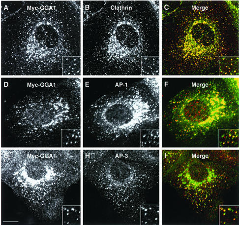
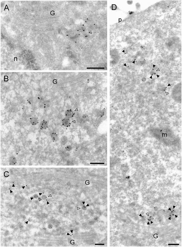
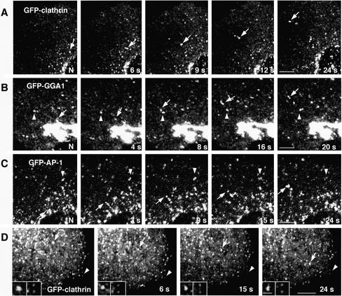
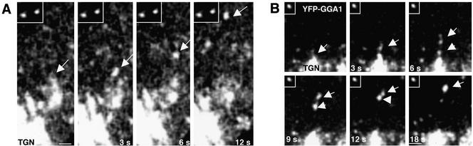
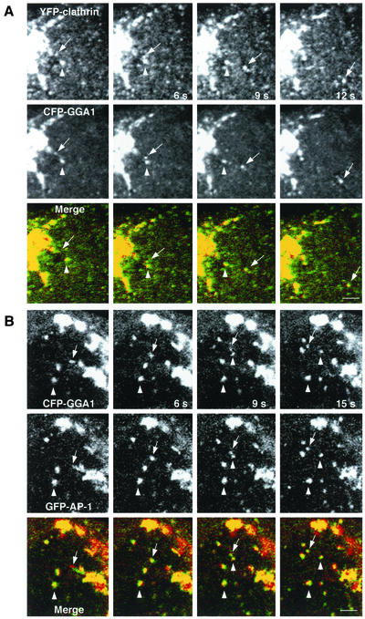
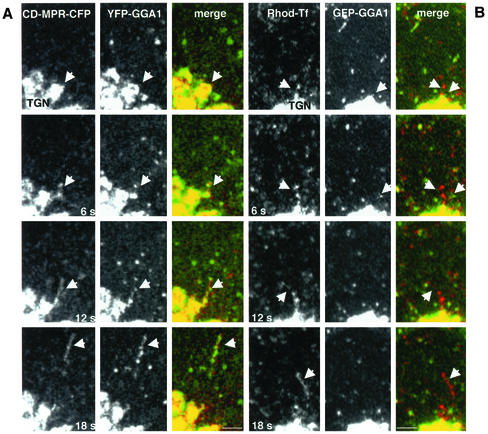
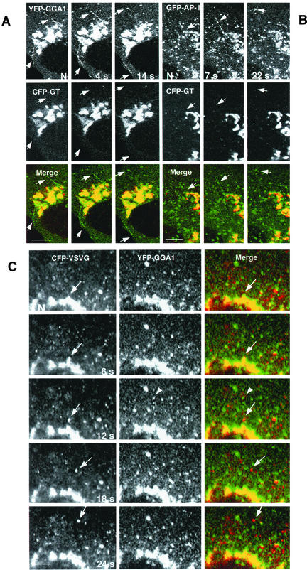
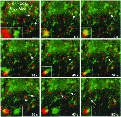
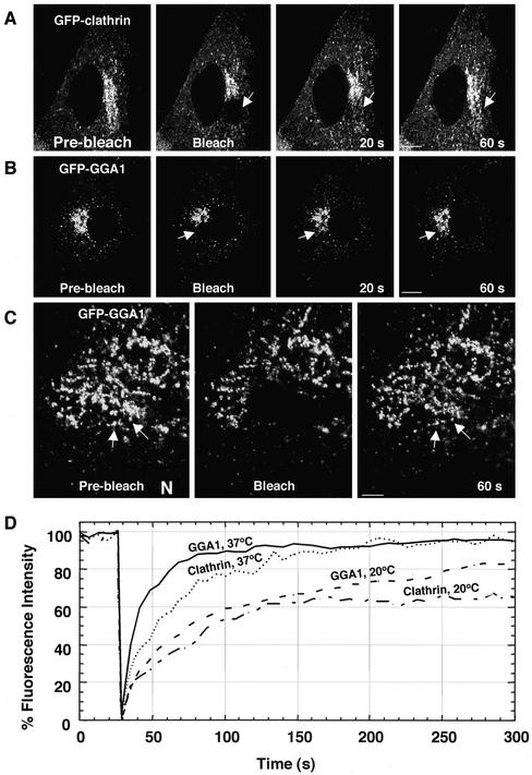
Sorting of transmembrane proteins and their ligands at various compartments of the endocytic and secretory pathways is mediated by selective incorporation into clathrin-coated intermediates. Previous morphological and biochemical studies have shown that these clathrin-coated intermediates consist of spherical vesicles with a diameter of 60-100 nm. Herein, we report the use of fluorescent imaging of live cells to demonstrate the existence of a different type of transport intermediate containing associated clathrin coats. Clathrin and the adaptors GGA1 and adaptor protein-1, labeled with different spectral variants of the green fluorescent protein, are shown to colocalize to the trans-Golgi network and to a population of vesicles and tubules budding from it. These intermediates are highly pleiomorphic and move toward the peripheral cytoplasm for distances of up to 10 microm with average speeds of approximately 1 microm/s. The labeled clathrin and GGA1 cycle on and off membranes with half-times of 10-20 s, independently of vesicle budding. Our observations indicate the existence of a novel type of trans-Golgi network-derived carriers containing associated clathrin, GGA1 and adaptor protein-1 that are larger than conventional clathrin-coated vesicles, and that undergo long-range translocation in the cytoplasm before losing their coats.


Figures

Figure 1
Figure 1
Intracellular distribution of Myc-GGA1 in MDCK cells analyzed by immunofluorescence microscopy. MDCK cells stably expressing Myc-GGA1 were fixed, permeabilized, and double labeled by incubation with antibodies to GGA1 (A, red channel), the Myc epitope (D, red channel; G, green channel), clathrin (B, green channel), the γ1-adaptin subunit of AP-1 (E, green channel), and the β3A subunit of the AP-3 complex (H, red channel), as indicated in the figure, followed by the corresponding fluorescently conjugated secondary antibodies. Stained cells were examined by confocal fluorescence microscopy. Merging the images in the red and green channels generated the third picture on each row (C, F, and I); yellow indicates overlapping localization. Insets show twofold-magnified views of peripheral cytoplasmic regions. Bar, 10 μm.
Figure 2
Figure 2
Immunoelectron microscopy localization of GGA1, Myc-GGA1, clathrin and AP-1. (A) Localization of endogenous GGA1 in monocyte-derived dendritic cells was analyzed by cryoimmunogold electron microscopy using antibodies to GGA1 (10-nm gold). (B–D) MDCK cells stably expressing Myc-GGA1 were analyzed by cryoimmunogold electron microscopy using antibodies to the Myc epitope (15-nm gold) and clathrin (10-nm gold) (B), GGA1 (15-nm gold) and the γ1 subunit of AP-1 (10-nm gold) (C and D). Open arrowheads in B indicate coated buds containing both Myc-GGA1 and clathrin, whereas closed arrowheads point to 15-nm gold particles marking the location of Myc-GGA1. Open arrowheads in C and D point to coated buds containing both Myc-GGA1 (or GGA1) and AP-1, whereas closed arrowheads point to 15-nm gold particles marking the location of Myc-GGA1 (or GGA1). G, Golgi complex; n, nucleus; p, plasma membrane; m, mitochondrion. Bars, 200 nm.
Figure 3
Figure 3
Characterization of TGN-derived carriers containing clathrin, GGA1, and AP-1. MDCK cells were grown on LabTek chambers and transiently transfected with constructs encoding GFP-clathrin light chain b (A and D), GFP-GGA1 (B), or GFP-γ1-adaptin (GFP-AP-1; C). At 15 h after transfection, cells expressing moderate levels of the fluorescent proteins were analyzed by time-lapse microscopy and images acquired at the indicated intervals. Insets in D show twofold-magnified comparisons of clathrin-containing carriers budding from the TGN with clathrin-coated pits at the plasma membrane. Arrows point to pleiomorphic carriers budding from the TGN and moving toward the cell periphery, whereas arrowheads indicate immobile structures. N, nucleus. Bars, 5 μm (A–C), 4 μm (D), 1 μm (D insets). QuickTime videos of the experiments shown in A, B, and C can be seen in Supplemental Materials (videos 1, 2, and 3, respectively).
Figure 4
Figure 4
Comparison of the size of GGA1-coated intermediaries to that of fluorescent beads. MDCK cells expressing YFP-GGA1 were incubated with 0.2-μm-diameter fluorescent beads before analysis by time-lapse confocal microscopy. Arrow in A points to a YFP-GGA1–containing intermediate moving from the TGN toward the cell periphery. Notice that this carrier seems larger (∼0.35 μm) than the beads shown in the inset (∼0.2 μm), even though these structures have similar fluorescent intensities (37.7 and 38.6 arbitrary units, respectively). In some cases, large intermediates seem to arise from clustering of smaller vesicles, as shown in B (fluorescent bead is shown in the inset). Bars, 0.5 μm (A), 1 μm (B).
Figure 5
Figure 5
Presence of clathrin and AP-1 on GGA1-containing carriers. MDCK cells were simultaneously transfected with constructs encoding YFP-clathrin and CFP-GGA1 (A) or GFP-γ1 (GFP-AP-1) and CFP-GGA1 (B). Confocal microscopy images were acquired every 3 s. Arrows indicate vesicular carriers budding from the TGN, whereas arrowheads point to immobile structures. The yellow color in the merged images indicates colocalization between clathrin and GGA1 (A) or GGA1 and AP-1 (B). Bars, 2 μm.
Figure 6
Figure 6
Cargo specificity of GGA1-containing intermediates. (A) Confocal microscopy imaging of live MDCK cells coexpressing CD-MPR-CFP and YFP-GGA1. Images were collected at 6-s intervals. Arrows indicate the formation and detachment of long tubular structures containing CD-MPR-CFP from the TGN. Notice that YFP-GGA1 associates with those tubular structures in a discontinuous pattern. (B) MDCK cells expressing GFP-GGA1 were incubated with rhodamine-transferrin for 20 min to load recycling endosomes and then examined by video microscopy. Images show that transferrin-containing intermediates in transit from the juxtanuclear area of the cell are devoid of GFP-GGA1. Bars, 3 μm.
Figure 7
Figure 7
Exclusion of galactosyl transferase and VSV-G protein from GGA1 carriers budding from the TGN. (A and B) Golgi-resident protein galactosyl transferase (GT) is not incorporated into vesicular carriers budding from the TGN. MDCK cells were doubly transfected with a trans-Golgi resident protein (CFP-GT) and with YFP-GGA1 (A) or GFP-γ1 (GFP-AP-1) (B). Time-lapse microscopy showed the absence of CFP-GT (red channel) from rows of coated intermediates budding from the TGN (green channel, arrows). (C) VSV-G and GGA-1 localize to different transport intermediaries. MDCK cells, transfected with YFP-GGA1 and CFP-VSV-G (ts045 mutant) were incubated for 24 h at 40°C, shifted to 20°C for 1 h to accumulate cargo in the TGN, and then warmed to 32°C before visualization. Consecutive video images from time-lapse series show a VSV-G–enriched vesicle budding from the TGN (arrow, red channel). Note the absence of GGA1 (green channel) in this structure. The arrowhead points to a row of YFP-GGA1-containing vesicles rapidly budding from the TGN. N, nucleus. Bars, 10 μm (A and B), 5 μm (C).
Figure 8
Figure 8
Interaction of GGA1-containing carriers with endosomes. MDCK cells were transfected with GFP-GGA1. At 15 h after transfection, the cells were loaded with rhodamine-albumin for 15 min before analysis by time-lapse confocal microscopy. Merged images for GFP-GGA1 (green) and rhodamine-albumin (red) are shown. Arrows point to vesicular structures containing rhodamine-albumin and GFP-GGA1. The insets show fourfold magnification of the structures pointed by the arrows. Bars, 2 and 0.5 μm (inset).
Figure 9
Figure 9
Dynamics of clathrin and GGA1 analyzed by fluorescence recovery after photobleaching. (A–C) MDCK cells were transiently transfected with constructs encoding GFP-clathrin (A) or GFP-GGA1 (B and C). Fluorescence associated with the Golgi complex was photobleached with high-intensity laser light and subsequent recovery of fluorescence was monitored by scanning the whole cell at low laser power at 5-s intervals for 5 min. The arrows in C indicate that the GFP-GGA1 recovers on the same TGN structures where it was located before photobleaching. N, nucleus. Bars, 10 μm (A and B), 5 μm (C). (D) Kinetics of GFP-clathrin and GFP-GGA1 recovery after photobleaching at 37 and 20°C. MDCK cells expressing GFP-clathrin or GFP-GGA1 were photobleached and then scanned at low laser power for 5 min. The graph shows the combined results of 10 determinations per condition. QuickTime videos of the experiments shown in panes A, B, and C can be seen in Supplemental Materials (videos A, B, and C).

Similar articles

Cited by

References

    1. Aridor M, Bannykh SI, Rowe T, Balch WE. Sequential coupling between COPII and COPI vesicle coats in endoplasmic reticulum to Golgi transport. J Cell Biol. 1995;131:875–893. - PMC - PubMed
    1. Boman AL. GGA proteins: new players in the sorting game. J Cell Sci. 2001;114:3413–3418. - PubMed
    1. Brodsky FM, Chen CY, Knuehl C, Towler MC, Wakeham DE. Biological basket weaving: formation and function of clathrin-coated vesicles. Annu Rev Cell Dev Biol. 2001;17:517–568. - PubMed
    1. Costaguta G, Stefan CJ, Bensen ES, Emr SD, Payne GS. Yeast GGA coat proteins function with clathrin in Golgi to endosome transport. Mol Biol Cell. 2001;12:1885–96. - PMC - PubMed
    1. Damer CK, O'Halloran TJ. Spatially regulated recruitment of clathrin to the plasma membrane during capping and cell translocation. Mol Biol Cell. 2000;11:2151–2159. - PMC - PubMed

MeSH terms