. 2003 Apr;14(4):1652-63.
doi: 10.1091/mbc.e02-08-0456.

Catabolite degradation of fructose-1,6-bisphosphatase in the yeast Saccharomyces cerevisiae: a genome-wide screen identifies eight novel GID genes and indicates the existence of two degradation pathways

Affiliations

Catabolite degradation of fructose-1,6-bisphosphatase in the yeast Saccharomyces cerevisiae: a genome-wide screen identifies eight novel GID genes and indicates the existence of two degradation pathways

Jochen Regelmann et al. Mol Biol Cell. 2003 Apr.

Abstract

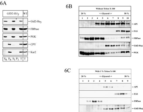
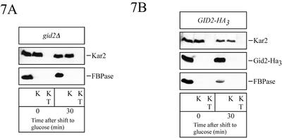
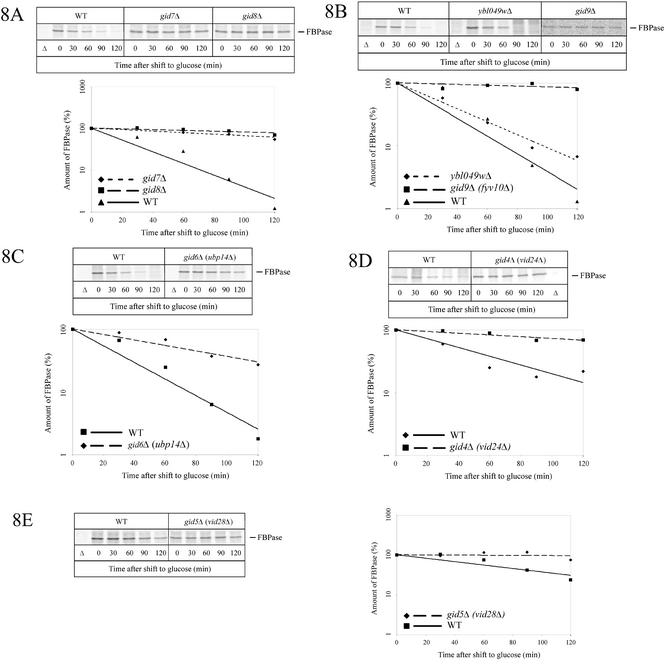
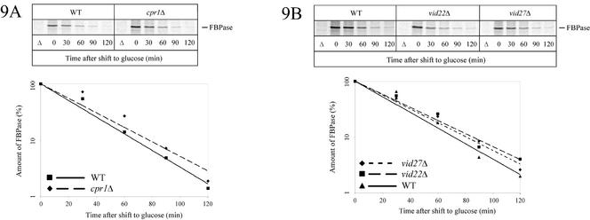
Metabolic adaptation of Saccharomyces cerevisiae cells from a nonfermentable carbon source to glucose induces selective, rapid breakdown of the gluconeogenetic key enzyme fructose-1,6-bisphosphatase (FBPase), a process called catabolite degradation. Herein, we identify eight novel GID genes required for proteasome-dependent catabolite degradation of FBPase. Four yeast proteins contain the CTLH domain of unknown function. All of them are Gid proteins. The site of catabolite degradation has been controversial until now. Two FBPase degradation pathways have been described, one dependent on the cytosolic ubiquitin-proteasome machinery, and the other dependent on vacuolar proteolysis. Interestingly, three of the novel Gid proteins involved in ubiquitin-proteasome-dependent degradation have also been reported by others to affect the vacuolar degradation pathway. As shown herein, additional genes suggested to be essential for vacuolar degradation are unnecessary for proteasome-dependent degradation. These data raise the question as to whether two FBPase degradation pathways exist that share components. Detailed characterization of Gid2p demonstrates that it is part of a soluble, cytosolic protein complex of at least 600 kDa. Gid2p is necessary for FBPase ubiquitination. Our studies have not revealed any involvement of vesicular intermediates in proteasome-dependent FBPase degradation. The influence of Ubp14p, a deubiquitinating enzyme, on proteasome-dependent catabolite degradation was further uncovered.


Figures

Figure 1
Figure 1
Identification of the GID2 gene. (A) After transformation with a YCp50-based genomic library, complemented gid2-1 colonies (WAY.5-4A/B2) are detected in a colony screen. Cells were transferred onto nitrocellulose sheets as outlined in MATERIALS AND METHODS and probed with antibodies against FBPase. Wild-type (WAY.5-4A) colonies (arrowheads) and gid2-1 colonies, carrying a complementing genomic fragment (arrow) are able to degrade FBPase and look white, due to their low level of FBPase. (B) Subcloning identifies YDR255c (GID2) as the complementing ORF. (C) Pulse-chase analysis of glucose-induced degradation of FBPase. (D) Quantification of C shows a significant stabilization of FBPase in gid2Δ (YTS1) cells. Control, wild-type (W303-1B) and UBC8 deleted (YTS2) cells.
Figure 2
Figure 2
Identification of the GID1 gene. (A) gid1-3 (WAY.5-4A/D1) cells were transformed with a YCp50-based genomic library, and after transfer of the colonies to nitrocellulose they were processed as described in MATERIALS AND METHODS. The level of FBPase was then detected with antibodies. Complemented colonies (arrow) and wild-type (WAY.5-4A) colonies (arrowheads) look white, due to degradation of FBPase. (B) Pulse-chase analysis of gid1Δ (vid30Δ) (AE7-6c) cells confirms the defect in glucose-induced degradation of FBPase. (C) Quantification of B. Control, wild-type cells (JK9-3da).
Figure 3
Figure 3
(A) Glucose-induced ubiquitination of FBPase is affected in gid2Δ (YTS1) cells. Crude extracts from wild-type (W303-1B) and gid2Δ (YTS1) cells overexpressing ubiquitin or Ha-tagged ubiquitin were immunoprecipitated with antibodies against FBPase. Proteins were separated by SDS-PAGE, blotted, and probed with antibodies against Ha. (B) In gid2Δ (YTS1) cells degradation of the N-end rule substrate Arg-β-gal is not affected. After pulse labeling, aliquots were withdrawn at the indicated times and the amount of Arg-β-gal was determined with a PhosphorImager after immunoprecipitation with antibodies against β-galactosidase.
Figure 4
Figure 4
Ha-tagged Gid2p expressed from the chromosome with its native promoter is functional during catabolite inactivation of FBPase (strain YTS3). After pulse labeling, the cells were chased in glucose-containing medium. After immunoprecipitation with antibodies against FBPase and SDS-PAGE, the amount of FBPase was detected with a PhosphorImager (A) and quantified (B). (C) Steady-state levels of Ha-tagged Gid2p. Crude extracts of cells (YTS3) grown in glucose- (G) or ethanol (E)-containing media were analyzed on immunoblots with antibodies against Ha. Ethanol-grown cells were further shifted to glucose media and at the indicated times aliquots were analyzed. As a control, wild-type (W303-1B) cells lacking an Ha-tag were included.
Figure 5
Figure 5
Sucrose density gradient fractionation of cells (YTS3) expressing Ha-tagged Gid2p from the chromosome. After 16 h of growth on ethanol medium, the cells were shifted to glucose medium. The cells were analyzed before (A) and 30 min after shift to glucose medium (B). From the sucrose gradient, fractions were collected from the top of the gradient (fraction 1) and analyzed by immunoblotting with antibodies against Ha (Ha-tagged Gid2p), cytosolic PGK, Fas (cytosolic fatty acid synthase), CPY (vacuolar carboxypeptidase yscY); vacuolar API, and ER lumenal Kar2p. The fractions were further tested for their Guanosine diphosphatase (Golgi) activity. Further details are given in the text.
Figure 6
Figure 6
(A) FBPase and Ha-tagged Gid2p are found in the soluble fraction 30 min after shift to glucose medium. Cells expressing Ha-tagged Gid2p from the chromosome (YTS3) were converted to spheroplasts. After mild hypotonic lysis, the cell lysate was fractionated by centrifugation. S6 and P6 refer to the 6000 × g supernatant and pellet, respectively; S1, 100,000 × g supernatant; P1, 100,000 × g pellet. (B and C) Glycerol gradient fractionation of cell lysates from cells expressing Ha-tagged Gid2p from the chromosome (YTS3). Fractions were immunoblotted with antibodies against API, Fas (fatty acid synthase), FBPase, Gid2-Ha3, and PGK. Glycerol gradient fractionation was done without (B) and with 1% Triton X-100 (C).
Figure 7
Figure 7
FBPase is not engulfed in vesicles under the inactivation conditions used (see MATERIALS AND METHODS). At the indicated times after shift to glucose medium, cells were spheroplasted, hypotonically lysed, and incubated with proteinase K (K) and proteinase K with Triton X-100 (KT). Samples were then immunoblotted and analyzed with antibodies against ER lumenal Kar2p, Ha, and FBPase. In A, gid2Δ (YTS1) cells and in B cells (YTS3) chromosomally expressing Ha-tagged Gid2p were used.
Figure 8
Figure 8
Pulse-chase analysis of FBPase degradation in novel gid mutant cells. gid7Δ (Y33446), gid8Δ (Y36576) (A), gid9Δ (fyv10Δ) (Y31488), ybl049wΔ (Y33075) (B), gid6Δ (ubp14Δ) (Y33195) (C), gid4Δ (vid24Δ) (YFJ1) (D), and gid5Δ (vid28Δ) (Y31410) (E) cells. Details are given in the text. In the experiment shown for gid5Δ, labeling was extended to 16 h to enhance the amount of labeled protein.
Figure 9
Figure 9
Vid27p, Vid22p, and Cpr1p are not essential for proteasome-dependent catabolite degradation of FBPase. Pulse-chase analysis of FBPase degradation was measured in cpr1Δ (Y33513) (A), vid22Δ (Y35282), and vid27Δ (Y32000) (B) cells.

References

    1. Abeijon C, Orlean P, Robbins PW, Hirschberg CB. Topography of glycosylation in yeast: characterization of GDPmannose transport and lumenal guanosine diphosphatase activities in Golgi-like vesicles. Proc Natl Acad Sci USA. 1989;86:6935–6939. - PMC - PubMed
    1. Amerik A, Swaminathan S, Krantz BA, Wilkinson KD, Hochstrasser M. In vivo disassembly of free polyubiquitin chains by yeast Ubp14 modulates rates of protein degradation by the proteasome. EMBO J. 1997;16:4826–4838. - PMC - PubMed
    1. Ausubel FM, Kingston RE, Seidman FG, Struhl K, Moore DD, Brent R, Smith FA. Current Protocols in Molecular Biology. New York: John Wiley & Sons; 1992.
    1. Bachmair A, Finley D, Varshavsky A. In vivo half-life of a protein is a function of its amino-terminal residue. Science. 1986;234:179–86. - PubMed
    1. Barth H, Thumm M. A genomic screen identifies AUT8 as a novel gene essential for autophagy in the yeast Saccharomyces cerevisiae. Gene. 2001;274:151–156. - PubMed

Publication types

MeSH terms