. 2003 May;72(5):1315-22.
doi: 10.1086/375122. Epub 2003 Apr 8.

Mutations of MYO6 are associated with recessive deafness, DFNB37

Affiliations

Mutations of MYO6 are associated with recessive deafness, DFNB37

Zubair M Ahmed et al. Am J Hum Genet. 2003 May.

Abstract

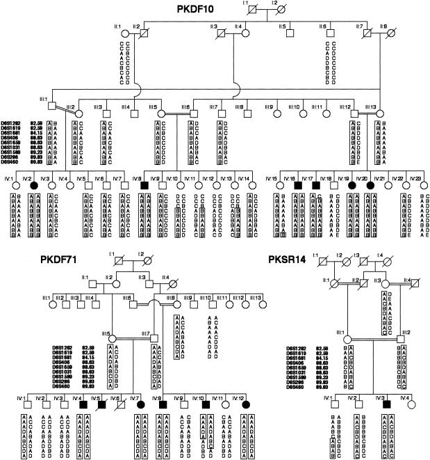
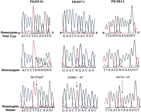
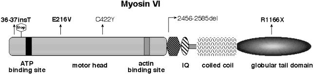
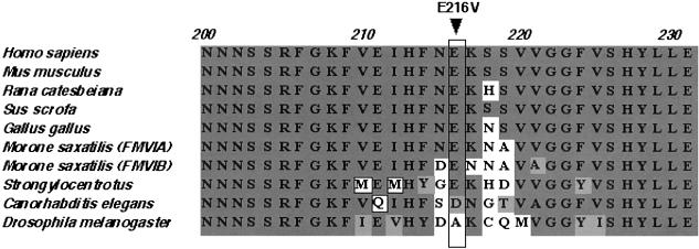
Cosegregation of profound, congenital deafness with markers on chromosome 6q13 in three Pakistani families defines a new recessive deafness locus, DFNB37. Haplotype analyses reveal a 6-cM linkage region, flanked by markers D6S1282 and D6S1031, that includes the gene encoding unconventional myosin VI. In families with recessively inherited deafness, DFNB37, our sequence analyses of MYO6 reveal a frameshift mutation (36-37insT), a nonsense mutation (R1166X), and a missense mutation (E216V). These mutations, along with a previously published missense allele linked to autosomal dominant progressive hearing loss (DFNA22), provide an allelic spectrum that probes the relationship between myosin VI dysfunction and the resulting phenotype.


Figures

Figure  1
Figure 1
Haplotypes of markers showing linkage to DFNB37 at 6q13 for three families segregating profound, sensorineural, recessive hearing loss. Affected individual IV:17 in family PKDF10 provided the proximal recombination breakpoint at marker D6S1282 (82.59 cM). The distal recombination is provided by affected individual IV:10 of family PKDF71 at marker D6S1031 (88.63 cM). National Institutes of Health (OH93-N-016) and National Centre of Excellence in Molecular Biology (CEMB) institutional review board approval and written informed consent were obtained from all participating subjects. DNA was extracted from either peripheral blood samples or buccal swabs and was amplified using fluorescent-labeled primers for STR markers linked to reported nonsyndromic recessive deafness (DFNB) loci. Amplimers were visualized by gel electrophoresis on ABI 377 DNA sequencers, and genotypes were determined using Genescan and Genotyper software (Applied Biosystems). The genetic linkage distances are from the Center for Medical Genetics Web server.
Figure  2
Figure 2
MYO6 mutations segregating in three Pakistani families. Left, Electropherograms of amplimers from genomic DNA templates, illustrating homozygosity for a single-base-pair insertion mutation in an affected individual, heterozygosity in an obligate carrier, and homozygosity for the wild-type allele in an unaffected individual from family PKDF10. An arrow indicates the site of the 36-37insT in the second exon. Center, Electropherograms illustrating genotypes of a homozygous wild-type allele, a 3496C→T heterozygote, and a person homozygous for 3496C→T of family PKDF71. Right, Electropherograms are shown for transversion mutation 647A→T, a carrier, and the wild-type allele of family PKSR14. All the mutations described here are numbered from start codon ATG (GenBank accession number AB002387).
Figure  3
Figure 3
A drawing of myosin VI, showing the locations of the mutations causing deafness in humans and mice. The three mutant alleles reported in this study are shown in bold letters, whereas C442Y and 2456-2585del are from the studies by Melchionda et al. (2001) and Avraham et al. (1995), respectively. Shown also is the predicted stop codon after twelve out-of-frame amino acids due to 36-37insT.
Figure  4
Figure 4
Alignment of a portion of myosin VI proteins from various species, showing conservation of the glutamate residue at position 216 in the motor domain among seven myosin VI proteins from Homo sapiens, Mus musculus, Rana catesbeiana, Sus scrofa, Gallus gallus, Morone saxatilis, and Strongylocentrotus. The conserved amino acids are shown with dark gray background, similar amino acids are shown with a light gray background, and the nonconserved amino acids are shown with a white background.

Similar articles

Cited by

References

Electronic-Database Information

    1. Center for Medical Genetics, Marshfield Medical Research Foundation, http://research.marshfieldclinic.org/genetics/
    1. Hereditary Hearing Loss Homepage, http://www.uia.ac.be/dnalab/hhh/
    1. GenBank, http://www.ncbi.nlm.nih.gov/Genbank/ (for MYO6 [accession number AB002387])
    1. Online Mendelian Inheritance in Man (OMIM), http://www.ncbi.nlm.nih.gov/Omim/ (for DFNA22) - PubMed
    1. Primer3 Web-Based Server, http://www.genome.wi.mit.edu/cgi-bin/primer/primer3_www.cgi

References

    1. Ahituv N, Sobe T, Robertson NG, Morton CC, Taggart RT, Avraham KB (2000) Genomic structure of the human unconventional myosin VI gene. Gene 261:269–275 - PubMed
    1. Ahmed ZM, Riazuddin S, Bernstein SL, Ahmed Z, Khan S, Griffith AJ, Morell RJ, Friedman TB, Wilcox ER (2001) Mutations of the protocadherin gene PCDH15 cause Usher syndrome type 1F. Am J Hum Genet 69:25–34 - PMC - PubMed
    1. Avraham KB, Hasson T, Sobe T, Balsara B, Testa JR, Skvorak AB, Morton CC, Copeland NG, Jenkins NA (1997) Characterization of unconventional MYO6, the human homologue of the gene responsible for deafness in Snell’s waltzer mice. Hum Mol Genet 6:1225–1231 - PubMed
    1. Avraham KB, Hasson T, Steel KP, Kingsley DM, Russell LB, Mooseker MS, Copeland NG, Jenkins NA (1995) The mouse Snell’s waltzer deafness gene encodes an unconventional myosin required for structural integrity of inner ear hair cells. Nat Genet 11:369–375 - PubMed
    1. Berg JS, Powell BC, Cheney RE (2001) A millennial myosin census. Mol Biol Cell 12:780–794 - PMC - PubMed

Publication types

Associated data