. 2003 May;52(5):720-8.
doi: 10.1136/gut.52.5.720.

Increases in free radicals and cytoskeletal protein oxidation and nitration in the colon of patients with inflammatory bowel disease

Affiliations

Increases in free radicals and cytoskeletal protein oxidation and nitration in the colon of patients with inflammatory bowel disease

A Keshavarzian et al. Gut. 2003 May.

Abstract

Background: Overproduction of colonic oxidants contributes to mucosal injury in inflammatory bowel disease (IBD) but the mechanisms are unclear. Our recent findings using monolayers of intestinal cells suggest that the mechanism could be oxidant induced damage to cytoskeletal proteins. However, oxidants and oxidative damage have not been well characterised in IBD mucosa.

Aims: To determine whether there are increases in oxidants and in tissue and cytoskeletal protein oxidation in IBD mucosa.

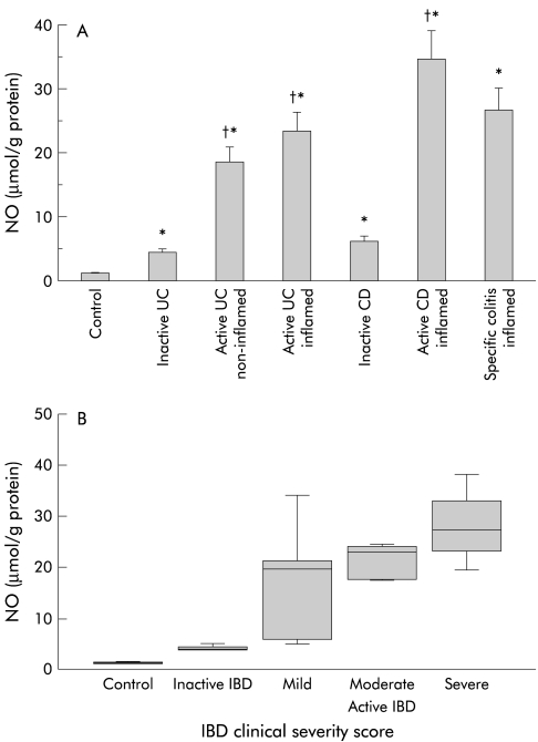
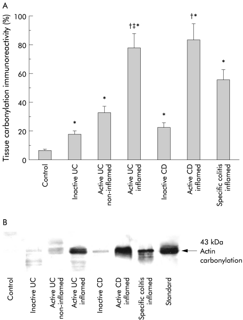
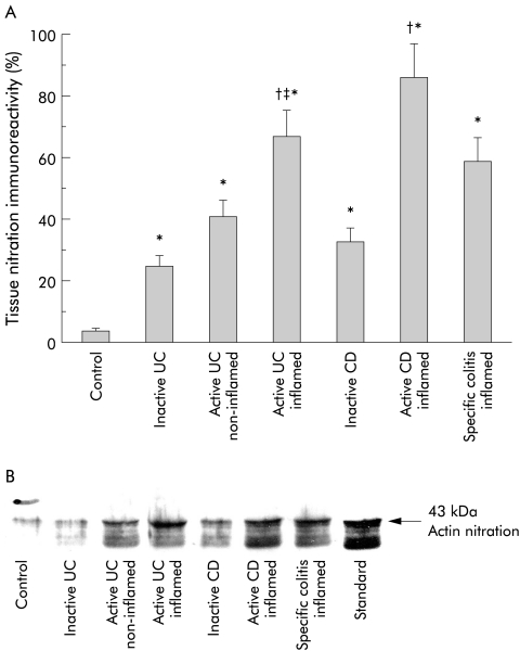
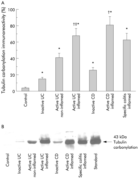
Methods: We measured nitric oxide (NO) and markers of oxidative injury (carbonylation and nitrotyrosination) to tissue and cytoskeletal proteins in colonic mucosa from IBD patients (ulcerative colitis, Crohn's disease, specific colitis) and controls. Outcomes were correlated with IBD severity score.

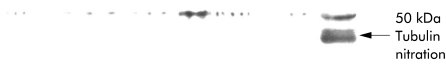
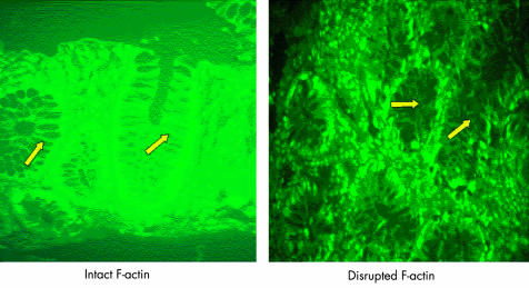
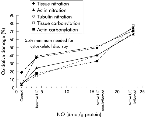
Results: Inflamed mucosa showed the greatest increases in oxidants and oxidative damage. Smaller but still significant increases were seen in normal appearing mucosa of patients with active and inactive IBD. Tissue NO levels correlated with oxidative damage. Actin was markedly (>50%) carbonylated and nitrated in inflamed tissues of active IBD, less so in normal appearing tissues. Tubulin carbonylation occurred in parallel; tubulin nitration was not observed. NO and all measures of oxidative damage in tissue and cytoskeletal proteins in the mucosa correlated with IBD severity. Disruption of the actin cytoarchitecture was primarily within the epithelial cells and paracellular area.

Conclusions: Oxidant levels increase in IBD along with oxidation of tissue and cytoskeletal proteins. Oxidative injury correlated with disease severity but is also present in substantial amounts in normal appearing mucosa of IBD patients, suggesting that oxidative injury does not necessarily lead to tissue injury and is not entirely a consequence of tissue injury. Marked actin oxidation (>50%)-which appears to result from cumulative oxidative damage-was only seen in inflamed mucosa, suggesting that oxidant induced cytoskeletal disruption is required for tissue injury, mucosal disruption, and IBD flare up.


Figures

Figure 1
Figure 1
(A) Concentrations of nitric oxide (NO) in homogenates from intestinal mucosal biopsies from: controls (n=10); patients with inactive ulcerative colitis (UC) (n=7); normal appearing non-inflamed mucosa of patients with active UC (n=6); inflamed ulcerated mucosa of patients with active UC (n=15); inactive Crohn’s disease (CD) (n=5); active CD (n=6); and inflamed mucosa of patients with specific colitis (n=14). NO levels were assessed by chemiluminescence. Data are mean (SEM); *p<0.05 versus controls, †p<0.05 versus inactive inflammatory bowel disease (IBD). (B) Correlation between mucosal NO levels and IBD disease severity score. Correlations are from: controls (n=5); patients with inactive IBD (UC=7, CD=5); and active IBD (UC=14, CD=5) (non-parametric correlation, Spearman correlation coefficient 0.81; p<0.0001).
Figure 2
Figure 2
(A) Slot immunoblotting analysis of levels of anti-nitrotyrosine (nitration) immunoreactivity of proteins in mucosal pinch biopsies. (B) Representative slot immunoblot of the nitrated mucosal proteins. Mucosal homogenates were analysed by slot blotting and processed for autoradiography and densitometry. Nitration immunoreactivity was expressed as follows: nitrotyrosine formation (optical density) in the patient group divided by the nitrated tissue standards, expressed as a percentage. Controls (n=10); patients with inactive ulcerative colitis (UC) (n=7); normal appearing non-inflamed mucosa of patients with active UC (n=6); inflamed ulcerated mucosa of patients with active UC (n=15); inactive Crohn’s disease (CD) (n=5); active CD (n=6); and inflamed mucosa of patients with specific colitis (n=14). *p<0.05 versus controls, †p<0.05 versus inactive inflammatory bowel disease (IBD), ‡p<0.05 versus non-inflamed UC. (C) Correlation between mucosal nitrotyrosine levels and nitric oxide (NO) (r=0.65, r2=0.43, p<0.0001). (D) Correlation between mucosal nitrotyrosine and IBD disease severity score (non-parametric correlation, Spearman correlation coefficient 0.84; p<0.0001). Number of subjects used for analysis as in previous figures.
Figure 3
Figure 3
(A) Slot immunoblotting analysis of leves of carbonylation (anti-dinitrophenylhydrazone) immunoreactivity of proteins in mucosal pinch biopsies. (B) Representative slot blot of the carbonylated tissue proteins. Oxidation was expressed as carbonyl formation (that is, optical density) in the patient group divided by the oxidised tissue standards, expressed as a percentage. From controls (n=10); patients with inactive ulcerative colitis (UC) (n=7); normal appearing non-inflamed mucosa of patients with active UC (n=6); inflamed ulcerated mucosa of patients with active UC (n=15); inactive Crohn’s disease (CD) (n=5); active CD (n=6); and inflamed mucosa of patients with specific colitis (n=14). *p<0.05 versus controls, †p<0.05 versus inactive inflammatory bowel disease (IBD), ‡p<0.05 versus non-inflamed UC. Correlation between mucosal carbonylation levels and mucosal nitric oxide (NO) levels (r=0.72, r2=0.51, p<0.0001) (C), between mucosal carbonylation levels and nitrotyrosine levels (r=0.96, r2=0.93, p<0.0001) (D), and between mucosal carbonylation levels and IBD disease severity score (non-parametric correlation, Spearman correlation coefficient 0.81; p<0.0001) (E). Number of subjects used for analysis was as above.
Figure 4
Figure 4
(A) Immunoblotting analysis of carbonylation (anti-dinitrophenylhydrazone (DNP)) immunoreactivity of the actin cytoskeleton from intestinal mucosa. A representative blot for actin carbonylation is shown in (B). Western blots of mucosal homogenates were processed for actin fractionation sodium dodecyl sulphate-polyacrylamide gel electrophoresis and processed sequentially using monoclonal anti-DNP and horseradish peroxidase conjugated secondary antibodies. The region of gel shown is between the M 42 000 and 63 000 pre-stained molecular weight standards, which were run in adjacent lanes. Carbonlyation of actin was expressed as carbonyl formation (that is, optical density) in the appropriate group divided by the oxidised actin standard. Controls (n=10); patients with inactive ulcerative colitis (UC) (n=7); normal appearing non-inflamed mucosa of patients with active UC (n=6); inflamed ulcerated mucosa of patients with active UC (n=15); inactive Crohn’s disease (CD) (n=5); active CD (n=6); and inflamed mucosa of patients with specific colitis (n=14). *p<0.05 versus controls, †p<0.05 versus inactive inflammatory bowel disease, ‡p<0.05 versus non-inflamed UC.
Figure 5
Figure 5
(A) Immunoblotting analysis of anti-nitrotyrosine immunoreactivity of actin from the intestinal mucosa and a representative immunoblot of this actin nitration (B). Nitration of actin was expressed as nitrotyrosine formation (optical density) in the patient group divided by the nitrated actin standard. Western blots were processed as in fig 4 ▶ except that monoclonal anti-nitrotyrosine was used as the primary antibody. Representative blot from controls (n=10); patients with inactive ulcerative colitis (UC) (n=7); normal appearing non-inflamed mucosa of patients with active UC (n=6); inflamed ulcerated mucosa of patients with active UC (n=15); inactive Crohn’s disease (CD) (n=5); active CD (n=6); and inflamed mucosa of patients with specific colitis (n=14). *p<0.05 versus controls, †p<0.05 versus inactive inflammatory bowel disease, ‡p<0.05 versus non-inflamed UC.
Figure 6
Figure 6
(A) Quantitative immunoblotting analysis of anti-dinitrophenylhydrazone immunoreactivity of the tubulin cytoskeleton from mucosal biopsies. A representative blot showing the oxidation of tubulin is shown in (B). Tubulin fractions from mucosal homogenates were separated by sodium dodecyl sulphate-polyacrylamide gel electrophoresis and analysed by autoradiography and densitometry. The region of gel shown is between the M 50 000 and 70 000 pre-stained molecular weight standards, which were run in adjacent lanes. Representatives blot from controls (n=10); patients with inactive ulcerative colitis (UC) (n=7); normal appearing non-inflamed mucosa of patients with active UC (n=6); inflamed ulcerated mucosa of patients with active UC (n=15); inactive Crohn’s disease (CD) (n=5); active CD (n=6); and inflamed mucosa of patients with specific colitis (n=14). *p<0.05 versus controls, †p<0.05 versus inactive inflammatory bowel disease, ‡p<0.05 versus non-inflamed UC.
Figure 7
Figure 7
Representative immunoblotting of anti-nitrotyrosine immunoreactivity of the tubulin fraction from mucosal biopsies. Note the absence of tubulin nitration in any group (same groups as in fig 6B ▶). Tubulin fractions from mucosal homogenates were processed for sodium dodecyl sulphate-polyacrylamide gel electrophoresis and immunoblotted using monoclonal anti-nitrotyrosine and horseradish peroxidase conjugated secondary antibodies. The region of gel shown is between the M 50 000 and 70 000 pre-stained molecular weights, which were run in adjacent lanes. Representative blot from controls (n=10); patients with inactive ulcerative colitis (UC) (n=7); normal appearing non-inflamed mucosa of patients with active UC (n=6); inflamed ulcerated mucosa of patients with active UC (n=15); inactive Crohn’s disease (CD) (n=5); active CD (n=6); and inflamed mucosa of patients with specific colitis (n=14).
Figure 8
Figure 8
Fluorescent staining of F-actin cytoskeleton as captured by ultra high resolution laser scanning confocal microscopy revealing its intracellular distribution in intestinal mucosa from a patient with inflammatory bowel disease (IBD) and a normal control. Normal mucosa (A) exhibits an intact, continuous, and smooth distribution of F-actin. In contrast, the F-actin in the colonic mucosa of a patient with active ulcerative colitis (UC) (B) appears to be fragmented, disorganised, and collapsed (arrows). Bar=100 μm. Representative photos from n=4 IBD (three active UC, one active Crohn’s disease) and three controls.
Figure 9
Figure 9
Analysis of oxidative damage versus nitric oxide (NO) levels from the sigmoid mucosa of patients with ulcerative colitis (UC). Note the parallel increases in several measures of oxidative damage (for example, tissue nitration and carbonylation, actin nitration and carbonylation, and tubulin carbonylation) and NO overproduction. Measures of oxidative damage are lower than 55% in normal appearing mucosa of patients with inactive UC and in the non-inflamed mucosa of patients with active left sided UC. In contrast, inflamed ulcerated mucosa of patients with active disease show increased oxidative damage well above the 55% levels.

References

    1. Simmonds NJ, Allen RE, Stevens TRJ, et al. Chemiluminescence assay of mucosal active oxygen metabolites in inflammatory bowel disease. Gastroenterology 1989;103:186–96. - PubMed
    1. Keshavarzian A, Sedghi S, Kanofsky J, et al. Excessive production of reactive oxygen metabolites by inflamed colon: Analysis by chemiluminescence probe. Gastroenterology 1992;103:177–85. - PubMed
    1. Harris ML, Schiller HJ, Reilly PM, et al. Free radicals and other reactive oxygen metabolites in inflammatory bowel disease: cause, consequence or epiphenomenon? Pharmacol Ther 1992;53:375–408. - PubMed
    1. McKenzie SJ, Baker MS, Buffinton GD, et al. Evidence of oxidant-induced injury to epithelial cells during inflammatory bowel disease. J Clin Invest 1996;98:136–41. - PMC - PubMed
    1. Grisham MB, Yamada T. Neutrophils, nitrogen oxides and inflammatory bowel disease. Ann N Y Acad Sci 1992;664:103–15. - PubMed

Publication types

MeSH terms