Review
. 2003 Apr;16(2):273-307.
doi: 10.1128/CMR.16.2.273-307.2003.

Acanthamoeba spp. as agents of disease in humans

Affiliations
Review

Acanthamoeba spp. as agents of disease in humans

Francine Marciano-Cabral et al. Clin Microbiol Rev. 2003 Apr.

Abstract

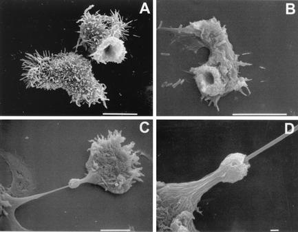
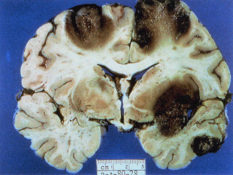
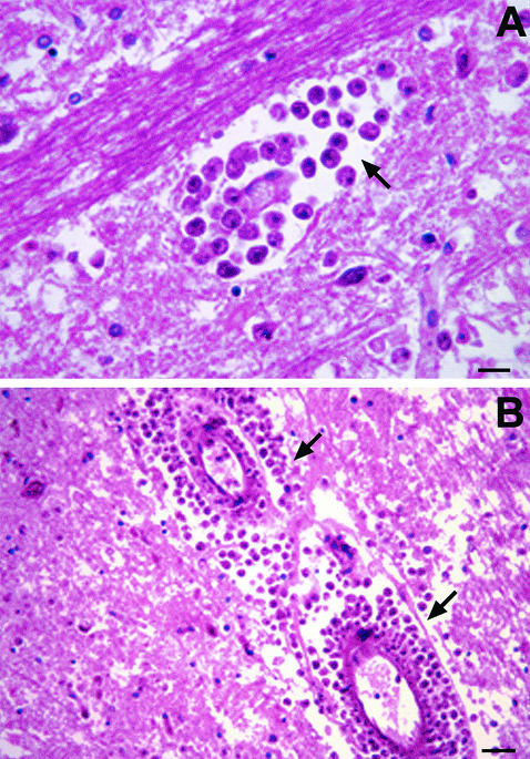
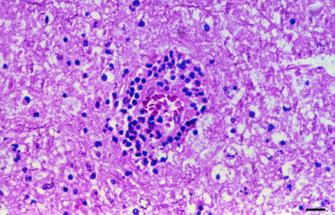
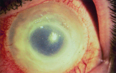
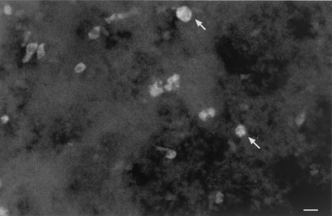
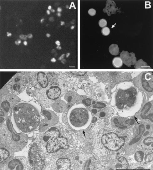
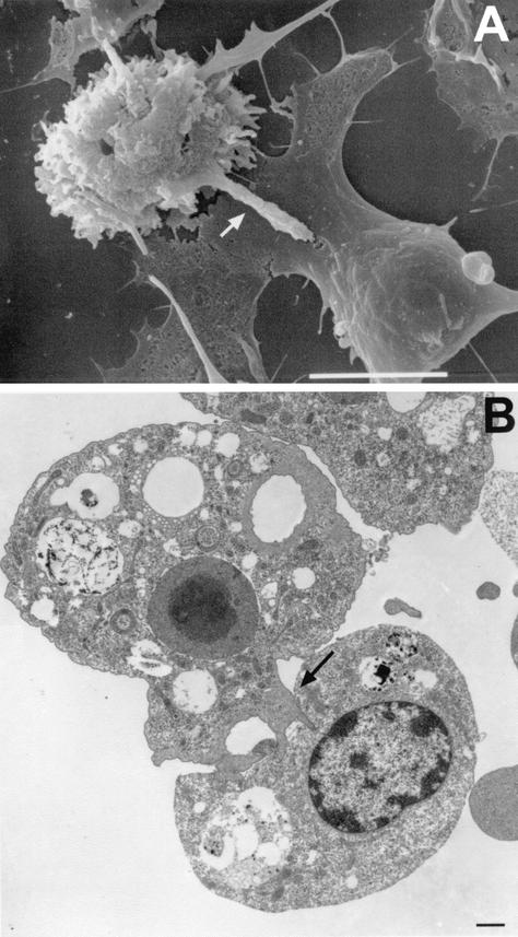
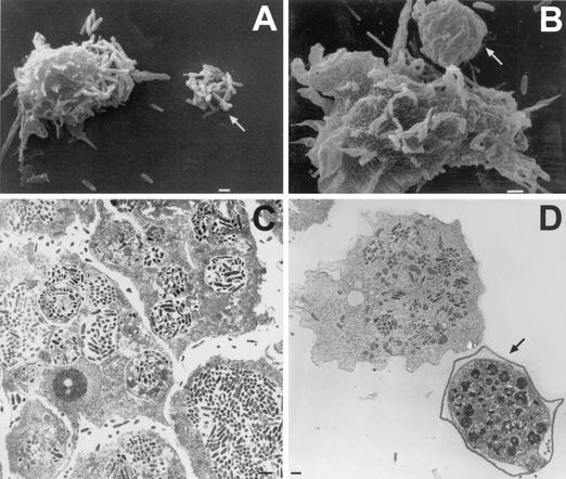
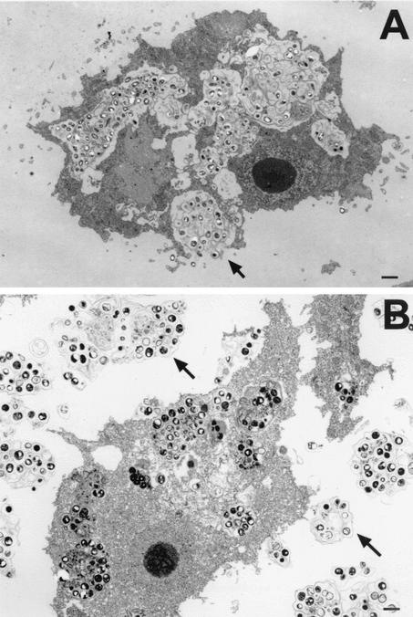
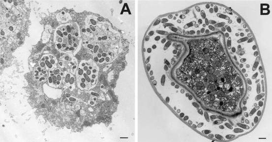
Acanthamoeba spp. are free-living amebae that inhabit a variety of air, soil, and water environments. However, these amebae can also act as opportunistic as well as nonopportunistic pathogens. They are the causative agents of granulomatous amebic encephalitis and amebic keratitis and have been associated with cutaneous lesions and sinusitis. Immuno compromised individuals, including AIDS patients, are particularly susceptible to infections with Acanthamoeba. The immune defense mechanisms that operate against Acanthamoeba have not been well characterized, but it has been proposed that both innate and acquired immunity play a role. The ameba's life cycle includes an active feeding trophozoite stage and a dormant cyst stage. Trophozoites feed on bacteria, yeast, and algae. However, both trophozoites and cysts can retain viable bacteria and may serve as reservoirs for bacteria with human pathogenic potential. Diagnosis of infection includes direct microscopy of wet mounts of cerebrospinal fluid or stained smears of cerebrospinal fluid sediment, light or electron microscopy of tissues, in vitro cultivation of Acanthamoeba, and histological assessment of frozen or paraffin-embedded sections of brain or cutaneous lesion biopsy material. Immunocytochemistry, chemifluorescent dye staining, PCR, and analysis of DNA sequence variation also have been employed for laboratory diagnosis. Treatment of Acanthamoeba infections has met with mixed results. However, chlorhexidine gluconate, alone or in combination with propamidene isethionate, is effective in some patients. Furthermore, effective treatment is complicated since patients may present with underlying disease and Acanthamoeba infection may not be recognized. Since an increase in the number of cases of Acanthamoeba infections has occurred worldwide, these protozoa have become increasingly important as agents of human disease.


Figures

FIG. 1.
FIG. 1.
Phylogenetic scheme of Acanthamoeba, Balamuthia, and Naegleria. Modified from references and .
FIG. 2.
FIG. 2.
Scanning electron micrograph of an Acanthamoeba trophozoite. Spiny surface structures called acanthopodia (arrows) distinguish Acanthamoeba from other free-living amebae that infect humans, such as B. mandrillaris, N. fowleri, and Sappinia diploidea. Bar, 1 μm.
FIG. 3.
FIG. 3.
Light micrographs of cultures depicting life cycle stages of Acanthamoeba spp. (A and C) Unstained preparations of cultures of A. astronyxis trophozoites (A) and cysts (C). (B and D) H-&-E-stained preparations of A. castellanii trophozoites (B) and cysts (D). Bars, represent 50 μm (A and C) and 25 μm (B and D).
FIG. 4.
FIG. 4.
Scanning (A and C) and transmission (B and D) electron micrographs depicting the life cycle stages of Acanthamoeba spp. (A) A. polyphaga trophozoite; (B) trophozoite of A. castellanii showing the prominent central nucleolus (Nu), mitochondria (m), and cytoplasmic food vacuoles (v); (C) wrinkled cyst of A. polyphaga; (D) double-walled cyst of A. castellanii. Bars, 10 μm (A and D) and 1 μm (B and C).
FIG. 5.
FIG. 5.
Scanning electron micrographs of trophozoites illustrating the presence of surface structures termed food cups. (A) Food cups present on the surface of a trophozoite of A. culbertsoni are temporary structures that form and reform for the intake of bacteria, yeast, or cellular debris. (B) Food cup present on the surface of A. astronyxis trophozoite used to ingest bacteria. (C) Food cup present on the surface of an A. castellanii trophozoite in the apparent process of ingesting a cultured nerve cell. (D) Higher magnification of the trophozoite in panel C to illustrate the food cup structure in the apparent process of ingestion. Bars, 10 μm (A to C), and 1 μm (D).
FIG. 6.
FIG. 6.
Coronal section of the cerebral hemispheres with cortical and subcortical necrosis from a fatal human case of GAE. (Courtesy of A. J. Martinez; reprinted from reference 277 with permission of the publisher.)
FIG. 7.
FIG. 7.
H & E stain of brain tissue from a human with GAE. (A). Numerous trophozoites can be identified within the tissue (arrow). (B) Trophozoites identified within vascular walls (arrow). Bars, 150 μm (A) and 300 μm (B). Photographs courtesy of A. J. Martinez.
FIG. 8.
FIG. 8.
H-&-E-stained section of paraffin-embedded brain tissue demonstrating granuloma formation in Acanthamoeba infection. Bar, 200 μm.
FIG. 9.
FIG. 9.
Stromal infiltrate in AK. Photograph of a human with AK provided by P. C. Maudgal, Katholieke Universiteit Leuven, Leuven, Belgium.
FIG. 10.
FIG. 10.
Calcofluor white fluorescent staining of a mouse brain section to identify trophozoites and cysts in infected tissues. Calcofluor white has been used for the identification of cysts (arrow) in cases of AK and can be used to identify cysts in brain tissue or cutaneous lesions. Bar, 50 μm.
FIG. 11.
FIG. 11.
Micrographs illustrating diagnostic methods used to identify Acanthamoeba in infected tissues. (A) Immunofluorescence of trophozoites using anti-ameba antibodies; (B) calcofluor white staining of trophozoites and cysts (arrow) of Acanthamoeba. (C) Electron micrograph of material from infected mice. Electron microscopy has been used to identify cysts (arrows) and trophozoites more readily in infected tissues. Bars, 50 μm (A), 25 μm (B) and 10 μm (C).
FIG. 12.
FIG. 12.
Western immunoblot of Acanthamoeba whole-cell lysates reacted with normal human serum, demonstrating immunoreactivity against Acanthamoeba antigens. Serum samples from two asymptomatic individuals served as a source of natural antibodies to four species of Acanthamoeba. Whole-cell lysates of A. astronyxis (lanes Aa), A. castellanii (lanes Acn), A. culbertsoni (lanes Ac), and A. polyphaga (lanes Ap) were prepared and used as the antigen source. Acanthamoeba protein (50 μg) was added to each well of a sodium dodecyl sulfate-12% polyacrylamide gel. Separated proteins were electrophoretically transferred to a nitrocellulose membrane and were incubated with normal human serum as the source of primary antibody and horseradish peroxidase-labeled goat anti-human IgG as the secondary antibody. The blots were then subjected to enhanced chemiluminescence.
FIG. 13.
FIG. 13.
Transmission electron micrographs demonstrating cellular events in an experimental mouse model of infection with Acanthamoeba. (A) Focal area of infection containing amebae after a 24-h exposure to A. castellanii. (B) Focal area of infection containing amebae after a 48-h exposure to A. castellanii. Neutrophils (N) are prominent in focal areas containing amebae (arrow) during the early phase of infection. Bars, 10 μm.
FIG. 14.
FIG. 14.
Transmission electron micrograph illustrating the accumulation of macrophages in focal areas containing amebae in an experimental mouse model of A. castellanii infection. Macrophages migrate to sites containing amebae during the later phase of infection. (A) Accumulation of macrophages around an A. castellanii cyst (72 h postinfection). (B) Accumulation of macrophages at a site containing amebae (96 h postinfection). Note the presence of an ingested cyst within the macrophage (arrow). Bars, 10 μm (A) and 1 μm (B).
FIG. 15.
FIG. 15.
Scanning electron micrograph of brain microglial cells (Mi) cocultured with A. castellanii (Acn). Microglial cells, the resident macrophages in the brain, are capable of injuring A. castellanii by cell contact-dependent lysis. Bar, 1 μm.
FIG. 16.
FIG. 16.
RNase protection assay illustrating multiple mRNAs elicited by microglia in response to Acanthamoeba. Primary microglial cells obtained from brain cerebral cortices of newborn rat were cocultured with A. castellanii, A. culbertsoni, or bacterial lipopolysaccharide (LPS) for 6 h. The cultures then were harvested with Trizol, and the RNA was subjected to RNase protection assay analysis for assessment of inducible cytokine gene expression. Microglial cells alone (lane MIC), microglia incubated with LPS (lane MIC + LPS), microglia incubated with A. culbertsoni (lane MIC + Ac), and microglia incubated with A. castellanii (lane MIC + Ac50494) are shown. GAPDH, glyceraldehyde-3-phosphate dehydrogenase.
FIG. 17.
FIG. 17.
Scanning (A) and transmission (B) electron micrographs of Acanthamoeba cocultured with rat B103 neuroblastoma cells. (A) A. castellanii with extended digipodium (arrow) into the cytoplasm of the target B103 cell. (B) A. culbertsoni cocultured with B103 cells, demonstrating fingerlike projections extending into the target cell. Cells targeted by digipodia (arrow) eventually die by apoptosis or necrosis. Bars, 10 μm (A) and 1 μm (B).
FIG. 18.
FIG. 18.
Scanning (A and B) and transmission (C and D) electron micrographs of co-cultures of A. astronyxis and L. pneumophila. (A) Trophozoite of A. astronyxis extending a fingerlike projection toward a “clump” of Legionella (arrow). (B) Trophozoite of A. astronyxis infected with L. pneumophila and in the apparent process of expelling a vesicle filled with bacteria (arrow). (C) A. astronyxis trophozoites harboring numerous L. pneumophila cells in cytoplasmic vesicles following coculture in vitro. (D) Cyst of A. astronyxis containing L. pneumophila organisms (arrow). Bars, 1 μm.
FIG. 19.
FIG. 19.
Electron micrographs of cocultures of A. astronyxis and Helicobacter pylori. (A) Trophozoite replete with bacteria and in the apparent process of releasing (arrow) a “bolus” of bacteria (96 h coincubation in vitro). (B) Trophozoite containing numerous intracellular vesicles replete with bacteria and surrounded by bacterium-filled vesicles which apparently have been released from the ameba (96 h coincubation in vitro). Bars, 1 μm.
FIG. 20.
FIG. 20.
Transmission electron micrographs of A. astronyxis cocultured with Pseudomonas aeruginosa. (A) Trophozoite filled with bacteria (72 h of coculture). (B) Cyst of A. astronyxis filled with Pseudomonas (6 days of coculture). Bars, 1 μm.

References

    1. Abu Kwaik, Y., L. Y. Gao, B. J. Stone, and O. S. Harb. 1998. Invasion of mammalian and protozoan cells by Legionella pneumophila. Bull. Inst. Pasteur 96:237-247.
    1. Abu Kwaik, Y., L. Y. Gao, B. J. Stone, C. Venkataraman, and O. S. Harb. 1998. Invasion of protozoa by Legionella pneumophila and its role in bacterial ecology and pathogenesis. Appl. Environ. Microbiol. 64:3127-3133. - PMC - PubMed
    1. Adeleke, A., J. Pruckler, R. Benson, T. Rowbotham, M. Halablab, and B. Fields. 1996. Legionella-like amebal pathogens—phylogenetic status and possible role in respiratory disease. Emerg. Infect. Dis. 2:225-230. - PMC - PubMed
    1. Alfieri, S. C., C. E. Correia, S. A. Motegi, and E. M. Pral. 2000. Proteinase activities in total extracts and in medium conditioned by Acanthamoeba polyphaga trophozoites. J. Parasitol. 86:220-227. - PubMed
    1. Alizadeh, H., S. Apte, M. S. El Agha, L. Li, M. Hurt, K. Howard, H. D. Cavanagh, J. P. McCulley, and J. Y. Niederkorn. 2001. Tear IgA and serum IgG antibodies against Acanthamoeba in patients with Acanthamoeba keratitis. Cornea 20:622-627. - PubMed

Publication types

LinkOut - more resources