

Cloning of the full-length rhesus cytomegalovirus genome as an infectious and self-excisable bacterial artificial chromosome for analysis of viral pathogenesis
- PMID: 12692210
- PMCID: PMC153942
- DOI: 10.1128/jvi.77.9.5073-5083.2003
Cloning of the full-length rhesus cytomegalovirus genome as an infectious and self-excisable bacterial artificial chromosome for analysis of viral pathogenesis
Abstract
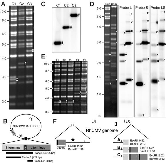
Rigorous investigation of many functions encoded by cytomegaloviruses (CMVs) requires analysis in the context of virus-host interactions. To facilitate the construction of rhesus CMV (RhCMV) mutants for in vivo studies, a bacterial artificial chromosome (BAC) containing an enhanced green fluorescent protein (EGFP) cassette was engineered into the intergenic region between unique short 1 (US1) and US2 of the full-length viral genome by Cre/lox-mediated recombination. Infectious virions were recovered from rhesus fibroblasts transfected with pRhCMV/BAC-EGFP. However, peak virus yields of cells infected with reconstituted progeny were 10-fold lower than wild-type RhCMV, suggesting that inclusion of the 9-kb BAC sequence impeded viral replication. Accordingly, pRhCMV/BAC-EGFP was further modified to enable efficient excision of the BAC vector from the viral genome after transfection into mammalian cells. Allelic exchange was performed in bacteria to substitute the cre recombinase gene for egfp. Transfection of rhesus fibroblasts with pRhCMV/BAC-Cre resulted in a pure progeny population lacking the vector backbone without the need of further manipulation. The genomic structure of the BAC-reconstituted virus, RhCMV-loxP(r), was identical to that of wild-type RhCMV except for the residual loxP site. The presence of the loxP sequence did not alter the expression profiles of neighboring open reading frames. In addition, RhCMV-loxP(r) replicated with wild-type kinetics both in tissue culture and seronegative immunocompetent macaques. Restriction analysis of the viral genome present within individual BAC clones and virions revealed that (i) RhCMV exhibits a simple genome structure and that (ii) there is a variable number of a 750-bp iterative sequence present at the S terminus.
Figures


Similar articles
-
A recombinant rhesus cytomegalovirus expressing enhanced green fluorescent protein retains the wild-type phenotype and pathogenicity in fetal macaques.J Virol. 2002 Sep;76(18):9493-504. doi: 10.1128/jvi.76.18.9493-9504.2002. J Virol. 2002. PMID: 12186931 Free PMC article.
-
Reevaluation of the coding potential and proteomic analysis of the BAC-derived rhesus cytomegalovirus strain 68-1.J Virol. 2012 Sep;86(17):8959-73. doi: 10.1128/JVI.01132-12. Epub 2012 Jun 20. J Virol. 2012. PMID: 22718821 Free PMC article.
-
Construction of a self-excisable bacterial artificial chromosome containing the human cytomegalovirus genome and mutagenesis of the diploid TRL/IRL13 gene.J Virol. 2002 Mar;76(5):2316-28. doi: 10.1128/jvi.76.5.2316-2328.2002. J Virol. 2002. PMID: 11836410 Free PMC article.
-
Cloning of herpesviral genomes as bacterial artificial chromosomes.Rev Med Virol. 2003 Mar-Apr;13(2):111-21. doi: 10.1002/rmv.380. Rev Med Virol. 2003. PMID: 12627394 Review.
-
[BAC system: A novel method for manipulation of herpesvirus genomes based on bacterial genetics].Uirusu. 2004 Dec;54(2):255-64. doi: 10.2222/jsv.54.255. Uirusu. 2004. PMID: 15745165 Review. Japanese.
Cited by
-
Targeted Mutagenesis of Guinea Pig Cytomegalovirus Using CRISPR/Cas9-Mediated Gene Editing.J Virol. 2016 Jul 11;90(15):6989-6998. doi: 10.1128/JVI.00139-16. Print 2016 Aug 1. J Virol. 2016. PMID: 27226370 Free PMC article.
-
Utilizing a TLR5-Adjuvanted Cytomegalovirus as a Lentiviral Vaccine in the Nonhuman Primate Model for AIDS.PLoS One. 2016 May 16;11(5):e0155629. doi: 10.1371/journal.pone.0155629. eCollection 2016. PLoS One. 2016. PMID: 27182601 Free PMC article.
-
Recent advances in cloning herpesviral genomes as infectious bacterial artificial chromosomes.Cell Cycle. 2011 Feb 1;10(3):434-40. doi: 10.4161/cc.10.3.14708. Epub 2011 Feb 1. Cell Cycle. 2011. PMID: 21245660 Free PMC article. Review.
-
Cytomegalovirus-vaccine-induced unconventional T cell priming and control of SIV replication is conserved between primate species.Cell Host Microbe. 2022 Sep 14;30(9):1207-1218.e7. doi: 10.1016/j.chom.2022.07.013. Epub 2022 Aug 17. Cell Host Microbe. 2022. PMID: 35981532 Free PMC article.
-
A sequence-independent in vitro transposon-based strategy for efficient cloning of genomes of large DNA viruses as bacterial artificial chromosomes.Nucleic Acids Res. 2009 Jan;37(1):e2. doi: 10.1093/nar/gkn890. Epub 2008 Nov 6. Nucleic Acids Res. 2009. PMID: 18988631 Free PMC article.
References
-
- Asher, D. M., C. J. Gibbs, Jr., D. J. Lang, D. C. Gajdusek, and R. M. Chanock. 1974. Persistent shedding of cytomegalovirus in the urine of healthy rhesus monkeys. Proc. Soc. Exp. Biol. Med. 145:794-801. - PubMed
Publication types
MeSH terms
Grants and funding
LinkOut - more resources
Full Text Sources
Other Literature Sources
