. 2003 May;77(9):5098-108.
doi: 10.1128/jvi.77.9.5098-5108.2003.

Mutations in the N-terminal region of influenza virus PB2 protein affect virus RNA replication but not transcription

Affiliations

Mutations in the N-terminal region of influenza virus PB2 protein affect virus RNA replication but not transcription

Pablo Gastaminza et al. J Virol. 2003 May.

Abstract

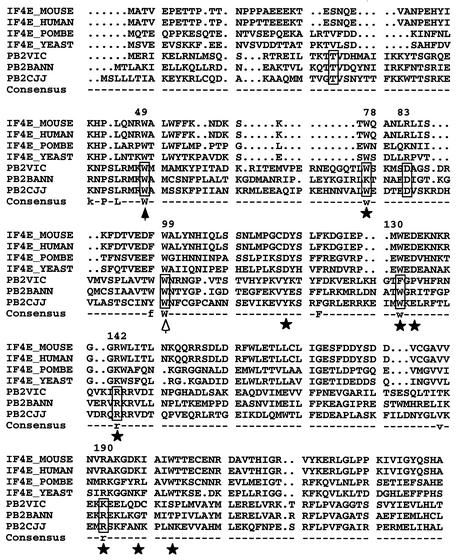
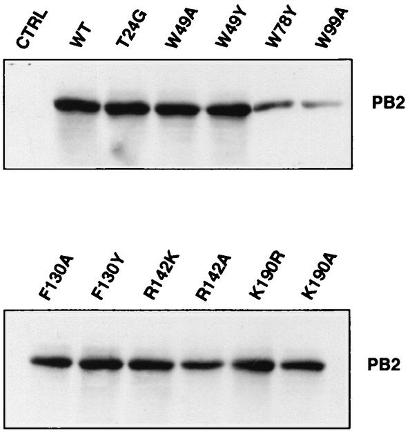
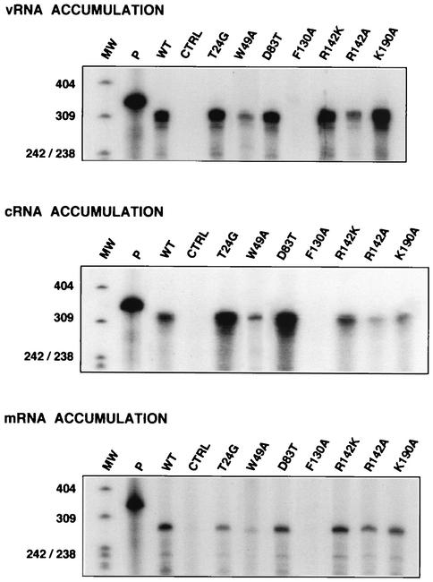
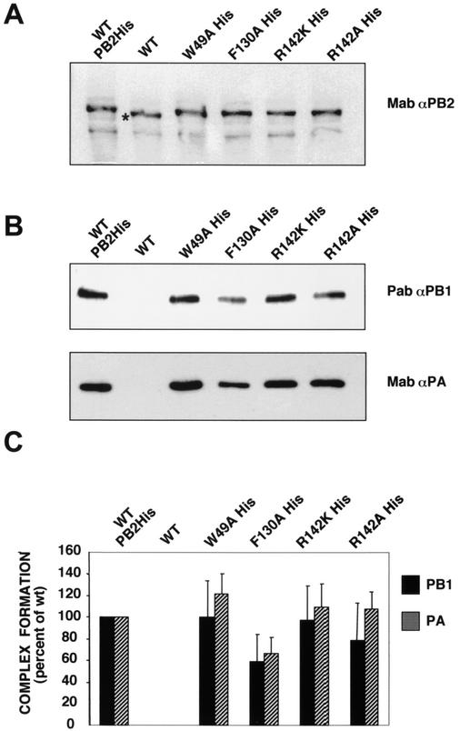
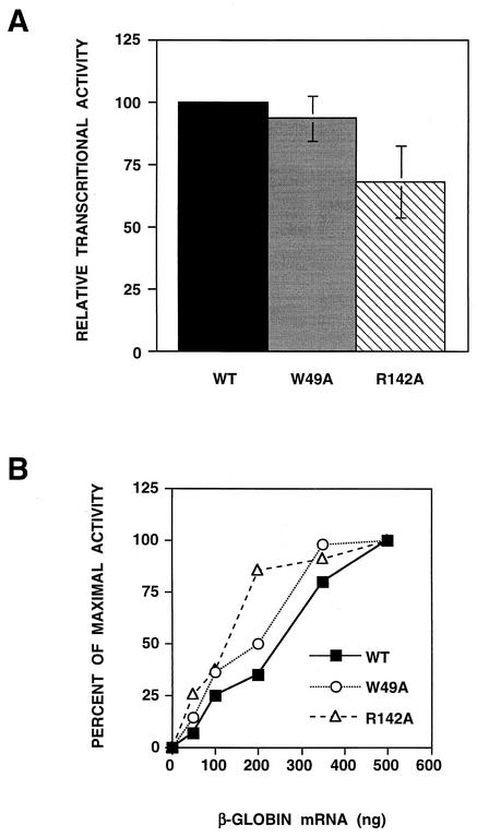
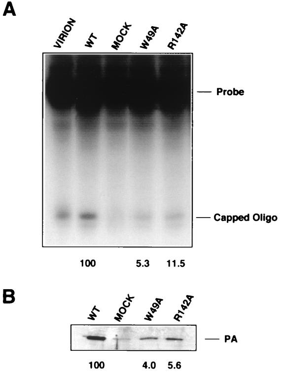
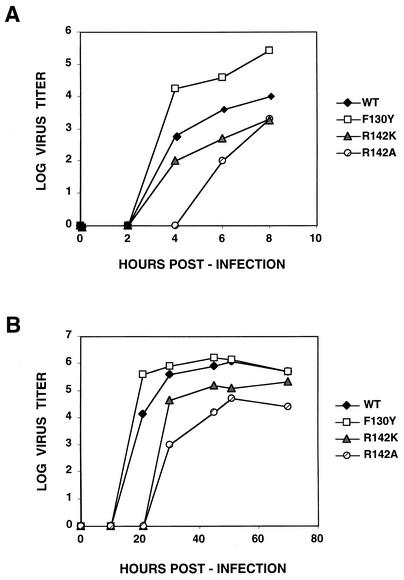
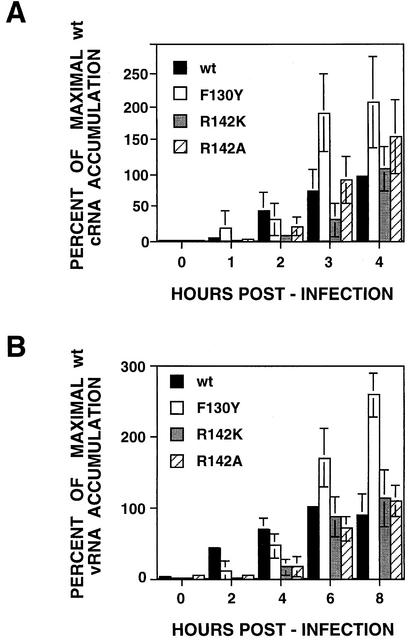
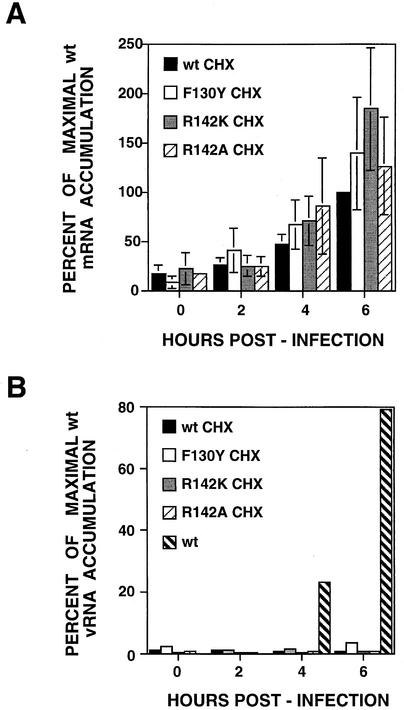
PB2 mutants of influenza virus were prepared by altering conserved positions in the N-terminal region of the protein that aligned with the amino acids of the eIF4E protein, involved in cap recognition. These mutant genes were used to reconstitute in vivo viral ribonucleoproteins (RNPs) whose biological activity was determined by (i) assay of viral RNA, cRNA, and mRNA accumulation in vivo, (ii) cap-dependent transcription in vitro, and (iii) cap snatching with purified recombinant RNPs. The results indicated that the W49A, F130A, and R142A mutations of PB2 reduced or abolished the capacity of mutant RNPs to synthesize RNA in vivo but did not substantially alter their ability to transcribe or carry out cap snatching in vitro. Some of the mutations (F130Y, R142A, and R142K) were rescued into infectious virus. While the F130Y mutant virus replicated faster than the wild type, mutant viruses R142A and R142K showed a delayed accumulation of cRNA and viral RNA during the infection cycle but normal kinetics of primary transcription, as determined by the accumulation of viral mRNA in cells infected in the presence of cycloheximide. These results indicate that the N-terminal region of PB2 plays a role in viral RNA replication.


Figures

FIG. 1.
FIG. 1.
Alignment of PB2 and eIF4E protein sequences. The figure shows the alignment of PB2 proteins from influenza A, B, and C type viruses with a series of eIF4E proteins from various origins (POMBE, Schizosaccharomyces pombe; YEAST, Saccharomyces cerevisiae). The positions of PB2 protein included in boxes were mutagenized as indicated in the text. Stars mark conserved residues of eIF4E involved in interaction with the cap structure (40). The solid arrowhead indicates a position conserved in eIF4E whose mutation altered cap recognition, although it does not contact the cap structure. The open arrowhead indicates a position conserved in eIF4E but not involved in cap recognition. The numbers at the top of the sequence indicate the positions in the A/Victoria/3/75 sequence. The consensus line indicates residues conserved among PB2 and eIF4E sequences.
FIG. 2.
FIG. 2.
Accumulation of wild-type (WT) and PB2 mutant proteins upon reconstitution of RNPs in vivo. Viral RNPs were reconstituted in vivo by transfection of plasmids encoding PB1, PB2, PA, and NP together with a ribozyme-containing plasmid that expresses a short vRNA template. Equal amounts of total cell extracts were assayed for accumulation of PB2 protein by Western blot with PB2-specific monoclonal antibodies. As a control (CTRL), RNP reconstitution was carried out in the absence of PB2-encoding plasmid.
FIG. 3.
FIG. 3.
Replication and transcription in vivo of viral RNPs containing wild-type or mutant PB2 subunits. The accumulation of mRNA and cRNA was determined by RNase protection assay of polyadenylated and nonpolyadenylated RNA, respectively, derived from reconstitution experiments in which vRNA-like template was provided in vivo. Likewise, vRNA accumulation was determined from reconstitution experiments in which cRNA-like template was provided in vivo. Control (CTRL) experiments lacked PB2-encoding plasmid in the reconstitution. P, probe used for RNase protection; MW, size markers (nucleotides).
FIG. 4.
FIG. 4.
Polymerase complex formation in vivo with wild-type and mutant PB2 proteins. The formation of polymerase complexes was determined by pulldown experiments from extracts of cells transfected with pGPB1, pGPA, pT7NSΔCAT-RT, and either wild-type (WT) or mutant pGPB2His plasmids, with Ni2+-nitrilotriacetic acid resin. As a control for binding specificity to the resin, extracts transfected with pT7NSΔCAT-RT, pGPB1, pGPB2, and pGPA plasmids were used. (A) Accumulation of PB2 proteins as revealed by Western blot with PB2-specific monoclonal antibodies. The star indicates the faster mobility of wild-type, untagged PB2 protein. (B) Complex formation as detected by Western blot of the proteins retained in the resin, with either PA-specific monoclonal antibodies (Mab) or PB1-specific serum (Pab). (C) Quantitation of three independent experiments performed as indicated in the panels above. The bars indicate the average and standard deviation of Western blot signals quantitated in a Bio-Rad Chemidoc, with the values obtained for wild-type PB2His complex used as a standard.
FIG. 5.
FIG. 5.
Transcriptional activity in vitro of purified RNPs containing wild-type (WT) or mutant PB2 subunits. Recombinant RNPs were generated by reconstitution in vivo as indicated above and purified by two cycles of glycerol gradient centrifugation. Purified RNPs were assayed for transcription in vitro with globin mRNA as the primer donor, and the results were corrected for the amount of RNPs used, as determined by Western blot with anti-PA monoclonal antibodies (see Fig. 6). (A) Maximal transcriptional activity was assayed with saturating amounts of primer donor. The results are presented as a percentage of the activity of wild-type RNPs and represent the average and range of two independent experiments. (B) Dose-response of primer donor. The transcriptional activity was measured with increasing amounts of primer donor. The results are presented as a percentage of maximal activity.
FIG. 6.
FIG. 6.
Cap-snatching activity in vitro of purified RNPs containing wild-type (WT) or mutant PB2 subunits. Recombinant RNPs, prepared and purified as indicated in the legend to Fig. 5, and virion-derived RNPs were tested for cap-snatching activity with a labeled probe capped and O-methylated in vitro. No cap snatching was observed in the absence of either capping or O-methylation of the probe. (A) The reaction products were separated in a 18% polyacrylamide-urea gel. The positions of the labeled probe and capped oligonucleotide product are indicated. (B) Western blot of parallel aliquots of the same RNPs used for cap snatching, with anti-PA monoclonal antibodies. MOCK, material obtained after purification of RNPs reconstituted in the absence of RNA template. The values are the percentage of capped oligonucleotide bands (A) or PA bands (B), with the wild-type value set at 100%.
FIG. 7.
FIG. 7.
Kinetics of virus multiplication of wild-type (WT) and mutant viruses. Cultures of MDCK cells were infected at a multiplicity of 5 to 10 PFU/cell (A) or 10−2 PFU/cell (B). Aliquots of the culture supernatants were collected at the times indicated, and the virus titer was determined on MDCK cells.
FIG. 8.
FIG. 8.
Kinetics of viral protein synthesis in wild-type (WT) and mutant virus-infected cells. Cultures of MDCK cells were infected at a multiplicity of 5 to 10 PFU/cell and labeled with [35S]methionine-cysteine. At the times indicated, total cell extracts were prepared in Laemmli sample buffer. The extracts were analyzed by polyacrylamide gel electrophoresis and autoradiography. The mobility of the polymerase subunits (POL) and the NP, NS1, and M1 proteins are indicated to the left. Numbers above each panel indicate the time (hours) after infection at which the extracts were prepared. C, control.
FIG. 9.
FIG. 9.
Kinetics of cRNA and viral vRNA in wild-type (wt) and mutant virus-infected cells. Cultures of MDCK cells were infected at a multiplicity of 5 to 10 PFU/cell. At the times indicated, total cell RNA was isolated. The accumulation of NP cRNA (A) and vRNA (B) was determined by real-time RT-PCR as indicated in Materials and Methods. The results are presented as a percentage of the maximal accumulation for wild-type virus-infected cells and are the averages and standard deviations of three to four determinations of the kinetics of infection.
FIG. 10.
FIG. 10.
Kinetics of viral primary transcription in wild-type (wt) and mutant virus-infected cells. Cultures of MDCK cells were infected at a multiplicity of 5 to 10 PFU/cell. The cultures were maintained in the presence of cycloheximide (CHX, 100 μg/ml) from the time of virus adsorption. At the times indicated, total cell RNA was isolated. The accumulation of NP mRNA (A) and vRNA (B) was determined by real-time RT-PCR as indicated in Materials and Methods. The results are presented as a percentage of the maximal accumulation for wild-type virus-infected cells and are the averages and standard deviations of four determinations of the kinetics of infection. The hatched bars in B indicate the accumulation of vRNA in a parallel infection carried out in the absence of cycloheximide.

Similar articles

Cited by

References

    1. Asano, Y., and A. Ishihama. 1997. Identification of two nucleotide binding domains on the PB1 subunit of influenza virus RNA polymerase. J. Biochem. 122:627-634. - PubMed
    1. Asano, Y., K. Mizumoto, T. Maruyama, and A. Ishihama. 1995. Photoaffinity labeling of influenza virus RNA polymerase PB1 subunit with 8-azido GTP. J. Biochem. Tokyo 117:677-682. - PubMed
    1. Bárcena, J., M. Ochoa, S. de la Luna, J. A. Melero, A. Nieto, J. Ortín, and A. Portela. 1994. Monoclonal antibodies against influenza virus PB2 and NP polypeptides interfere with the initiation step of viral mRNA synthesis in vitro. J. Virol. 68:6900-6909. - PMC - PubMed
    1. Biswas, S. K., P. L. Boutz, and D. P. Nayak. 1998. Influenza virus nucleoprotein interacts with influenza virus polymerase proteins. J. Virol. 72:5493-5501. - PMC - PubMed
    1. Biswas, S. K., and D. P. Nayak. 1994. Mutational analysis of the conserved motifs of influenza A virus polymerase basic protein 1. J. Virol. 68:1819-1826. - PMC - PubMed

Publication types

MeSH terms

LinkOut - more resources