. 2003 May;77(9):5127-35.
doi: 10.1128/jvi.77.9.5127-5135.2003.

Minimal cis-acting elements required for adenovirus genome packaging

Affiliations

Minimal cis-acting elements required for adenovirus genome packaging

Philomena Ostapchuk et al. J Virol. 2003 May.

Abstract

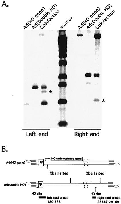
The design of drugs for treatment of virus infections and the exploitation of viruses as drugs for treatment of diseases could be made more successful by understanding the molecular mechanisms of virus-specific events. The process of assembly, and more specifically packaging of the genome into a capsid, is an obligatory step leading to future infections. To enhance our understanding of the molecular mechanism of packaging, it is necessary to characterize the viral components necessary for the event. In the case of adenovirus, sequences between nucleotides 200 and 400 at the left end of the genome are essential for packaging. This region contains a series of redundant bipartite sequences, termed A repeats, that function in packaging. Synthetic packaging sequences made of multimers of a single A repeat substitute for the authentic adenovirus packaging domain. A repeats are binding sites for the CCAAT displacement protein and the viral protein IVa2. Several lines of evidence implicate these proteins in the packaging process. It was not known, however, whether other cis-acting elements play a role in the packaging process as well. We utilized an in vivo approach to address the role of the inverted terminal repeats and the covalently linked terminal proteins in packaging of the adenovirus genome. Our results show that these elements are not necessary for efficient packaging of the viral genome. A significant implication of these results applicable to gene therapy vector design is that the linkage of the adenovirus packaging domain to heterologous DNA sequences should suffice for targeting to the viral capsid.


Figures

FIG. 1.
FIG. 1.
Schematic diagram of the structures of the Ad used in this study. Strands of DNA are indicated by double black lines. Grey boxes indicate the ITRs, ovals indicate the covalently linked terminal protein, and the white boxes indicate the packaging sequences (ψ). The curved lines indicate that the diagram is not drawn to scale. Ad(ITR:HO:ψ) has a 135-bp fragment containing a cleavage site for S. cerevisiae HO endonuclease inserted at nt 106. Ad(Double HO) contains the HO site at nt 106 as well as a second HO site in the E3 region inserted at nt 29602. The HO gene virus has a cassette containing the cytomegalovirus enhancer/promoter sequences, a synthetic splice site, and the S. cerevisiae HO gene sequences inserted in place of Ad5 nt 355 to 2965.
FIG. 2.
FIG. 2.
Ad genomes lacking the left ITR package efficiently. (A) 293 cells were infected with the Ad(ITR:HO:Ψ) virus or the Ad(HO gene) virus or were coinfected with both viruses. Forty-eight hours postinfection a fraction of the cells was used to isolate nuclear DNA. Viral particles were isolated from the remaining fraction, and viral DNA was prepared. Shown are the results of a Southern blot analysis of the DNAs digested with XbaI and detected by using a probe corresponding to Ad nt 180 to 828. The lane labeled Marker indicates the molecular size standards. XbaI digestion of the Ad(ITR:HO:Ψ) genome gave rise to a DNA fragment of 1,480 bp. Upon cleavage with HO endonuclease, this fragment was reduced to two fragments of 1,320 (indicated by an asterisk) and 190 bp. XbaI digestion of the Ad(HO gene) genome gave rise to a DNA fragment of ∼1,000 bp. (B) The structures of the Ad(ITR:HO:Ψ) and Ad(HO gene) viral genomes. The HO cleavage site, relevant XbaI sites, and the probe positions are indicated.
FIG. 3.
FIG. 3.
Elimination of the packaging domain in addition to the left ITR results in the loss of packaging of the right-end fragment. (A) 293 cells were infected with either Ad(HO gene) or Ad(E1 HO site) or were coinfected with both viruses. Forty-eight hours postinfection nuclear DNA (labeled Nuclear DNA) was isolated from cells from the three different infections. Viral particles were isolated from cells coinfected with both viruses, and viral DNA was purified (labeled Viral DNA). Shown are the results of a Southern blot of DNAs digested with SmaI and detected by using a probe corresponding to Ad nt 2982 to 3655. SmaI digestion of the Ad(E1 HO site) virus DNA yields a left-end fragment of 1,850 nt. Upon cleavage with HO endonuclease, this fragment was reduced to two fragments of 1,400 and 450 bp. The uncut fragment of 1,850 bp and the 1,400-bp HO-cut fragment (indicated by an asterisk) hybridize to the probe. The 450-bp left end was not recognized by the probe used. (B) The structures of the genomes of the Ad(HO gene) and Ad(E1 HO site) viral genomes. The HO cleavage site, relevant SmaI sites, and the probe position are indicated. Both viruses are derived from an E1 deletion of nt 356 to 2965 and carry different-size inserts, hence the relative difference in the sizes of the left ends.
FIG. 4.
FIG. 4.
The left-end 160-bp fragment from the HO-cleaved Ad(ITR:HO:Ψ) genome is not packaged. 293 cells were infected with Ad(HO gene) or Ad(ITR:HO:Ψ) or were coinfected with both viruses. Shown are the results of a Southern blot analysis of nuclear DNAs and viral particle DNAs prepared after virus infection. Also shown is a titration of a plasmid standard. The plasmid used for the standard was pEB-HO (see Materials and Methods), a plasmid that contains Ad nt 1 to 1340 with the HO site inserted at nt 106. Digestion of pEB-HO with EcoRI and BglII yielded three fragments due to partial cleavage at the BglII site. A fragment of 249 bp corresponds to Ad sequences 1 to 106 plus the 135-bp HO site insertion; the fragment of 108 bp includes Ad nt 1 to 106. The 700-bp probe used was derived from pEB-HO and includes Ad nt 1 to 194 and 506 nt of adjacent plasmid sequences. The largest fragment corresponds to the remaining plasmid and Ad sequences. The number of molecules of plasmid loaded in each lane of the standard is indicated. Molecules (∼9 × 10⁹) of HO-cleaved DNA were loaded in the nuclear DNA (labeled Coinfection). Molecules (∼1 × 10¹⁰) of HO-cleaved DNA were loaded in the viral particle lane (labeled Coinfection). Shown below is a longer exposure of the lower region of the Southern blot encompassing the smaller fragments of 249 and 108 bp. The asterisk indicates the 160-nt fragment unique to the coinfection, i.e., the HO endonuclease-cleaved DNA fragment.
FIG. 5.
FIG. 5.
Left- and right-end HO sites in the Ad(Double HO) genome are cleaved by HO endonuclease. (A) Nuclear DNAs were isolated 24 h postinfection from infections of N52.E6 cells with either Ad(HO gene), Ad(Double HO), or a coinfection of both viruses. Results of Southern blot analysis of XbaI-digested DNAs hybridized with either a left-end probe (Ad nt 180 to 828) or right-end probe (Ad nt 28667 to 29169) are shown. XbaI digestion of the Ad(Double HO) genome gave rise to a DNA fragment of 2,067 bp. Upon cleavage by HO endonuclease, two DNA fragments of 1,016 and 1,051 bp were generated. The asterisks indicate the XbaI fragments that are unique to the coinfection and recognized by the probe. (B) Illustrated are the structures of the Ad(Double HO) and Ad(HO gene) virus genomes. The HO-cut sites, relevant XbaI sites, and the probe's positions are indicated.
FIG. 6.
FIG. 6.
Both right-end-cut and left-end-cut genomes are found in viral particles from a coinfection with Ad(Double HO) and Ad(HO gene). (A) The results of Southern blot analysis of DNA isolated from infected cell nuclei (labeled Nuclear DNA) and from CsCl gradient purified viral particles (labeled Viral DNA) are shown. DNAs were digested with XbaI and were hybridized with either a probe of Ad nt 180 to 828 (labeled Left end) or Ad nt 28667 to 29169 (labeled Right end). The bands in the hybridization that are unique to the coinfection are indicated by asterisks. (B) Schematic diagrams for the four different genomic structures of Ad(Double HO) that could be generated by cleavage with HO endonuclease at either or both of the HO sites.
FIG. 7.
FIG. 7.
Viral particles with genomes lacking the right end are enriched from particles with full-length genomes on a CsCl equilibrium gradient. 293 cells were coinfected with Ad(Double HO) and Ad(HO gene) viruses. Viral particles were enriched for the lighter-density fraction on a CsCl equilibrium gradient and were repurified by using a second gradient (see Materials and Methods). Fractions were collected from the top of the gradient, and DNA was purified from the particles in each fraction. The XbaI-digested DNAs were hybridized with the right-end probe, Ad nt 28667 to 29169, as described in the legend to Fig. 4.

Similar articles

Cited by

References

    1. Banan, M., I. C. Rojas, W. H. Lee, H. L. King, J. V. Harriss, R. Kobayashi, C. F. Webb, and P. D. Gottlieb. 1997. Interaction of the nuclear matrix-associated region (MAR)-binding proteins, SATB1 and CDP/Cux, with a MAR element (L2a) in an upstream regulatory region of the mouse CD8a gene. J. Biol. Chem. 272:18440-18452. - PubMed
    1. Bjornsti, M. A., B. E. Reilly, and D. L. Anderson. 1983. Morphogenesis of bacteriophage phi29 of Bacillus subtilis: oriented and quantized in vitro packaging of DNA protein gp3. J. Virol. 45:383-396. - PMC - PubMed
    1. Bridge, E., S. Medghalchi, S. Ubol, M. Leesong, and G. Ketner. 1993. Adenovirus early region 4 and viral DNA synthesis. Virology 193:794-801. - PubMed
    1. Carmo-Fonseca, M. 2002. The contribution of nuclear compartmentalization to gene regulation. Cell 108:513-521. - PubMed
    1. Cremer, T., and C. Cremer. 2001. Chromosome territories, nuclear architecture and gene regulation in mammalian cells. Nat. Rev. Genet. 2:292-301. - PubMed

Publication types

LinkOut - more resources