. 2003 May;77(9):5253-65.
doi: 10.1128/jvi.77.9.5253-5265.2003.

The Papillomavirus E2 protein binds to and synergizes with C/EBP factors involved in keratinocyte differentiation

Affiliations

The Papillomavirus E2 protein binds to and synergizes with C/EBP factors involved in keratinocyte differentiation

Dirk Hadaschik et al. J Virol. 2003 May.

Abstract

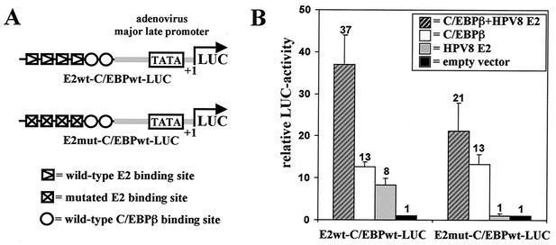
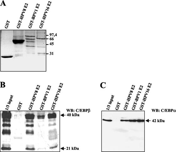
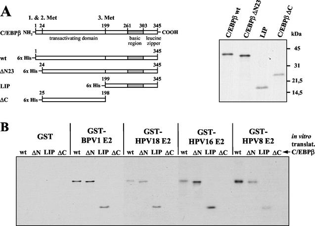
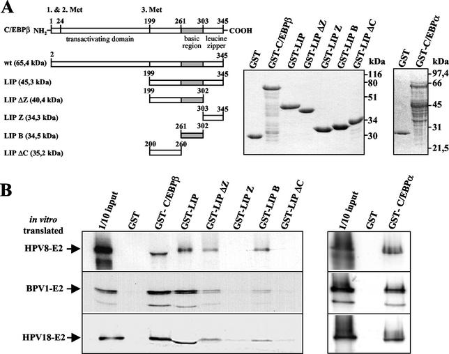
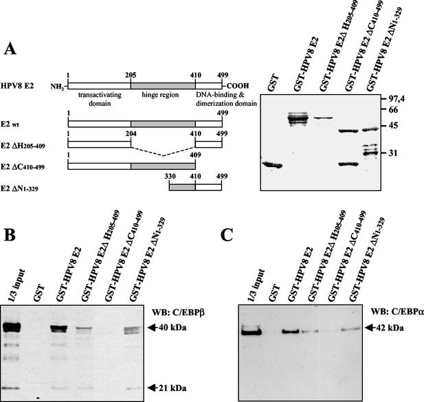
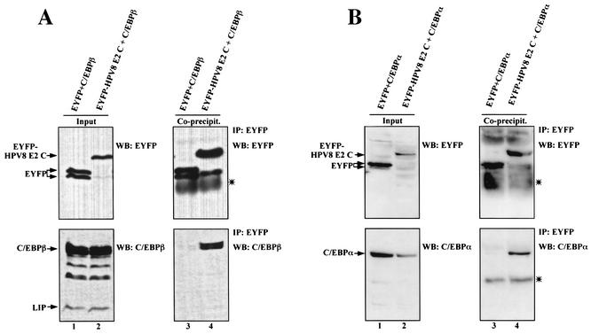
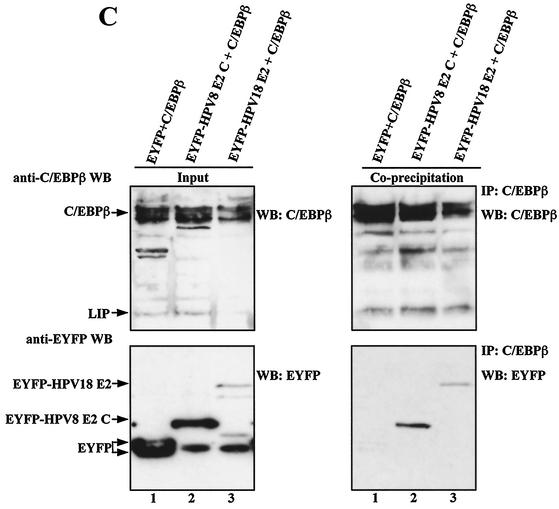
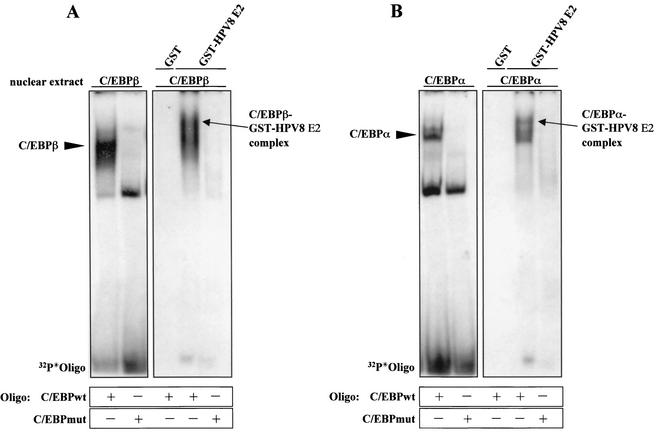
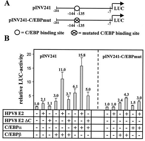
The papillomavirus life cycle is closely linked to the differentiation program of the host keratinocyte. Thus, late gene expression and viral maturation are restricted to terminally differentiated keratinocytes. A variety of cellular transcription factors including those of the C/EBP family are involved in the regulation of keratinocyte differentiation. In this study we show that the papillomavirus transcription factor E2 cooperates with C/EBPalpha and -beta in transcriptional activation. This synergism was independent of an E2 binding site. E2 and C/EBP factors synergistically transactivated a synthetic promoter construct containing classical C/EBPbeta sites and the C/EBPalpha-responsive proximal promoter of the involucrin gene, which is naturally expressed in differentiating keratinocytes. C/EBPalpha or -beta coprecipitated with E2 proteins derived from human papillomavirus type 8 (HPV8), HPV16, HPV18, and bovine papillomavirus type 1 in vitro and in vivo, indicating complex formation by the cellular and viral factors. The interaction domains could be mapped to the C terminus of E2 and amino acids 261 to 302 located within the bZIP motif of C/EBPbeta. Our data suggest that E2, via its interaction with C/EBP factors, may contribute to enhancing keratinocyte differentiation, which is suppressed by the viral oncoproteins E6 and E7 in HPV-induced lesions.


Figures

FIG. 1.
FIG. 1.
HPV8 E2 and C/EBPβ synergize in transcriptional activation. (A) Schematic presentation of reporter plasmids containing the adenovirus major late promoter and two C/EBP and four intact or mutated E2 binding sites upstream of the luciferase gene. (B) C33A cells were seeded in six-well plates. The next day the reporter plasmid E2wt-C/EBPwt-LUC or E2mut-C/EBPwt-LUC (0.5 μg each) was transfected with or without plasmids expressing C/EBPβ (0.8 μg) and HPV8 E2 (0.4 μg). The total amount of DNA was adjusted with empty pcDNA3.1+ vector. After 48 h luciferase activities were determined and normalized with the protein concentration of the respective luciferase extract. The value of the control (empty pcDNA3.1+ vector only) was set for 1. Transfections were conducted in duplicate, and the indicated values were averaged from at least three independent experiments.
FIG. 2.
FIG. 2.
E2 physically interacts with C/EBPα and C/EBPβ. Nuclear extracts were prepared from C33A cells overexpressing C/EBPα or C/EBPβ. In coprecipitation assays GST or E2 proteins from different papillomavirus types fused to GST (A) were bound to glutathione-Sepharose and incubated with these extracts. After washing, the bound proteins were subjected to SDS-PAGE. C/EBPβ (B) and C/EBPα (C) were identified by Western blotting with anti-C/EBPβ or anti-C/EBPα antibodies. In panel B the larger (40-kDa) and the shorter (21-kDa) forms of C/EBPβ were detected. WB, Western blot. Numbers at right of panel A are molecular masses in kilodaltons.
FIG. 3.
FIG. 3.
E2 binds to the C terminus of C/EBPβ. (A) Schematic presentation of C/EBPβ deletion mutants translated in vitro (left panel). Equal amounts of these proteins were resolved by SDS-PAGE (right panel). (B) In pull-down assays GST, GST-BPV1 E2, GST-HPV18 E2, GST-HPV16 E2, and GST-HPV8 E2 were bound to glutathione-Sepharose and incubated with in vitro-translated C/EBPβ and its deletion mutants. C/EBPβ proteins were visualized by SDS-PAGE and autoradiography.
FIG. 4.
FIG. 4.
Fine mapping of the E2-interaction domain within C/EBPβ. (A) Schematic presentation of C/EBPβ deletion mutants fused to GST (left panel). Bacterially expressed and purified C/EBPβ or C/EBPα GST fusion proteins (middle and right panels, respectively) were used to precipitate in vitro-translated E2 proteins from HPV8, HPV18, and BPV1 (B). After washing, the bound 35S-labeled E2 proteins were separated by SDS-PAGE and detected by autoradiography.
FIG. 5.
FIG. 5.
C/EBP factors interact with the C-terminal part of HPV8 E2. (A) Schematic presentation of HPV8 E2 deletion mutants fused to GST (left panel). The hinge region is marked as a gray box, and all amino acid positions are indicated. In pull-down assays GST and the GST-HPV8 E2 fusion proteins (right panel) were coupled to glutathione-Sepharose and incubated with nuclear extracts from C33A cells overexpressing C/EBPβ (B) or C/EBPα (C). All bound proteins were separated by SDS-PAGE. The C/EBP factors were identified by Western blot (WB) analysis with the respective antibodies. Numbers at right of panel A are molecular masses in kilodaltons.
FIG. 6.
FIG. 6.
Coimmunoprecipitation of C/EBPβ or C/EBPα and the HPV E2 protein in vivo. Plasmids encoding EYFP-HPV8 E2 C (10 μg) or equimolar amounts of EYFP vector were cotransfected with C/EBPβ (A) or C/EBPα (B) expression plasmids (20 μg) into 293T cells (150-cm2 dishes). For panel C 20 μg of EYFP-HPV8 E2 C or EYFP-HPV18 E2 or equimolar amounts of EYFP vector and 10 μg of C/EBPβ plasmid were used. The amount of DNA was adjusted with empty pBluescript SKII(+) vector. After 24 h extracts were incubated with rabbit polyclonal EYFP-specific antibodies (A and B) or rabbit polyclonal C/EBPβ-specific antibodies (C) and protein G-Sepharose. The precipitates were washed and resolved by SDS-PAGE (lanes 3 and 4 in panels A and B and right panels in panel C). Left panels of each panel show 1/33 (A, lanes 1 and 2), 1/70 (B, lanes 1 and 2), or 1/100 (C, lanes 1 to 3) of the input extracts used for coprecipitation. EYFP fusion proteins, C/EBPβ, or C/EBPα was detected by Western blotting with rabbit anti-EYFP peptide antibody (A and B), mouse anti-EYFP antibody (C), mouse anti-C/EBPβ antibody, or goat anti-C/EBPα antibody. The asterisks indicate the light chains of immunoglobulins used for immunoprecipitation which were detected by the secondary antibodies in Western blot analysis. WB, Western blot; IP, immunoprecipitation.
FIG. 6.
FIG. 6.
Coimmunoprecipitation of C/EBPβ or C/EBPα and the HPV E2 protein in vivo. Plasmids encoding EYFP-HPV8 E2 C (10 μg) or equimolar amounts of EYFP vector were cotransfected with C/EBPβ (A) or C/EBPα (B) expression plasmids (20 μg) into 293T cells (150-cm2 dishes). For panel C 20 μg of EYFP-HPV8 E2 C or EYFP-HPV18 E2 or equimolar amounts of EYFP vector and 10 μg of C/EBPβ plasmid were used. The amount of DNA was adjusted with empty pBluescript SKII(+) vector. After 24 h extracts were incubated with rabbit polyclonal EYFP-specific antibodies (A and B) or rabbit polyclonal C/EBPβ-specific antibodies (C) and protein G-Sepharose. The precipitates were washed and resolved by SDS-PAGE (lanes 3 and 4 in panels A and B and right panels in panel C). Left panels of each panel show 1/33 (A, lanes 1 and 2), 1/70 (B, lanes 1 and 2), or 1/100 (C, lanes 1 to 3) of the input extracts used for coprecipitation. EYFP fusion proteins, C/EBPβ, or C/EBPα was detected by Western blotting with rabbit anti-EYFP peptide antibody (A and B), mouse anti-EYFP antibody (C), mouse anti-C/EBPβ antibody, or goat anti-C/EBPα antibody. The asterisks indicate the light chains of immunoglobulins used for immunoprecipitation which were detected by the secondary antibodies in Western blot analysis. WB, Western blot; IP, immunoprecipitation.
FIG. 7.
FIG. 7.
HPV8 E2 forms complexes with C/EBPβ and C/EBPα in a DNA-bound state. Nuclear extracts were prepared from 293T cells overexpressing C/EBPβ (A) or C/EBPα (B). Sixty micrograms of each extract was incubated with wild-type or mutated C/EBP oligonucleotides for 40 min at 4°C. Ten percent of these reaction mixtures were investigated by EMSA (left panels). GST and the GST-HPV8 E2 fusion proteins (Fig. 2C) were coupled to glutathione-Sepharose and added to 90% of these mixtures for 2 h in LSDB buffer containing 100 mM KCl at 4°C. After washing, complexes were eluted in 25 μl of EMSA buffer containing 25 mM glutathione for 1 h at 4°C and then resolved by EMSA (right panels). Oligo, oligonucleotide.
FIG. 8.
FIG. 8.
HPV8 E2 and C/EBP factors synergistically transactivate the involucrin promoter. (A) Schematic presentation of the reporter plasmid pINV241 comprising the proximal promoter fragment of the involucrin promoter upstream of the luciferase gene with wild-type or mutated C/EBP binding sites. (B) pINV241 (left panel) or pINV241-C/EBPmut (right panel) (0.5 μg each) was transfected in RTS3b cells with or without plasmids expressing C/EBPβ (0.8 μg), C/EBPα (0.01 μg), and HPV8 E2 full-length (0.4 μg) or equimolar amounts of HPV8 E2 ΔC lacking the C terminus of E2. The total amount of DNA was adjusted with empty pcDNA3.1+ vector. After 48 h luciferase activities were determined and normalized with the protein concentration of the respective luciferase extract. The value of the control (empty pcDNA3.1+ vector only) was set for 1. Transfections were conducted in duplicate. The indicated values were averaged from three independent experiments.

Similar articles

Cited by

References

    1. Agarwal, C., T. Efimova, J. F. Welter, J. F. Crish, and R. L. Eckert. 1999. CCAAT/enhancer-binding proteins. A role in regulation of human involucrin promoter response to phorbol ester. J. Biol. Chem. 274:6190-6194. - PubMed
    1. Akira, S., H. Isshiki, T. Sugita, O. Tanabe, S. Kinoshita, Y. Nishio, T. Nakajima, T. Hirano, and T. Kishimoto. 1990. A nuclear factor for IL-6 expression (NF-IL6) is a member of a C/EBP family. EMBO J. 9:1897-1906. - PMC - PubMed
    1. Benson, J. D., R. Lawande, and P. M. Howley. 1997. Conserved interaction of the papillomavirus E2 transcriptional activator proteins with human and yeast TFIIB proteins. J. Virol. 71:8041-8047. - PMC - PubMed
    1. Breiding, D. E., F. Sverdrup, M. J. Grossel, N. Moscufo, W. Boonchai, and E. J. Androphy. 1997. Functional interaction of a novel cellular protein with the papillomavirus E2 transactivation domain. Mol. Cell. Biol. 17:7208-7219. - PMC - PubMed
    1. Crish, J. F., T. M. Zaim, and R. L. Eckert. 1998. The distal regulatory region of the human involucrin promoter is required for expression in epidermis. J. Biol. Chem. 273:30460-30465. - PubMed

Publication types

LinkOut - more resources