. 2003 May;77(9):5370-7.
doi: 10.1128/jvi.77.9.5370-5377.2003.

Conformational changes, plasma membrane penetration, and infection by human rhinovirus type 2: role of receptors and low pH

Affiliations

Conformational changes, plasma membrane penetration, and infection by human rhinovirus type 2: role of receptors and low pH

Marianne Brabec et al. J Virol. 2003 May.

Abstract

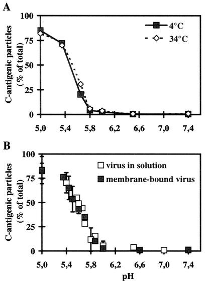
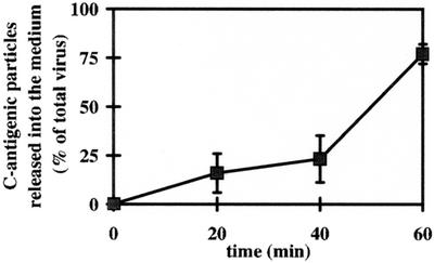
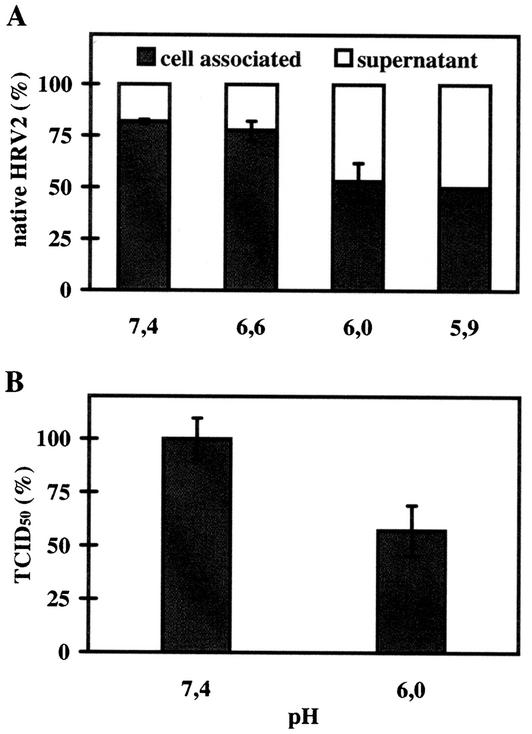
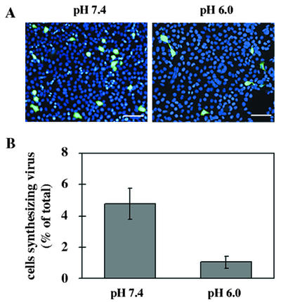
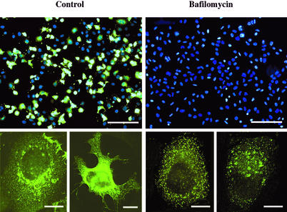
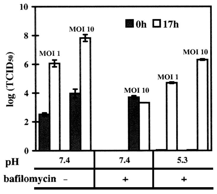
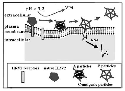
Human rhinovirus type 2 (HRV2) is internalized by members of the low-density lipoprotein (LDL) receptor (LDLR) family. It then progresses into late endosomes, where it undergoes conversion from D- to C-antigenicity at pH < 5.6. Upon uncoating, the viral RNA is transferred into the cytoplasm across the endsosomal membrane. However, C-antigenic particles fail to attach to LDLR; this raised the question of whether the virus remains attached to the receptors and is carried to late compartments or rather falls off at the higher pH in early endosomes. We therefore determined the pH dependence of virus-receptor dissociation and virus conversion to C-antigen under conditions preventing endocytosis. (35)S-HRV2 was attached to HeLa cells at 4 degrees C and incubated in buffers of pH 7.4 to 5.0; levels of native virus and C-antigenic particles remaining cell associated or having been released into the medium were determined by immunoprecipitation. At pH 6.0, HRV2 was readily released from plasma membrane receptors in its native form, whereas at pH < or = 5.4, it was entirely converted to C-antigen, which, however, only dissociated from the surface upon prolonged incubation. The antigenic conversion occurred at the same pH regardless of whether HRV2 was free in solution or bound to its receptors. These data suggest that, in vivo, the virus is no longer bound to its receptors when the antigenic conversion and uncoating occur in more acidic late endosomes. When virus was bound to HeLa cells at 4 degrees C, converted into C-antigen by exposure to pH 5.3, and subsequently warmed to 34 degrees C in the presence of bafilomycin (to prevent endosomal uncoating), viral de novo synthesis was detected. This study demonstrates for the first time that a nonenveloped virus such as HRV2 can infect from the plasma membrane when artificially exposed to low pH. This implies that the viral RNA can gain access to the cytoplasm from the plasma membrane.


Figures

FIG. 1.
FIG. 1.
Influence of temperature and receptor binding on the pH-dependent conformational change of HRV2 in solution. (A) 35S-labeled HRV2 (1.5 × 10⁴ cpm) was incubated in 500 μl of isotonic buffers at the pHs indicated for 20 min at 4 or 34°C. The percentage of C-antigenic particles formed was then determined by sequential immunoprecipitation with MAb 2G2 and rabbit antiserum against HRV2 and scintillation counting. The data are expressed as the percentage of total 35S-labeled HRV2. The data represent the mean ± standard deviation for three independent experiments. (B) 35S-labeled HRV2 (2 × 10⁴ cpm) was bound to 106 HeLa cells at 4°C for 60 min, and nonbound virus was washed away with cold PBS. Cell-bound virus or free virus (1.5 × 10⁴ cpm) was then incubated at 4°C for 20 min in isotonic buffers at the pH indicated. The percentage of C-antigen (cell associated plus supernatant in the case of cell-bound virus [for details, see Materials and Methods]) was determined as described for panel A. The data represent the mean ± standard deviation for 11 independent experiments carried out in triplicate.
FIG. 2.
FIG. 2.
Kinetics of dissociation of C-antigenic HRV2 from HeLa cells. 35S-labeled HRV2 (2 × 10⁴ cpm) was bound to 106 HeLa cells at 4°C for 60 min, nonbound virus was washed away with cold PBS, and the cells were incubated in 500 μl of isotonic buffer at pH 5.0 and 7.4 (control [data not shown]) at 4°C. At the times indicated, the supernatant was removed, and the cells were lysed. C-antigen present in the supernatant and in the cell lysate was determined as described for Fig. 1B. Data represent the mean ± standard deviation for three independent experiments, presented as a percentage of total virus bound to the cells at time 0. Note that only 80% of total C-antigen can be immunoprecipitated, corresponding to 100% conversion (Fig. 1A).
FIG. 3.
FIG. 3.
Dissociation of native HRV2 from the cell surface. (A) 35S-HRV2 (2 × 10⁴ cpm) was bound to 106 HeLa cells for 1 h at 4°C, unbound virus was removed, and cells were incubated in isotonic buffers at different pHs for 20 min at 4°C. Native virus in the cell pellet or in the supernatant was determined by immunoprecipitation. The average of three independent experiments carried out in triplicate ± standard deviation is shown. (B) HeLa cells grown in 24-well plates were preincubated in infection medium for 30 min at 34°C. HRV2 at an MOI of 10 was added and allowed to attach for 1 h on ice. Cells were washed and exposed to isotonic buffers at pHs 6.0 and 7.4 (control, 100%) for 1 h on ice; cell-associated infectious virus was then determined as TCID50. The data represent the mean ± standard deviation for three independent experiments.
FIG. 4.
FIG. 4.
Infection at pH 6 is less efficient. HRV2 was preincubated for 20 min and subsequently bound to HeLa cells (MOI of 1) at pH 7.4 (control) and at 6.0 for 1 h at 4°C. Cells were washed, unbound virus was removed, and cells were further incubated in infection medium at 34°C for 17 h. (A) Cells were fixed, and HRV2 was stained with MAb 8F5 followed by antimouse Alexa 488-conjugated IgG. Nuclei were revealed with Hoechst stain. Bars, 100 μm. (B) The percentage of cells synthesizing viral antigen was determined in two different experiments with a total of 1,500 cells counted.
FIG. 5.
FIG. 5.
HRV2 synthesis is blocked by 20 nM bafilomycin: Cells were preincubated without (control) and with 20 nM bafilomycin for 30 min, followed by incubation with an MOI of HRV2 of 10 at 4°C for 1 h (± bafilomycin). Cells were washed and further incubated for 17 h ± bafilomycin at 34°C. Cells were fixed, viral antigen was stained with MAb 8F5 followed by antimouse IgG conjugated to Alexa 488, and nuclei were stained with Hoechst dye. The upper panels show an overlay of nuclear staining and staining for viral antigen (bars, 200 μm). The lower panels show higher magnification of representative single cells with Alexa 488 staining only (bars, 10 μm).
FIG. 6.
FIG. 6.
HRV2 infection can occur from the plasma membrane at low pH. Cells grown in 24-well plates were preincubated without and with bafilomycin (40 nM) for 30 min, challenged with HRV2 at an MOI of 1 or 10 at 4°C, and exposed to buffers at pH 7.4 (control) or 5.3. After further incubation in infection medium for 17 h, the viral titer was compared to that at time 0. Where indicated, bafilomycin was maintained at a concentration of 20 nM. The data represent the mean ± standard deviation for three independent experiments.
FIG. 7.
FIG. 7.
Potential mechanism of HRV2 uncoating at the plasma membrane upon exposure to low pH. HRV2 binds to its receptors close to the top of the fivefold axis. Upon low-pH treatment, hydrophobic domains in the N terminus of VP1 become externalized, giving rise to C-antigenic particles that are subsequently released from the receptors. Insertion of these domains into the plasma membrane can now occur, resulting in pore formation and RNA release.

References

    1. Bayer, N., E. Prchla, M. Schwab, D. Blaas, and R. Fuchs. 1999. Human rhinovirus HRV14 uncoats from early endosomes in the presence of bafilomycin. FEBS Lett. 463:175-178. - PubMed
    1. Bayer, N., D. Schober, E. Prchla, R. F. Murphy, D. Blaas, and R. Fuchs. 1998. Effect of bafilomycin A1 and nocodazole on endocytic transport in HeLa cells: implications for viral uncoating and infection. J. Virol. 72:9645-9655. - PMC - PubMed
    1. Belnap, D. M., B. M. McDermott, Jr., D. J. Filman, N. Cheng, B. L. Trus, H. J. Zuccola, V. R. Racaniello, J. M. Hogle, and A. C. Steven. 2000. Three-dimensional structure of poliovirus receptor bound to poliovirus. Proc. Natl. Acad. Sci. USA 97:73-78. - PMC - PubMed
    1. Blake, K., and S. O'Connell. 1993. Virus culture, p. 81-122. In D. R. Harper (ed.), Virology Labfax. Blackwell Scientific Publications, London, United Kingdom.
    1. Brown, M. S., J. Herz, and J. L. Goldstein. 1997. LDL receptor structure: calcium cages, acid baths and recycling receptors. Nature 388:629-630. - PubMed

Publication types

LinkOut - more resources