. 2003 May;77(9):5389-400.
doi: 10.1128/jvi.77.9.5389-5400.2003.

Disulfide bonding among micro 1 trimers in mammalian reovirus outer capsid: a late and reversible step in virion morphogenesis

Affiliations

Disulfide bonding among micro 1 trimers in mammalian reovirus outer capsid: a late and reversible step in virion morphogenesis

Amy L Odegard et al. J Virol. 2003 May.

Abstract

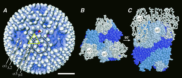
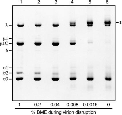
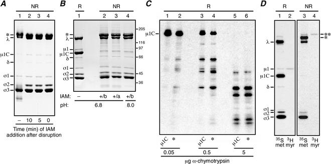
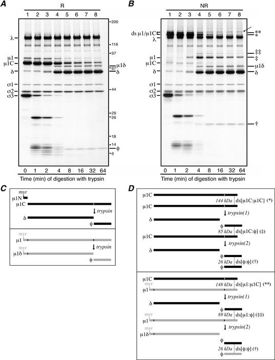
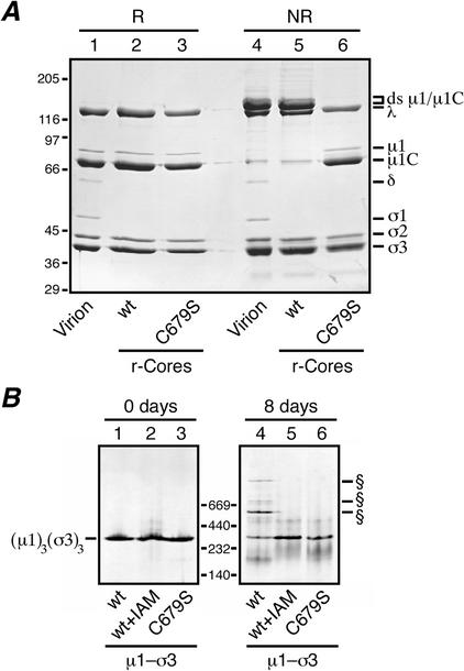
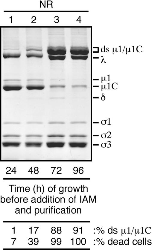
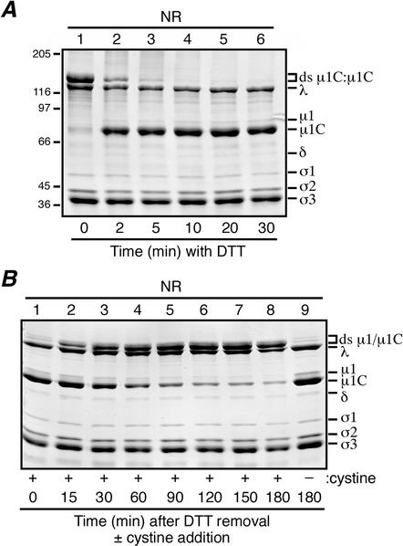
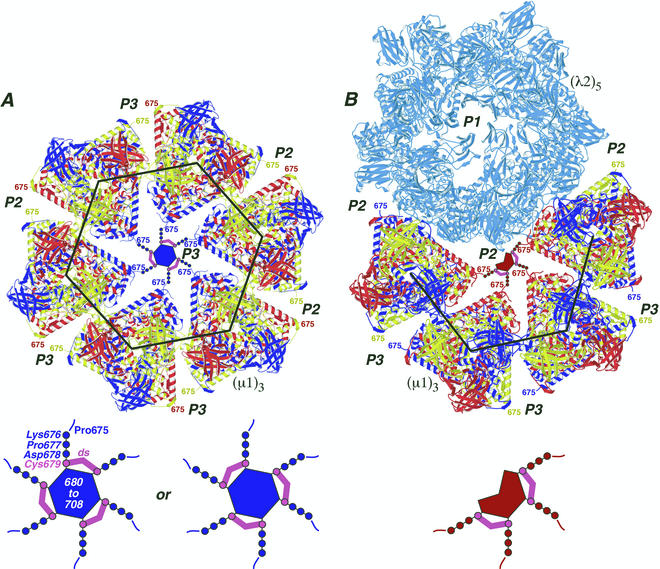
We examined how a particular type of intermolecular disulfide (ds) bond is formed in the capsid of a cytoplasmically replicating nonenveloped animal virus despite the normally reducing environment inside cells. The micro 1 protein, a major component of the mammalian reovirus outer capsid, has been implicated in penetration of the cellular membrane barrier during cell entry. A recent crystal structure determination supports past evidence that the basal oligomer of micro 1 is a trimer and that 200 of these trimers surround the core in the fenestrated T=13 outer capsid of virions. We found in this study that the predominant forms of micro 1 seen in gels after the nonreducing disruption of virions are ds-linked dimers. Cys679, near the carboxyl terminus of micro 1, was shown to form this ds bond with the Cys679 residue from another micro 1 subunit. The crystal structure in combination with a cryomicroscopy-derived electron density map of virions indicates that the two subunits that contribute a Cys679 residue to each ds bond must be from adjacent micro 1 trimers in the outer capsid, explaining the trimer-dimer paradox. Successful in vitro assembly of the outer capsid by a nonbonding mutant of micro 1 (Cys679 substituted by serine) confirmed the role of Cys679 and suggested that the ds bonds are not required for assembly. A correlation between micro 1-associated ds bond formation and cell death in experiments in which virions were purified from cells at different times postinfection indicated that the ds bonds form late in infection, after virions are exposed to more oxidizing conditions than those in healthy cells. The infectivity measurements of the virions with differing levels of ds-bonded micro 1 showed that these bonds are not required for infection in culture. The ds bonds in purified virions were susceptible to reduction and reformation in situ, consistent with their initial formation late in morphogenesis and suggesting that they may undergo reduction during the entry of reovirus particles into new cells.


Figures

FIG. 1.
FIG. 1.
Reovirus outer capsid and μ1. (A) Surface view of the virion from cryoEM and 3D image reconstruction (16). Features attributable to outer capsid proteins σ1, λ2, σ3, and μ1 are visible and marked by arrows. The particle display was radially depth cued to enhance the recognition of symmetrical features (59). One μ1-σ3 heterohexamer, shown in crystallographic detail in panels B and C (34), is enclosed in an open yellow triangle. Six subunits of σ3, associated with six different μ1-σ3 heterohexamers, surround each P3 channel, one of which is enclosed in a black hexagon. A blob of density is visible within each of these channels (red arrowhead; see Discussion for more information) (15, 16). Four subunits of σ3, associated with four different μ1-σ3 heterohexamers, surround each P2 channel, one of which is enclosed in a black partial hexagon. Bar, 20 nm. (B and C) Top and side views, respectively, of the μ1-σ3 heterohexamer by X-ray crystallography (34). The heterohexamer is shown in the same orientation from the top as the one enclosed by the triangle in panel A (note the three white σ3 subunits at the triangle's corners). The σ3 and μ1 subunits are shown in backbone and space-filling formats, respectively. The three interwound subunits of μ1 within its trimer are shown in different shades of blue.
FIG. 2.
FIG. 2.
Effects of reduced concentrations of BME on the migration of reovirus proteins during SDS-PAGE. Purified virions of reovirus T3D were mixed with pH 6.8 sample buffers containing decreasing concentrations of BME and disrupted in a boiling water bath. The viral proteins were then resolved on a full-sized SDS-PAGE (10% acrylamide) gel and visualized by Coomassie staining. Known positions of the viral proteins are indicated by name. The major new high-Mr band observed at low BME concentrations is also indicated (*).
FIG. 3.
FIG. 3.
Effects of IAM addition on the migration of reovirus μ1 and μ1C proteins during SDS-PAGE. (A and B) Purified virions of reovirus T1L were used in these experiments. Viral proteins were resolved on a mini-sized SDS-PAGE [10% (A) or 8% (B) acrylamide] gel and visualized by Coomassie staining. The major new high-Mr band observed after nonreducing disruption is indicated (*). (A) Virions were disrupted in sample buffer that contained no reducing agent (NR), and IAM was either not added (−) or added at different times after disruption (0 to 10 min) as indicated. (B) Virions were disrupted in pH 6.8 or 8.0 sample buffer that contained no reducing agent but to which 50 mM IAM was added either before (b) or immediately after (a) disruption as indicated. A sample of virions disrupted in reducing sample buffer (R; 5 mM DTT) was analyzed on the same gel (lane 1). Positions of molecular mass markers also resolved on the gel are indicated in kilodaltons. (C) Purified [35S]methionine-cysteine-labeled virions of reovirus T3D were used in this experiment. The Coomassie-stained protein bands excised from the first gel (not shown; μ1C was obtained from samples of reduced virions and the high-Mr band [*] was obtained from samples of IAM-treated nonreduced virions) were subjected to reduction in the gel fragments and otherwise treated as described in the text. The resulting proteins and protein fragments were resolved on a second gel and visualized by fluorography. The position of the full-length μ1C monomer is indicated. The amount of chymotrypsin added to each gel piece atop the second gel is also indicated. (D) Purified [35S]methionine-cysteine- or [3H]myristate-labeled virions of reovirus T3D were mixed with reducing or nonreducing sample buffer and disrupted in a boiling water bath. The viral proteins were then resolved on a full-sized 5 to 20% acrylamide gradient SDS-PAGE gel and visualized by fluorography. The major (*) and minor (**) new high-Mr bands observed after nonreducing disruption of each sample are indicated.
FIG. 4.
FIG. 4.
Trypsin digestions of reovirus particles. (A and B) Purified [35S]methionine-cysteine-labeled virions of reovirus T1L were treated with trypsin at 32°C. At each specified time, digestion was stopped by adding trypsin inhibitor. For the 0-min aliquot (lanes A1 and B1), trypsin inhibitor was added before trypsin. Each aliquot was then split in two, with half being mixed with reducing sample buffer (R) and half being mixed with nonreducing sample buffer (NR). Following disruption, proteins from the reduced (A) and nonreduced (B) samples were separately resolved on full-sized 5 to 20% acrylamide gradient SDS-PAGE gels and visualized by fluorography. The putative φ:φ homodimer band is indicated in panel B (†). The putative μ1C:φ and μ1:φ heterodimer bands are also indicated in panel B (‡ and ‡‡, respectively). A band of unknown origin in panel B, lanes 4 to 8, is indicated by an arrow; this band was not routinely observed in other experiments. Positions of molecular mass markers also resolved on the gel are indicated in kilodaltons in panel A. (C and D) The μ1 cleavage products are diagrammed, including the position of the intermolecular ds bond. Predicted molecular masses of the ds-bonded species are shown in kilodaltons. (C, upper box) Trypsin cleavage of the reduced μ1C monomer to yield monomeric fragments δ and φ. The myristoylated (myr) μ1N fragment arising along with μ1C from cleavage of full-length μ1 is also shown. (C, lower box) Trypsin cleavage of the reduced μ1 monomer to yield monomeric fragments μ1δ and φ. (D, upper box) Trypsin cleavage of the ds-bonded μ1C:μ1C homodimer. The necessity for nonsimultaneous cleavages of the two μ1C chains within each dimer results in an intermediate, heterodimer product that is later cleaved again. The first trypsin cleavage [trypsin(1)] yields a monomeric δ fragment and a ds-bonded μ1C:φ heterodimer. The second trypsin cleavage [trypsin(2)] then resolves the heterodimer into another monomeric δ fragmentand a ds-bonded φ:φ homodimer. (D, lower box) Trypsin cleavage of the ds-bonded μ1:μ1C heterodimer. Similar to that in panel C, the necessity for nonsimultaneous cleavages of the two chains within the dimer results in an intermediate heterodimer product that is later cleaved again. In this example, the first trypsin cleavage [trypsin(1)] acts on the μ1C chain to yield a monomeric δ fragment and a ds-bonded μ1:φ heterodimer. The second trypsin cleavage [trypsin(2)] then resolves this new heterodimer into another monomeric μ1δ fragment and a ds-bonded φ:φ homodimer. Not shown is the case in which the first trypsin cleavage acts on the μ1 chain in the μ1:μ1C heterodimer. Also not shown is the pattern of trypsin cleavage of the μ1:μ1 homodimer.
FIG. 5.
FIG. 5.
Analyses for ds bond formation with μ1 mutant C679S. (A) Recoated cores containing T1L wt σ3 and either T1L wt μ1 or the T1L μ1 mutant C679S were generated and purified, and particle concentrations were determined by densitometry. Equal amounts of the wt (lanes 2 and 5) or C679S (lanes 3 and 6) recoated cores were mixed with reducing (R) or nonreducing (NR) sample buffer, disrupted by boiling, and resolved on a mini-sized SDS-PAGE (8% acrylamide) gel. Virions disrupted under reducing (lane 1) or nonreducing (lane 4) conditions were included for comparison. Viral proteins were visualized by Coomassie staining. (B) Purified μ1-σ3 heterohexamers stored frozen in buffer with 10 mM DTT were thawed, and a small amount of each was diluted into nonreducing sample buffer and analyzed on a 4 to 15% acrylamide gradient native gel (Amersham Pharmacia Biotech) (lanes 1 to 3; 0 days). The remainder of each sample was passed through a PD-10 column to remove DTT and then stored at 4°C for 8 days at ambient conditions. At the end of that time, a small amount of each was diluted into sample buffer and analyzed on another 4 to 15% native gel (lanes 4 to 6). Three types of μ1-σ3 preparations were included in this analysis: complexes containing wt T1L μ1 and σ3 (lanes 1 and 4), complexes containing wt T1L μ1 and σ3 that were treated with 10 mM IAM to block free cysteines before storage (lanes 2 and 5), and complexes containing T1L C679S μ1 and wt T1L σ3 (lanes 3 and 6). The position of the native μ1-σ3 heterohexamer is indicated to the left. The positions of higher-Mr forms specific to the complexes containing wt T1L μ1 and σ3 (lanes 1) are indicated (§). Positions of native gel markers (Amersham Pharmacia Biotech) are indicated in kilodaltons.
FIG. 6.
FIG. 6.
Formation of the μ1/μ1C ds bonds during the course of infection. T1L reovirus-infected cells were harvested at either 24, 48, 72, or 96 h postinfection. 50 mM IAM was added immediately upon resuspension of the infected cells in homogenization buffer. After purification, virions from each time point were mixed with nonreducing sample buffer (NR) and disrupted by boiling. The viral proteins were then resolved on a mini-sized SDS-8% PAGE gel. The amount of ds-bonded μ1/μ1C (% ds μ1/μ1C) relative to the total μ1, μ1C, and ds-bonded μ1/μ1C in each preparation was determined by densitometry of the Coomassie-stained gel. Immediately before harvesting, aliquots were taken from each time point and the proportion of dead/dying cells (% dead cells) was determined by trypan blue staining.
FIG. 7.
FIG. 7.
Reversibility of the virion-associated μ1/μ1C ds bonds. For each experiment, aliquots were removed from the reaction mixture at the indicated intervals, and the reaction was quenched with 50 mM IAM. When all samples had been collected for each experiment, they were mixed with nonreducing sample buffer (NR) and disrupted by boiling. Viral proteins were resolved on mini-sized SDS-8% PAGE gels and visualized by Coomassie staining. (A) DTT at a concentration of 5 mM was added to purified T1L virions and allowed to incubate at room temperature to effect in situ reduction of the ds bonds. The 0-min aliquot (lane 1) was removed before the addition of DTT. Positions of molecular mass markers also resolved on the gel are indicated to the left. (B) The ds bonds in a sample of T1L virions were reduced by DTT as in panel A, lane 6, after which the DTT was removed by dialysis. Cystine at a concentration of 5 mM was then added to promote in situ reformation of the ds bonds. The 0-min aliquot (lane 1) was removed before the addition of cystine. A sample to which cysteine was never added was also analyzed (lane 9).
FIG. 8.
FIG. 8.
Locations of the ds bonds in the μ1 outer capsid structure. The crystal structure of μ1 was extracted from the μ1-σ3 heterohexamer complex and positioned as it appears on the surface of the virion (34). Each μ1 trimer [(μ1)3] is colored blue, red, and yellow to represent the three individual μ1 subunits. The large black hexagon and partial hexagon indicate the respectively marked regions in Fig. 1 except that the σ3 subunits are missing and the region of the partial hexagon is rotated downward by approximately 60° relative to that in Fig. 1. (A) Six μ1 trimers are shown surrounding one of the 60 P3 channels. Surrounding channels are also labeled (P2 or P3). The C-terminal residue in the μ1 crystal structure, Pro675, faces into the channels and is labeled in each of the μ1 subunits. A model for μ1 residues 676 to 708, which were not visualized in the crystal structure of the μ1-σ3 heterohexamer (34), is shown within the P3 channel that is central in this image. Cys679 and its ds bonds are shown in magenta. Residues 676 to 678 and 680 to 708 are shown in blue to match the six surrounding μ1 subunits from which they extend. Residues 680 to 708 are proposed to form the central blob evident in Fig. 1 and discussed in the text and reference . A magnified view of these modeled features is shown at the bottom, with labels for residues 675 to 708 and a ds bond. The alternative arrangement of the three ds bonds is shown at bottom right. (B) Four μ1 trimers and a λ2 pentamer [(λ2)5] (light blue) are shown surrounding one of the 60 P2 solvent channels. The P1 channel, which is surrounded by the five λ2 subunits and plugged by σ1 in virions (not shown), is included and labeled in this view. A model for μ1 residues 676 to 708 is shown within the P2 channel that is central in this image. Other labeling and color coding is as described for panel A, except that residues 676 to 678 and 680 to 708 are shown in red to match the four surrounding μ1 subunits from which they extend.

Similar articles

Cited by

References

    1. Aslund, F., and J. Beckwith. 1999. Bridge over troubled waters: sensing stress by disulfide bond formation. Cell 96:751-753. - PubMed
    1. Attoui, H., Q. Fang, F. M. Jaafar, J. F. Cantaloube, P. Biagini, P. De Micco, and X. De Lamballerie. 2002. Common evolutionary origin of aquareoviruses and orthoreoviruses revealed by genome characterization of Golden shiner reovirus, Grass carp reovirus, Striped bass reovirus and golden ide reovirus (genus Aquareovirus, family Reoviridae). J. Gen. Virol. 83:1941-1951. - PubMed
    1. Barton, E. S., J. L. Connolly, J. C. Forrest, J. D. Chappell, and T. S. Dermody. 2000. Utilization of sialic acid as a coreceptor enhances reovirus attachment by multi-step adhesion-strengthening. J. Biol. Chem. 276:2200-2211. - PubMed
    1. Barton, E. S., J. C. Forrest, J. L. Connolly, J. D. Chappell, Y. Liu, F. J. Schnell, A. Nusrat, C. A. Parkos, and T. S. Dermody. 2001. Junction adhesion molecule is a receptor for reovirus. Cell 104:441-451. - PubMed
    1. Bodkin, D. K., M. L. Nibert, and B. N. Fields. 1989. Proteolytic digestion of reovirus in the intestinal lumens of neonatal mice. J. Virol. 63:4676-4681. - PMC - PubMed

Publication types

MeSH terms

LinkOut - more resources