Crystal structure of Swine vesicular disease virus and implications for host adaptation
- PMID: 12692248
- PMCID: PMC153985
- DOI: 10.1128/jvi.77.9.5475-5486.2003
Crystal structure of Swine vesicular disease virus and implications for host adaptation
Abstract
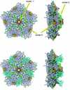
Swine vesicular disease virus (SVDV) is an Enterovirus of the family Picornaviridae that causes symptoms indistinguishable from those of foot-and-mouth disease virus. Phylogenetic studies suggest that it is a recently evolved genetic sublineage of the important human pathogen coxsackievirus B5 (CBV5), and in agreement with this, it has been shown to utilize the coxsackie and adenovirus receptor (CAR) for cell entry. The 3.0-A crystal structure of strain UK/27/72 SVDV (highly virulent) reveals the expected similarity in core structure to those of other picornaviruses, showing most similarity to the closest available structure to CBV5, that of coxsackievirus B3 (CBV3). Features that help to cement together and rigidify the protein subunits are extended in this virus, perhaps explaining its extreme tolerance of environmental factors. Using the large number of capsid sequences available for both SVDV and CBV5, we have mapped the amino acid substitutions that may have occurred during the supposed adaptation of SVDV to a new host onto the structure of SVDV and a model of the SVDV/CAR complex generated by reference to the cryo-electron microscopy-visualized complex of CBV3 and CAR. The changes fall into three clusters as follows: one lines the fivefold pore, a second maps to the CAR-binding site and partially overlaps the site for decay accelerating factor (DAF) to bind to echovirus 7 (ECHO7), and the third lies close to the fivefold axis, where the low-density lipoprotein receptor binds to the minor group of rhinoviruses. Later changes in SVDV (post-1971) map to the first two clusters and may, by optimizing recognition of a pig CAR and/or DAF homologue, have improved the adaptation of the virus to pigs.
Figures


Similar articles
-
Structure of swine vesicular disease virus: mapping of changes occurring during adaptation of human coxsackie B5 virus to infect swine.J Virol. 2003 Sep;77(18):9780-9. doi: 10.1128/jvi.77.18.9780-9789.2003. J Virol. 2003. PMID: 12941886 Free PMC article.
-
More recent swine vesicular disease virus isolates retain binding to coxsackie-adenovirus receptor, but have lost the ability to bind human decay-accelerating factor (CD55).J Gen Virol. 2005 May;86(Pt 5):1369-1377. doi: 10.1099/vir.0.80669-0. J Gen Virol. 2005. PMID: 15831949
-
The coxsackie-adenovirus receptor (CAR) is used by reference strains and clinical isolates representing all six serotypes of coxsackievirus group B and by swine vesicular disease virus.Virology. 2000 May 25;271(1):99-108. doi: 10.1006/viro.2000.0324. Virology. 2000. PMID: 10814575
-
Swine vesicular disease virus. Pathology of the disease and molecular characteristics of the virion.Anim Health Res Rev. 2000 Dec;1(2):119-26. doi: 10.1017/s1466252300000104. Anim Health Res Rev. 2000. PMID: 11708597 Review.
-
The coxsackievirus and adenovirus receptor.Curr Top Microbiol Immunol. 2008;323:67-87. doi: 10.1007/978-3-540-75546-3_4. Curr Top Microbiol Immunol. 2008. PMID: 18357766 Review.
Cited by
-
Characterization of a putative ancestor of coxsackievirus B5.J Virol. 2010 Oct;84(19):9695-708. doi: 10.1128/JVI.00071-10. Epub 2010 Jul 14. J Virol. 2010. PMID: 20631132 Free PMC article.
-
Structure of Seneca Valley Virus-001: an oncolytic picornavirus representing a new genus.Structure. 2008 Oct 8;16(10):1555-61. doi: 10.1016/j.str.2008.07.013. Structure. 2008. PMID: 18940610 Free PMC article.
-
Structures of Coxsackievirus A16 Capsids with Native Antigenicity: Implications for Particle Expansion, Receptor Binding, and Immunogenicity.J Virol. 2015 Oct;89(20):10500-11. doi: 10.1128/JVI.01102-15. Epub 2015 Aug 12. J Virol. 2015. PMID: 26269176 Free PMC article.
-
Catching a virus in the act of RNA release: a novel poliovirus uncoating intermediate characterized by cryo-electron microscopy.J Virol. 2010 May;84(9):4426-41. doi: 10.1128/JVI.02393-09. Epub 2010 Feb 24. J Virol. 2010. PMID: 20181687 Free PMC article.
-
Enteric viruses of humans and animals in aquatic environments: health risks, detection, and potential water quality assessment tools.Microbiol Mol Biol Rev. 2005 Jun;69(2):357-71. doi: 10.1128/MMBR.69.2.357-371.2005. Microbiol Mol Biol Rev. 2005. PMID: 15944460 Free PMC article. Review.
References
-
- Acharya, R., E. Fry, D. I. Stuart, G. Fox, D. Rowlands, and F. Brown. 1989. The three-dimensional structure of foot-and-mouth disease virus at 2.9A resolution. Nature 337:709-716. - PubMed
-
- Arnold, E., and M. G. Rossmann. 1990. Analysis of the structure of a common cold virus, human rhinovirus 14, refined at a resolution of 3.0A. J. Mol. Biol. 211:763-801. - PubMed
-
- Bergelson, J. M., J. A. Cunningham, G. Droguett, E. A. Kurt-Jones, A. Krithivas, J. S. Hong, M. S. Horwitz, R. L. Crowell, and R. W. Finberg. 1997. Isolation of a common receptor for coxsackie B viruses and adenoviruses 2 and 5. Science 275:1320-1323. - PubMed
Publication types
MeSH terms
Substances
Associated data
- Actions
LinkOut - more resources
Full Text Sources
Miscellaneous

