. 2003 Apr 29;100(9):5164-9.
doi: 10.1073/pnas.0834959100. Epub 2003 Apr 11.

Band 3 is a host receptor binding merozoite surface protein 1 during the Plasmodium falciparum invasion of erythrocytes

Affiliations

Band 3 is a host receptor binding merozoite surface protein 1 during the Plasmodium falciparum invasion of erythrocytes

Vikas K Goel et al. Proc Natl Acad Sci U S A. .

Erratum in

  • Proc Natl Acad Sci U S A. 2003 Sep 16;100(19):11184

Abstract

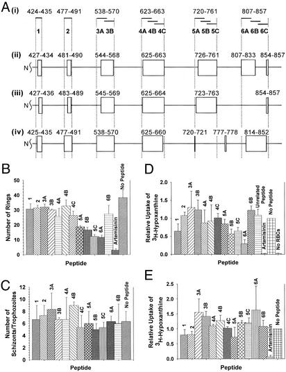
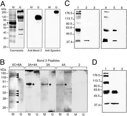
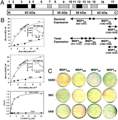
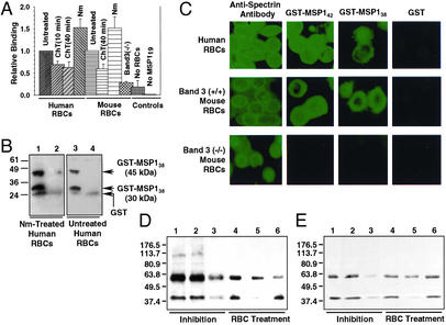
We report the molecular identification of a sialic acid-independent host-parasite interaction in the Plasmodium falciparum malaria parasite invasion of RBCs. Two nonglycosylated exofacial regions of human band 3 in the RBC membrane were identified as a crucial host receptor binding the C-terminal processing products of merozoite surface protein 1 (MSP1). Peptides derived from the receptor region of band 3 inhibited the invasion of RBCs by P. falciparum. A major segment of the band 3 receptor (5ABC) bound to native MSP1(42) and blocked the interaction of native MSP1(42) with intact RBCs in vitro. Recombinant MSP1(19) (the C-terminal domain of MSP1(42)) bound to 5ABC as well as RBCs. The binding of both native MSP1(42) and recombinant MSP1(19) was not affected by the neuraminidase treatment of RBCs, but sensitive to chymotrypsin treatment. In addition, recombinant MSP1(38) showed similar interactions with the band 3 receptor and RBCs, although the interaction was relatively weak. These findings suggest that the chymotrypsin-sensitive MSP1-band 3 interaction plays a role in a sialic acid-independent invasion pathway and reveal the function of MSP1 in the Plasmodium invasion of RBCs.


Figures

Figure 1
Figure 1
Band 3 peptides inhibit the P. falciparum invasion of RBCs. (A) Human band 3 peptides. (i) Overlapping 12- to 20-aa peptides shown in solid bars were prepared according to the inclusive ectodomain boundaries based on the following three topology models: the Casey model (ii; ref. 27), the Reithmeier model (iii; ref. 28), and the Sherman model (iv; ref. 29). Boxes denote ectodomains. (B) Invasion inhibition assay by the visual counting method. The number of ring-stage parasites counted from Giemsa-stained thin smears is plotted. No Peptide, negative control. (C) Effect of peptides on intraerythrocytic parasite maturation. The total number of schizonts and trophozoites was scored from the same smears in B. (D) Invasion inhibition assay by the [3H]hypoxanthine incorporation method. Effects relative to the control sample (no peptide) are shown. Unrelated peptide was derived from RBC dematin (negative control). (E) Growth inhibition assay by the [3H]hypoxanthine incorporation method. Effects on the growth of ring-stage parasites are shown relative to the control sample (no peptide). In all assays, artemisinin (25 μM) served as a negative control for parasite development in the culture. Parasitemia was calculated as the mean of three experiments (±SE), and Student's t test was used to compare the mean.
Figure 2
Figure 2
The binding of human band 3 and native P. falciparum merozoite proteins is shown. (A) Merozoite protein separation. Total proteins in isolated merozoites (M) and human RBC ghosts (G) are shown in the Coomassie gel. Residual human serum albumin (apparent mass 67 kDa) from the culture medium is marked with an asterisk. Western blotting using anti-band 3 and anti-spectrin antibodies (Sigma) is shown. (B) Blot overlay assay. Negative control peptides 3A and 4A showed only nonspecific bindings to human serum albumin (asterisk) in merozoite samples and α/β spectrin (240/220 kDa) in ghost samples. Peptide 2 also served as negative control. (C) Western blot using P. falciparum MSP1 T9/94 antiserum. Binding of native MSP1 to 5ABC is shown. Lanes: 1, parasite lysate (PL); 2, PL + GST-5ABC beads; 3, PL + GST beads; 4, P. falciparum culture supernatant (CS); 5, CS + GST-5ABC beads; and 6, CS + GST beads. (D) Western blot using mAb 5.2. Lanes: 1, CS; 2, CS + GST-5ABC beads; and 3, CS + GST beads.
Figure 3
Figure 3
The band 3–MSP1 interaction is shown. (A) P. falciparum MSP1. Conserved (black), semiconserved (gray), and polymorphic (white) regions are shown based on previous sequence diversity analyses (20, 21). Various MSP1 constructs derived from the FCB-1 strain were prepared for solution binding and yeast two-hybrid assays as illustrated. (B) Solution binding assay. 32P-labeled 5ABC (10, 20, 40, and 80 μM) and 5BC (21, 42, 84, and 168 μM) bound to GST-MSP142 (Top) and GST-MSP138 (Middle), respectively, on beads in a concentration-dependent manner. Biotinylated Trx-MSP119 (0.78, 1.56, 3.13, 6.25, 12.5, and 25.0 nM) bound to immobilized GST-5ABC in ELISA when GST alone did not (Bottom). Trx did not interact with GST-5ABC (not shown). (C) Yeast two-hybrid assay. The protein interaction was analyzed by a cotransformation method in yeast AH109 cells using SD/−Leu/−Trp selection plates. Activation of the MEL1 reporter gene on specific binding of the MSP1 domain to the band 3 peptide gave positive blue colonies when using the α-galactosidase assay.
Figure 4
Figure 4
The binding of P. falciparum MSP1 to RBCs is shown. (A) Relative binding of 32P-labeled MSP119 to various RBCs in suspension. Means (±SE) were plotted relative to the control (untreated wild type) and compared using Student's t test. Assays were repeated three to five times. Normalization: human RBC control, 1.0 = 9,387 counts per minute (cpm); mouse RBC control, 1.0 = 17,787 cpm. (B) Binding of MSP138 to RBCs. Anti-GST Western blotting shows that GST-MSP138 binds to Nm-treated (lane 1) and untreated (lane 3) human RBCs. GST was used as a control (lanes 2 and 4). (C) Indirect immunofluorescence assay. The binding of GST-MSP142 and GST-MSP138 is shown by using anti-GST antibody and FITC-conjugated secondary antibody. GST was used as a control. Anti-spectrin antibody staining confirmed that all RBC types were morphologically normal. (D) Western blotting using P. falciparum MSP1 T9/94 rabbit antiserum. The binding of native P. falciparum MSP1 to human RBCs is shown. Lanes: 1, culture supernatant (CS) + untreated RBCs; 2, CS + GST + RBCs; 3, CS + GST-5ABC + RBCs; 4, CS + RBCs treated with T (1.0 mg/ml); 5, CS + RBCs treated with ChT (0.5 mg/ml); and 6, CS + RBCs treated with Nm (3 milliunits/ml). (E) Western blot analysis using mAb 5.2 confirms the 42-kDa protein shown in D is MSP142. The lanes are identical to those in D.

References

    1. Mitchell G H, Hadley T J, McGinniss M H, Klotz F W, Miller L H. Blood. 1986;67:1519–1521. - PubMed
    1. Dolan S A, Miller L H, Wellems T E. J Clin Invest. 1990;86:618–624. - PMC - PubMed
    1. Orlandi P A, Klotz F W, Haynes J D. J Cell Biol. 1992;116:901–909. - PMC - PubMed
    1. Sim B K, Chitnis C E, Wasniowska K, Hadley T J, Miller L H. Science. 1994;264:1941–1944. - PubMed
    1. Miller L H, Haynes J D, McAuliffe F M, Shiroishi T, Durocher J R, McGinniss M H. J Exp Med. 1977;146:277–281. - PMC - PubMed

Publication types

MeSH terms

Substances

Associated data

LinkOut - more resources