

Addition of inhibition in the olivocerebellar system and the ontogeny of a motor memory
- PMID: 12692555
- PMCID: PMC1393286
- DOI: 10.1038/nn1042
Addition of inhibition in the olivocerebellar system and the ontogeny of a motor memory
Abstract
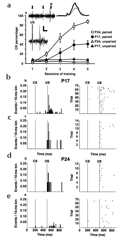
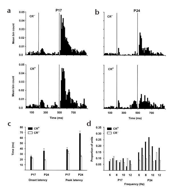
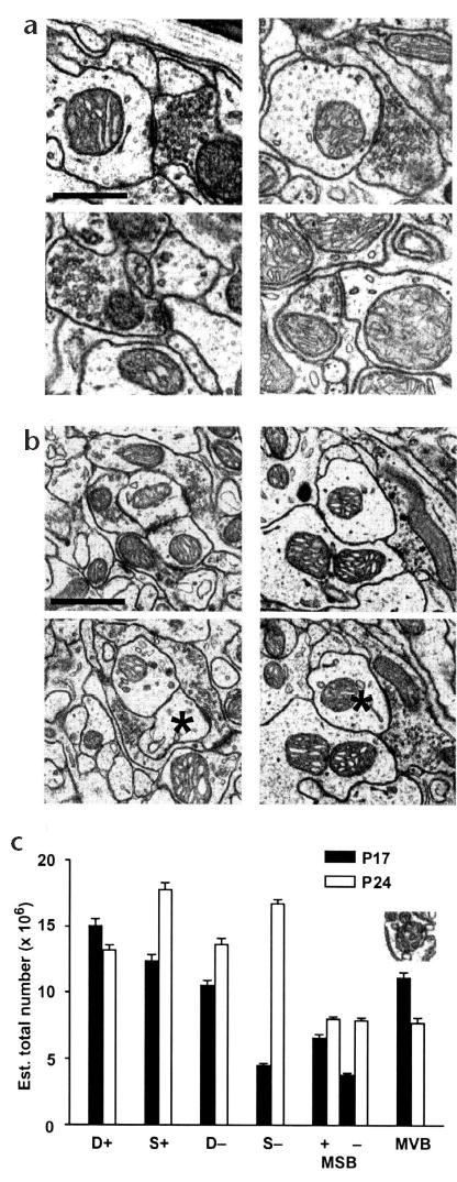
The developmental emergence of learning has traditionally been attributed to the maturation of single brain regions necessary for learning in adults, rather than to the maturation of synaptic interactions within neural systems. Acquisition and retention of a simple form of motor learning, classical conditioning of the eyeblink reflex, depends on the cerebellum and interconnected brainstem structures, including the inferior olive. Here, we combined unit recordings from Purkinje cells in eye regions of the cerebellar cortex and quantitative electron microscopy of the inferior olive to show that the developmental emergence of eyeblink conditioning in rats is associated with the maturation of inhibitory feedback from the cerebellum to the inferior olive. The results are consistent with previous work in adult animals and indicate that the maturation of cerebellar inhibition within the inferior olive may be a critical factor for the formation and retention of learning-specific cerebellar plasticity and eyeblink conditioning.
Conflict of interest statement
Competing interests statement
The authors declare that they have no competing financial interests.
Figures
Similar articles
-
Developmental changes in eyeblink conditioning and simple spike activity in the cerebellar cortex.Dev Psychobiol. 2004 Jan;44(1):45-57. doi: 10.1002/dev.10149. Dev Psychobiol. 2004. PMID: 14704989
-
Graded error signals in eyeblink conditioning.Neurobiol Learn Mem. 2020 Apr;170:107023. doi: 10.1016/j.nlm.2019.04.011. Epub 2019 Apr 24. Neurobiol Learn Mem. 2020. PMID: 31028891 Review.
-
Cerebellar dysfunction explains the extinction-like abolition of conditioned eyeblinks after NBQX injections in the inferior olive.J Neurosci. 2008 Jan 2;28(1):10-20. doi: 10.1523/JNEUROSCI.3403-07.2008. J Neurosci. 2008. PMID: 18171918 Free PMC article.
-
Extinction of conditioned blink responses by cerebello-olivary pathway stimulation.Neuroreport. 2007 Sep 17;18(14):1479-82. doi: 10.1097/WNR.0b013e3282e326e8. Neuroreport. 2007. PMID: 17712278
-
Ontogenetic changes in the neural mechanisms of eyeblink conditioning.Integr Physiol Behav Sci. 2001 Jan-Mar;36(1):15-35. doi: 10.1007/BF02733945. Integr Physiol Behav Sci. 2001. PMID: 11484994 Review.
Cited by
-
Integration of Purkinje cell inhibition by cerebellar nucleo-olivary neurons.J Neurosci. 2015 Jan 14;35(2):544-9. doi: 10.1523/JNEUROSCI.3583-14.2015. J Neurosci. 2015. PMID: 25589749 Free PMC article.
-
Cerebellar control of the inferior olive.Cerebellum. 2006;5(1):7-14. doi: 10.1080/14734220500462757. Cerebellum. 2006. PMID: 16527758 Review.
-
Movements during sleep reveal the developmental emergence of a cerebellar-dependent internal model in motor thalamus.Curr Biol. 2021 Dec 20;31(24):5501-5511.e5. doi: 10.1016/j.cub.2021.10.014. Epub 2021 Nov 1. Curr Biol. 2021. PMID: 34727521 Free PMC article.
-
Eyeblink conditioning in rats using pontine stimulation as a conditioned stimulus.Integr Physiol Behav Sci. 2004 Jul-Sep;39(3):180-91. doi: 10.1007/BF02734438. Integr Physiol Behav Sci. 2004. PMID: 15929500 Free PMC article.
-
Extinction, reacquisition, and rapid forgetting of eyeblink conditioning in developing rats.Learn Mem. 2014 Nov 17;21(12):696-708. doi: 10.1101/lm.036103.114. Print 2014 Dec. Learn Mem. 2014. PMID: 25403458 Free PMC article.
References
-
- Albus JS. A theory of cerebellar function. Math Biosci. 1971;10:25–61.
-
- Ito, M. The Cerebellum and Neural Control (Raven, New York, 1984).
-
- Carew TJ. Developmental assembly of learning in Aplysia. Trends Neurosci. 1989;12:389–394. - PubMed
-
- Thompson RF, Krupa DJ. Organization of memory traces in the mammalian brain. Annu Rev Neurosci. 1994;17:519–549. - PubMed
Publication types
MeSH terms
Grants and funding
LinkOut - more resources
Full Text Sources
Medical
