. 2003 Feb;84(1):17-30.
doi: 10.1046/j.1365-2613.2003.00236.x.

The histological features of microwave coagulation therapy: an assessment of a new applicator design

Affiliations

The histological features of microwave coagulation therapy: an assessment of a new applicator design

Benjamin Swift et al. Int J Exp Pathol. 2003 Feb.

Abstract

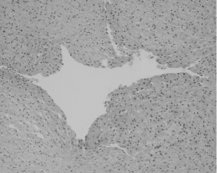
Microwave ablation of tumours within the liver may become an adjunct or alternative to resection in patients with primary or secondary cancers. This technique combines the benefits of a large, localized coagulative effect with a single insertion of the applicator, in a significantly shorter time than comparable treatments. A new range of microwave applicators were developed and tested in animal models and both ex-vivo and in-vivo specimens of human liver at resection. At laparotomy, the applicator tip was inserted into normal liver parenchyma and tumours, with each specimen subjected to irradiation for 180 s or more and at varying power outputs. On sectioning an area of spherical blanching was observed around the applicator cavity. Microscopically a zone of coagulative necrosis was seen adjacent to the site of probe insertion. Damage to blood vessels and bile ducts occurred distal to the probe cavity suggesting the passage of heated fluid, a finding that was diminished by temporary occlusion of the hepatic vasculature (a Pringle manoeuvre). Ultra-structural damage was confirmed within the burn zone and selected liver enzymes were shown to be functioning beyond this region. We suggest this indicates the surrounding liver parenchyma is functioning normally and therefore the volume of microwave-induced damage is controllable. We are confident that the new applicator design will allow the effective treatment of larger tumours in a safe and controlled manner with a single application of energy.


Figures

Figure 1
Figure 1
The appearance of a microwave ablation lesion within a porcine model at time 0 h. The lesion is well demarcated with the application probe cavity situated centrally.
Figure 2
Figure 2
Low power microscopic view of a lesion within a rat model at time 0 h. The zone of haemorrhage marking the edge of liver plate collapse is visible.
Figure 3
Figure 3
(a,b) The normal appearance of collagen (left) when viewed under plane polarized light displays a difference in colouration to that affected by thermal damage (right).
Figure 4
Figure 4
The appearance of an ablation lesion within a rat model at 24 h. The wedge of infarction noted occurred within the postablation period suggesting ischaemia related damage.
Figure 5
Figure 5
Microscopic view of the lesion edge at 6 h demonstrating early neutrophil infiltration of the infracted area. Magnification × 40
Figure 6
Figure 6
Microscopic view of a lesion at 24 h, within a rat model. Necrosis is evident producing a clear border with the adjacent normal liver. Magnification × 12.5
Figure 7
Figure 7
High power magnification of a lesion edge at 48 h (rat model). Inflammatory cells are present with early evidence of fibroblast proliferation. Magnification × 400
Figure 8
Figure 8
(a,b) The appearance of both rat (left) and porcine lesions at one week show similarities, with a fibrous capsule evolving around the circumference of the burn.
Figure 9
Figure 9
Microscopic examination of a rat lesion at one week confirms the presence of a fibrous connective tissue capsule forming a distinct boundary between normal hepatic parenchyma and necrotic hepatocytes centrally. The application cavity is still visible. Magnification × 12.5
Figure 10
Figure 10
High power magnification view of the lesion edge revealing benign bile duct proliferation within a fibrous capsule: one week, rat model. Magnification × 200
Figure 11
Figure 11
A porcine lesion at 60 days. The fibrous capsule is established and the lesion contents have decreased in size. The application cavity is visible at this time. Note that the adjacent large blood vessels are unaffected by the lesion, being beyond the burn radius.
Figure 12
Figure 12
View of a central vein showing lobule damage relating to the hot gas/liquid formation at time 0 h within a porcine model. Magnification × 100
Figure 13
Figure 13
The zonal damage is appreciable in this medium power magnification of a tumour ablation. The application cavity is sited to the right with marked coagulative necrosis adjacent. The second zone demonstrates nuclear smearing and blends with the last zone, which shows no appreciable damage. Magnification × 100
Figures 14
Figures 14
(a,b,c) Liver enzyme activity shows a demonstrable cessation of function within the confines of the burn lesion induced within normal liver. Human model at time 0 h. (a = acid phosphatase, b = phosphofructokinase, c = glucose-6-phosphatase) Magnification × 100
Figures 15
Figures 15
(a,b,c,d) Transmission electron microscopy reveals complete thermal disruption of normal liver tissue within the region adjacent to the applicator (Image a, magnification × 10000), though at a distance of 5 mm the original ultrastructural appearances can be recognized (Image b, magnification × 10000). At a distance of 10 mm the cellular architecture appears almost normal though vesicular swelling of the mitochondria is appreciable (Image c, magnification × 2600). Beyond the macroscopic burn radius (10 mm) the liver possesses a normal appearance. (Image d, magnification × 3300)

Similar articles

Cited by

References

    1. Boon ME, Kok LP. Microwave Cookbook for Pathology. 3. Leiden: Colomb Press; 1989.
    1. Cozzi PJ, Stewart GJ, Morris DL. Thrombocytopenia after hepatic cryotherapy for colorectal metastases: correlates with hepatocellular injury. World J. Surg. 1994;18:774–777. - PubMed
    1. Culling CFA. Handbook of histopathological Techniques. London: Butterworths Publishing; 1963. p. 244.
    1. Curley SA, Izzo F, Ellis LM, Vauthey JN, Vallone P. Radiofrequency ablation of hepatocellular cancer in 110 patients with cirrhosis. Ann. Surg. 2000;232:381–391. - PMC - PubMed
    1. Dodd GD, III, Soulen MC, Kane RA, Livraghi T, Lees WR, Yamashita Y, Gillams AR, Karahan OI, Rhim H. Minimally invasive treatment of malignant hepatic tumours: At the threshold of a major breakthrough 1. Radiographics. 2000;20:9–27. - PubMed

Publication types