. 2003 Feb;84(1):31-48.
doi: 10.1046/j.1365-2613.2003.00239.x.

A new model of busulphan-induced chronic bone marrow aplasia in the female BALB/c mouse

Affiliations

A new model of busulphan-induced chronic bone marrow aplasia in the female BALB/c mouse

Frances M Gibson et al. Int J Exp Pathol. 2003 Feb.

Abstract

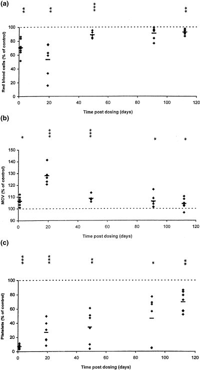
Aplastic anaemia (AA) is characterized by hypocellular marrow, pancytopenia, and risk of severe anaemia, haemorrhage and infection. AA is often idiopathic, but frequently occurs after exposure to drugs/chemicals. However, the pathogenesis of AA is not clearly understood, and there are no convenient animal models of drug-induced AA. We have evaluated regimens of busulphan (BU) administration in the mouse to produce a model of chronic bone marrow aplasia showing features of human AA. Mice were given 8 doses of BU at 0, 5.25 and 10.50 mg/kg over 23 days; marrow and blood samples were examined at 1, 19, 49, 91 and 112 days after dosing. At day 1 post dosing, in mice treated at 10.50 mg/kg, nucleated marrow cells, CFU-GM and Erythroid-CFU were reduced. Similarly, peripheral blood erythrocytes, leucocytes, platelets and reticulocytes were reduced. At day 19 and 49 post dosing, there was a trend for parameters to return towards normal. However, at day 91 and 112 post dosing, values remained significantly depressed, with a stabilized chronic bone marrow aplasia. At day 91 and 112 post dosing, marrow cell counts, CFU-GM and Erythroid-CFU were decreased; marrow nucleated cell apoptosis and c-kit+ cell apoptosis were increased; peripheral blood erythrocyte, leucocyte, and platelet counts were reduced. We conclude that this is a model of chronic bone marrow aplasia which has many interesting features of AA. The model is convenient to use and has potential in several areas, particularly for investigations on mechanisms of AA pathogenesis in man.


Figures

Figure 1
Figure 1
Hematological parameters from individual mice, expressed as a percentage of the mean control value at each time point, at 1, 19, 49, 91 and 112 days after BU dosing at 10.50 mg/kg (a), red blood cells; (b), mean cell volume; (c), platelets; (d), lymphocytes. Horizontal bars indicate group means; *P < 0.05, **P < 0.01, ***P < 0.001 are presented vertically above the data points (see Table 1).
Figure 1
Figure 1
Hematological parameters from individual mice, expressed as a percentage of the mean control value at each time point, at 1, 19, 49, 91 and 112 days after BU dosing at 10.50 mg/kg (a), red blood cells; (b), mean cell volume; (c), platelets; (d), lymphocytes. Horizontal bars indicate group means; *P < 0.05, **P < 0.01, ***P < 0.001 are presented vertically above the data points (see Table 1).
Figure 2
Figure 2
H&E stained sections of mouse sterna from control (vehicle-treated) and BU-treated (10.50 mg/kg) animals. (a) and (b), control mouse at day 1 after vehicle dosing showing normal marrow cellularity. (A, original magnification [OM] × 100; B, OM × 400). (c), BU-treated mouse at day 1 after dosing, showing marked hypocellularity, fatty replacement (arrow heads), small megakaryocytic foci (arrows), and a haemorrhagic area (H). (OM × 100). (d), mouse treated with BU at 19 days after dosing; there is marked hypocellularity with fatty replacement, a large cystic space, and an occasional myeloid focus (arrow) (OM × 100). (d) and (f), sternal marrow from a BU-treated mouse at 112 days after dosing; there is some hypocellularity, but with only slight erythroid activity (E, OM × 100; F, OM × 400).
Figure 3
Figure 3
May-GrünwaldBGiemsa stained mouse humeral marrow smears (a-f.) from control (vehicle-treated) and BU-treated (10.50mg/kg) animals. (a), control mouse at day 1 after vehicle dosing illustrating the normal distribution of myeloid (arrow), erythroid (open arrow) and lymphoid (arrow head) elements (OM × 1000). (b), BU-treated mouse at day 91 after dosing, showing hypocellularity, with fat cells and mast cells predominating; normal haemopoietic elements are absent (OM × 1000). (c), BU-treated mouse at day 112 post dosing; the marrow shows significant depletion of erythroid precursors (OM × 1000). (D), BU-treated mouse at day 19 post dosing; the marrow is hypocellular, with few myeloid cells, and many Howell-Jolly bodies (arrows) are present (OM × 1000). (e), day 19 after BU dosing: dyserythropoiesis; many late normoblasts (arrows) are multinucleated (OM × 1000). (f), day 19 after BU dosing: dyserythropoiesis; megaloblastic development (arrows) of intermediate and late normoblasts (OM × 1000).
Figure 4
Figure 4
Committed progenitor cell content of femoral marrow from mice treated with BU at 1, 19, 49, 91 and 112 days after BU dosing. CFU-GM (A) and erythroid colony numbers (B) in BU-treated mice (▴ BU, 5.25 mg/kg; • BU, 10.50 mg/kg) as percentages (mean ± SEM) of the control mice. *P < 0.05, **P < 0.01, ***P < 0.001.
Figure 5
Figure 5
Apoptosis of femoral marrow from mice treated with BU at 1, 19, 49, 91 and 112 days post dosing. Marrow nucleated cell apoptosis (A), proportion of c-kit+ cells (B), and apoptosis in c-kit+ cells (C) in BU-treated mice (▴ BU, 5.25 mg/kg; • BU, 10.50 mg/kg) as percentages (mean ± SEM)) of the control mice. Asterisks indicating degree of significance are as for Figure 4.

Similar articles

Cited by

References

    1. Andrews CM, Dash LM, Williams TC, Craig Gray J, Turton JA. Long-term effects of busulphan on lymphocyte subpopulations in female B6C3F1 mice. Comp. Haematol. Int. 1997;7:230–237.
    1. Andrews CM, Spurling NW, Turton JA. Characterisation of busulphan-induced myelotoxicity in B6C3F1 mice using flow cytometry. Comp. Haematol. Int. 1993;3:538–546.
    1. Andrews CM, Williams TC, Turton JA. Long-term haematological alterations in female B6C3F1 mice treated with busulphan. Comp. Haematol. Int. 1998;8:125–138.
    1. Appelbaum FR, Fefer A. The pathogenesis of aplastic anaemia. Sem. Haematol. 1981;18:241–257. - PubMed
    1. Barnes DWH, Mole RH. Aplastic anaemia in sublethally irradiated mice given allogeneic lymph node cells. Br. J. Haematol. 1967;13:482–491. - PubMed

Publication types