

Comparative Study
MR-guided catheter navigation of the intracranial subarachnoid space
Affiliations
- PMID: 12695192
- PMCID: PMC8148659
Item in Clipboard
Comparative Study
MR-guided catheter navigation of the intracranial subarachnoid space
AJNR Am J Neuroradiol.
2003 Apr.
Abstract
Percutaneous intraspinal navigation (PIN) is a new minimally invasive approach to the CNS. The authors studied the utility of MR-guided intracranial navigation following access to the subarachnoid compartment via PIN. The passive tracking technique was employed to visualize devices during intracranial navigation. Under steady-state free precession (SSFP) MR-guidance a microcatheter-microguidewire was successfully navigated to multiple brain foci in two cadavers. SSFP MR fluoroscopy possesses adequate contrast and temporal resolution to allow MR-guided intracranial navigation.
Figures
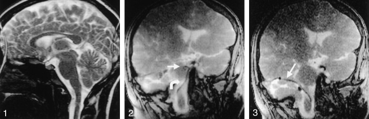
SSFP image in human volunteer. The image was produced in 253 milliseconds (four frames per second), with the following parameters: TR/TE, 4.4/2.2/90; flip angle, 192 × 256; section thickness, 10 mm; FOV, 200 mm; 80% reduced FOV; 60% image; and 50% keyhole. Note the paucity of CSF pulsation artifact or magnetic susceptibility effects at the skull base.
A 3F catheter (curved arrow) and 0.014-inch guidewire (straight arrow) are in the pontine cistern. Note that magnetic susceptibility artifact allows adequate visualization of the microguidewire.
The 3F microcatheter (arrow) traverses cerebellopontine angle.

The catheter-guidewire (curved arrows) is advanced through prepontine cistern; it impacts the posterior clinoid process (straight arrows) and buckles in the pontine cistern. The catheter-guidewire is then redirected to suprasellar cistern and advanced to the left sylvian fissure (arrowhead).
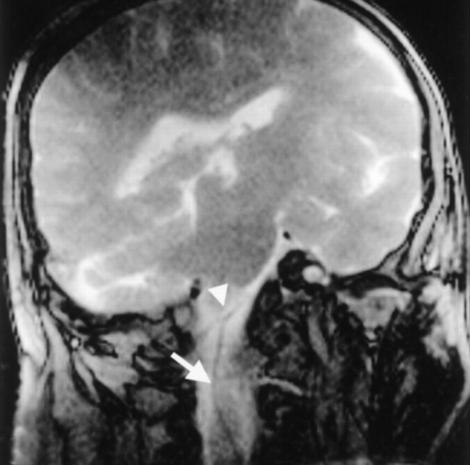
A 5F catheter (arrow) contacts the vertebral artery (arrowhead).
Comment in
-
Catheter navigation within the subarachnoid space.AJNR Am J Neuroradiol. 2004 Jun-Jul;25(6):1124. AJNR Am J Neuroradiol. 2004. PMID: 15205163 Free PMC article. No abstract available.
References
-
- Hamada J, Mizuno T, Kai Y, Morioka M, Ushio Y. Microcatheter intrathecal urokinase infusion into cisterna magna for prevention of cerebral vasospasm: preliminary report. Stroke 2000;31:2141–2148 - PubMed
-
- Mueller PR, Stark DD, Simeone JF, et al. MR-guided aspiration biopsy: needle design and clinical trials. Radiology 1986;161:605–609 - PubMed
-
- Mahfouz AE, Rahmouni A, Zylbersztejn C, Mathieu D. MR-guided biopsy using ultrafast T1- and T2- weighted reordered turbo fast low-angle shot sequences: feasibility and preliminary clinical applications. AJR Am J Roentgenol 1996;167:167–169 - PubMed
-
- Lewin JS, Connell CF, Duerk JL, et al. Interactive MRI-guided radiofrequency interstitial thermal ablation of abdominal tumors: clinical trial for evaluation of safety and feasibility. J Magn Reson Imaging 1998;8:40–47 - PubMed
Publication types
MeSH terms
LinkOut - more resources
Full Text Sources
Other Literature Sources
Medical