

Diameter of the superior ophthalmic vein in relation to intracranial pressure
- PMID: 12695206
- PMCID: PMC8148698
Diameter of the superior ophthalmic vein in relation to intracranial pressure
Abstract
Background and purpose: Bilateral engorged superior ophthalmic veins (SOV) have been reported in patients with diffuse brain swelling. We investigated the relationship between the diameter of the SOV on brain MR images and the intracranial pressure (ICP).
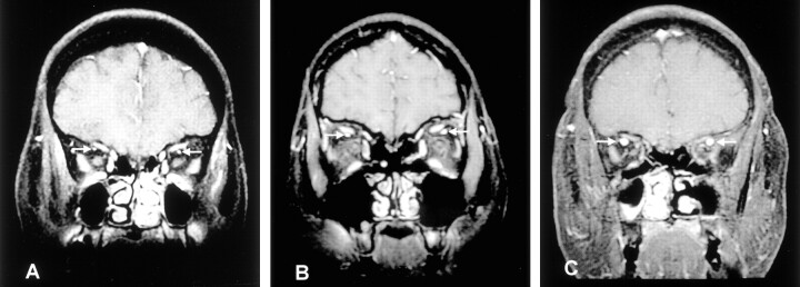
Methods: We reviewed the medical records of neurologic inpatients who had undergone both MR imaging of the brain and lumbar puncture. MR imaging had to have been performed before lumbar puncture, and the two studies had to have occurred within 2 days. The diameters of the SOV were measured on coronal contrast-enhanced fat-saturated T1-weighted MR images. For this, the image nearest the rear of the globe of the eye was chosen.
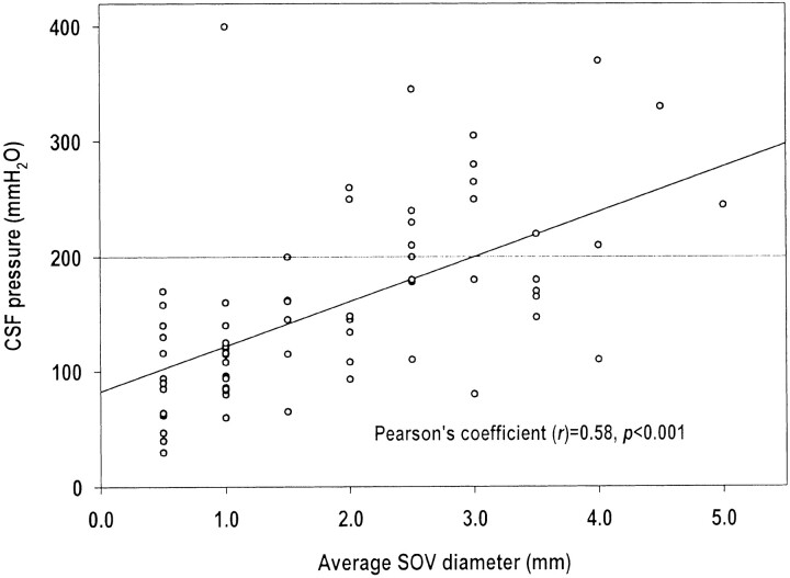
Results: Sixty-nine patients (32 male, 37 female; mean age, 46 years +/- 19) were included. The average diameters of the SOV and the ICP were positively correlated (r = 0.58, P <.001), if an SOV diameter of <1 mm was treated as 0.5 mm for calculations. In patients with increased ICP (CSF pressure >200 mm H(2)O), SOV diameters were larger than those of patients with a normal CSF pressure (3.0 vs 1.6 mm, P <.001). Frequencies of increased ICP were 3% among patients with an average SOV diameter of 0.5-1 mm, 15% for 1.5-2 mm, and 58% for 2.5-5 mm (P <.001).
Conclusion: This study showed that the SOV diameter, determined on the basis on MR imaging, was positively correlated with ICP. Dilatation of the SOV should alert physicians to the possibility of increased ICP.
Figures
References
-
- Bacon KT, Duchesneau PM, Weinstein MA. Demonstration of the superior ophthalmic vein by high resolution computed tomography. Radiology 1977;124:129–131 - PubMed
-
- Nugent RA, Belkin RI, Neigel JM, et al. Graves’ orbitopathy: correlation of CT and clinical findings. Radiology 1990;177:675–682 - PubMed
-
- Peyster RG, Savino PJ, Hoover ED, Schatz NJ. Differential diagnosis of the enlarged superior ophthalmic vein. J Comput Assist Tomogr 1984;8:103–107 - PubMed
-
- Khanna RK, Pham CJ, Malik GM, Spickler EM, Metha B, Rosenblum ML. Bilateral superior ophthalmic vein enlargement associated with diffuse cerebral swelling: report of 11 cases. J Neurosurg 1997;86:893–897 - PubMed
-
- Spektor S, Piontek E, Umansky F. Orbital venous drainage into the anterior cavernous sinus space: microanatomic relationships. Neurosurgery 1997;40:532–540 - PubMed
Publication types
MeSH terms
LinkOut - more resources
Full Text Sources
Other Literature Sources
Medical