

Contrast-enhanced fluid-attenuated inversion recovery imaging for leptomeningeal disease in children
- PMID: 12695212
- PMCID: PMC8148663
Contrast-enhanced fluid-attenuated inversion recovery imaging for leptomeningeal disease in children
Abstract
Background and purpose: To develop an MR imaging method that improves detection of leptomeningeal disease when compared with the current reference standard, contrast-enhanced T1-weighted imaging.
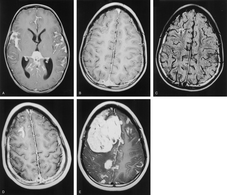
Methods: We investigated the cases of 10 children who were at high risk of intracranial leptomeningeal disease (Sturge-Weber syndrome and medulloblastoma). The cases of Sturge-Weber syndrome were investigated by using one MR imaging examination, and the cases of medulloblastoma were investigated by using four MR imaging examinations performed over 18 months. In all cases, contrast-enhanced fluid-attenuated inversion recovery (FLAIR) images were acquired in addition to the routine sequences. The parameters of the FLAIR sequence were chosen to maximize the T1 component of the signal intensity, to maximize detection of leptomeningeal enhancement. We made subjective and objective assessments of the presence and extent of leptomeningeal disease as shown on contrast-enhanced T1-weighted images and contrast-enhanced FLAIR images.
Results: In three of the four cases of Sturge-Weber syndrome, the T1 and FLAIR sequences showed comparable extent of leptomeningeal enhancement. For one child, FLAIR images showed unexpected bilateral disease and more extensive leptomeningeal enhancement on the clinically suspected side. In four of six cases of medulloblastoma, no leptomeningeal enhancement was shown on any examinations during the 18-month period. In two cases, FLAIR images showed more extensive leptomeningeal enhancement when compared with T1-weighted images.
Conclusion: Contrast-enhanced FLAIR imaging seems to improve detection of leptomeningeal disease when compared with routine contrast-enhanced T1-weighted imaging. This seems to be partly because of suppression of signal intensity from normal vascular structures on the surface of the brain by FLAIR, which allows easier visualization of abnormal leptomeninges. We think that these findings can be extrapolated to the investigation of leptomeningeal disease of all causes and at all ages.
Figures


References
-
- Rodesch G, Van Bogaert P, Mavroudakis N, et al. Neuroradiologic findings in leptomeningeal carcinomatosis: the value interest of gadolinium-enhanced MRI. Neuroradiology 1990;32:26–32 - PubMed
-
- Wilkinson ID, Griffiths PD, Hoggard N, Cleveland TJ, Gaines PA, Venables GS. Unilateral meningeal enhancement following carotid stent insertion detected by magnetic resonance imaging. Stroke 2000;31:848–851 - PubMed
-
- Griffiths PD. A neuroimaging protocol for imaging children with brain tumours. Clin Radiol 1999;54:558–562 - PubMed
-
- Deliganis AV, Fisher DJ, Lam AM, Maravilla KR. CSF signal intensity increase on FLAIR MR images in patients under general anesthesia: the role of supplemental oxygen. Radiology 2001;218:152–156 - PubMed
MeSH terms
LinkOut - more resources
Full Text Sources
Medical