. 2003 May 1;17(9):1090-100.
doi: 10.1101/gad.1054603. Epub 2003 Apr 14.

NF2 deficiency promotes tumorigenesis and metastasis by destabilizing adherens junctions

Affiliations

NF2 deficiency promotes tumorigenesis and metastasis by destabilizing adherens junctions

Dominique Lallemand et al. Genes Dev. .

Abstract

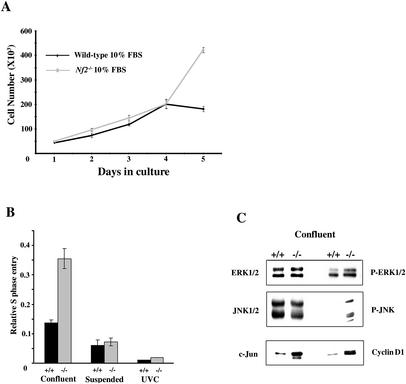
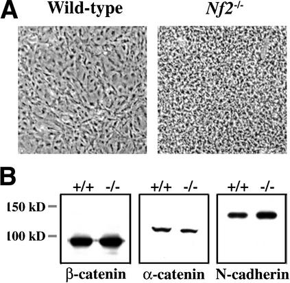
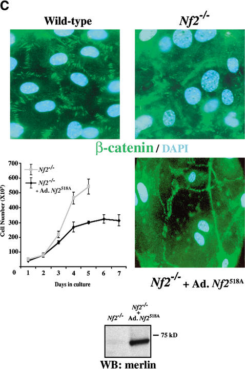
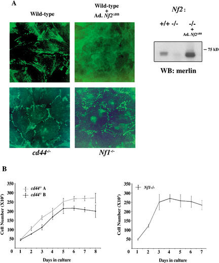
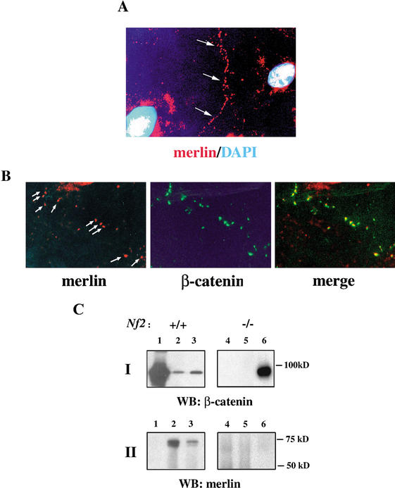
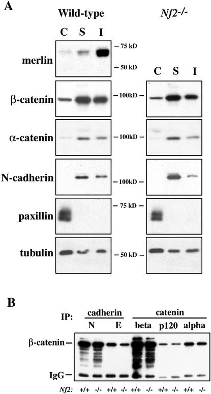
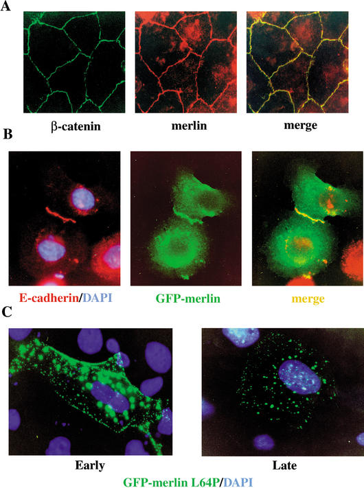
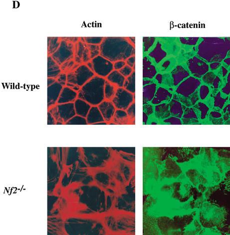
Mutation of the Neurofibromatosis 2 (NF2) tumor suppressor gene leads to cancer development in humans and mice. Recent studies suggest that Nf2 loss also contributes to tumor metastasis. The Nf2-encoded protein, merlin, is related to the ERM (ezrin, radixin, and moesin) family of membrane:cytoskeleton-associated proteins. However, the cellular mechanism whereby merlin controls cell proliferation from this location is not known. Here we show that the major cellular consequence of Nf2 deficiency in primary cells is an inability to undergo contact-dependent growth arrest and to form stable cadherin-containing cell:cell junctions. Merlin colocalizes and interacts with adherens junction (AJ) components in confluent wild-type cells, suggesting that the lack of AJs and contact-dependent growth arrest in Nf2(-/-) cells directly results from the absence of merlin at sites of cell:cell contact. Our studies indicate that merlin functions as a tumor and metastasis suppressor by controlling cadherin-mediated cell:cell contact.


Figures

Figure 1
Figure 1
Nf2−/− MEFs do not undergo contact-dependent inhibition of proliferation. (A) Wild-type and Nf2−/− MEFs cultured in the presence of growth factors were counted daily for 5 d. (B) Growth arrest of Nf2−/− MEFs by high density but not loss of adhesion or UVC-irradiation is impaired. Subconfluent, confluent, suspended, or UVC-treated wild-type and Nf2−/− MEFs were labeled with BrdU and analyzed by fluorescent-activated cell sorting (FACs) analysis. The graph displays the relative reduction in S phase of wild-type and Nf2−/− MEFs compared with subconfluent MEFs of the same genotype. Equivalent percentages of wild-type and Nf2−/− cells remained viable in these experiments (data not shown). FACs analysis also revealed that wild-type and Nf2−/− MEFs have equivalent cell volumes (data not shown). (C) The level and phosphorylation of key mitogenic signaling molecules were examined in confluent (unstarved) wild-type and Nf2−/− MEFs by Western blot. Elevated levels of cyclin D, c-jun, and phosphorylated (active) ERK1/2, JNK persist in confluent Nf2−/− MEFs.
Figure 2
Figure 2
Loss of AJs in Nf2−/− MEFs. (A) Wild-type and Nf2−/− MEFs were cultured in the presence of growth factors for 4 d after reaching confluence and photographed by phase contrast. (B) Expression of β-catenin, α-catenin, and N-cadherin in confluent wild-type and Nf2−/− MEFs was analyzed by Western blot analysis. (C) Wild-type and Nf2−/− MEFs were grown to confluence and starved for 48 h. Immunofluorescent localization of β-catenin revealed punctate staining along cell:cell boundaries in wild-type but not Nf2−/− MEFs. The lower panels show the rescue of both contact-dependent inhibition of proliferation (left) and AJ formation (right) by reintroduction of active merlin (Nf2S518A). Expression of Nf2S518A detected by Western blot is shown below.
Figure 2
Figure 2
Loss of AJs in Nf2−/− MEFs. (A) Wild-type and Nf2−/− MEFs were cultured in the presence of growth factors for 4 d after reaching confluence and photographed by phase contrast. (B) Expression of β-catenin, α-catenin, and N-cadherin in confluent wild-type and Nf2−/− MEFs was analyzed by Western blot analysis. (C) Wild-type and Nf2−/− MEFs were grown to confluence and starved for 48 h. Immunofluorescent localization of β-catenin revealed punctate staining along cell:cell boundaries in wild-type but not Nf2−/− MEFs. The lower panels show the rescue of both contact-dependent inhibition of proliferation (left) and AJ formation (right) by reintroduction of active merlin (Nf2S518A). Expression of Nf2S518A detected by Western blot is shown below.
Figure 3
Figure 3
Expression of dominant-negative merlin (Nf2ΔBB) disrupts β-catenin localization in wild-type MEFs. (A) Immunofluorescence staining of β-catenin in confluent, serum-starved cd44−/− MEFs, Nf1−/− MEFs, or wild-type MEFs infected with an adenovirus expressing Nf2ΔBB. Expression of dominant-negative merlin disrupts β-catenin localization in wild-type cells (top right). In contrast, β-catenin localizes normally in cd44−/− (bottom left) or Nf1−/− (bottom right) MEFs. Expression of Nf2ΔBB in Nf2−/− MEFs was demonstrated by Western blot using an anti-merlin antibody (right). (B) cd44−/− and Nf1−/− MEFs exhibit contact-dependent inhibition of growth. cd44−/− (two independently prepared populations, A and B) and Nf1−/− MEFs were seeded at a subconfluent density in the presence of growth factors and counted daily. After 4–5 d, MEFs of both genotypes reached saturation and stopped proliferating.
Figure 4
Figure 4
Merlin and β-catenin colocalize and associate with AJ components. (A) Localization of merlin (red) in confluent wild-type MEFs along a boundary of cell: cell contact (arrows; 20× magnification). Nuclei are stained with DAPI (blue). (B) Colocalization of merlin (red, arrows in left panel) and β-catenin (green, middle) in confluent wild-type MEFs at sites of cell:cell contact (merge, right panel; 40× magnification). This staining is not detectable in Nf2−/− MEFs (data not shown). (C) Immunoprecipitation of β-catenin (lane 1) and merlin (N-terminal epitope, lanes 2,4; C-terminal epitope, lanes 3,5) from the membrane soluble fraction of wild-type (left panel) and Nf2−/− (right panel) MEFs, followed by Western blot (WB) analysis using anti-β-catenin (upper panel I) or anti-merlin (lower panel II) antibodies. (Lane 6) Total membrane soluble fraction from Nf2−/− MEFs. In wild-type MEF membranes, β-catenin coimmunoprecipitates with merlin using both anti-merlin antibodies. As expected, β-catenin was not detected when immunoprecipitations from Nf2−/− membranes were performed using anti-merlin antibodies (lanes 4,5).
Figure 5
Figure 5
Core AJ complexes form in Nf2−/− MEF membranes. (A) Merlin and AJ component distribution in cytosolic (C), membrane soluble (S), and membrane insoluble (I) fractions from confluent, serum-starved wild-type (left) and Nf2−/− (right) MEFs. The distribution of AJ components is identical in wild-type and Nf2−/− MEFs. Merlin is largely insoluble and cofractionates with β-catenin but not paxillin. (B) Equivalent levels of β-catenin coimmunoprecipitate with N-cadherin, E-cadherin, β-catenin, p120 catenin, or α-catenin from confluent wild-type and Nf2−/− membranes.
Figure 6
Figure 6
Merlin localization in primary keratinocytes. (A) Confluent wild-type keratinocytes were cultured in the presence of 2 mM CaCl2 for 20 h to stimulate the formation of AJs. Immunostaining revealed perfect colocalization of endogenous merlin (red) and β-catenin (green; merge, yellow). (B) A GFP-Nf2 (wild-type) fusion protein (green; middle) colocalizes with E-cadherin (red) to a region of nascent cell:cell contact in wild-type primary keratinocytes (merge, yellow). Transfection of the GFP-Nf2-expressing plasmid and calcium induction were carried out simultaneously; 24 h later, cells were fixed, permeabilized, and processed for immunofluorescence. (C) A mutant GFP-Nf2L64P version of merlin exhibits limited localization to cell: cell boundaries at early times (12 h) after transfection, but at late times (48 h) after transfection, GFP-Nf2L64P exhibits only punctate, cytoplasmic localization. Nuclei are stained with DAPI (blue). (D) In a wild-type keratinocyte monolayer (upper panels), β-catenin localizes precisely to cell:cell boundaries (right panel). In contrast, in Nf2−/− keratinocytes (bottom panels), β-catenin is diffusely localized throughout the cell. In addition, rhodamine-phalloidin staining (left panels) reveals that the cortical actin ring is malformed and badly disorganized in Nf2−/− keratinocytes.
Figure 6
Figure 6
Merlin localization in primary keratinocytes. (A) Confluent wild-type keratinocytes were cultured in the presence of 2 mM CaCl2 for 20 h to stimulate the formation of AJs. Immunostaining revealed perfect colocalization of endogenous merlin (red) and β-catenin (green; merge, yellow). (B) A GFP-Nf2 (wild-type) fusion protein (green; middle) colocalizes with E-cadherin (red) to a region of nascent cell:cell contact in wild-type primary keratinocytes (merge, yellow). Transfection of the GFP-Nf2-expressing plasmid and calcium induction were carried out simultaneously; 24 h later, cells were fixed, permeabilized, and processed for immunofluorescence. (C) A mutant GFP-Nf2L64P version of merlin exhibits limited localization to cell: cell boundaries at early times (12 h) after transfection, but at late times (48 h) after transfection, GFP-Nf2L64P exhibits only punctate, cytoplasmic localization. Nuclei are stained with DAPI (blue). (D) In a wild-type keratinocyte monolayer (upper panels), β-catenin localizes precisely to cell:cell boundaries (right panel). In contrast, in Nf2−/− keratinocytes (bottom panels), β-catenin is diffusely localized throughout the cell. In addition, rhodamine-phalloidin staining (left panels) reveals that the cortical actin ring is malformed and badly disorganized in Nf2−/− keratinocytes.

References

    1. Adams CL, Chen YT, Smith SJ, Nelson WJ. Mechanisms of epithelial cell:cell adhesion and cell compaction revealed by high-resolution tracking of E-cadherin-green fluorescent protein. J Cell Biol. 1998;142:1105–1119. - PMC - PubMed
    1. Angres B, Barth A, Nelson WJ. Mechanism for transition from initial to stable cell–cell adhesion: Kinetic analysis of E-cadherin-mediated adhesion using a quantitative adhesion assay. J Cell Biol. 1996;134:549–557. - PMC - PubMed
    1. Bernards R, Weinberg RA. A progression puzzle. Nature. 2002;418:823. - PubMed
    1. Bretscher A, Edwards K, Fehon RG. ERM proteins and merlin: Integrators at the cell cortex. Nat Rev Mol Cell Biol. 2002;3:586–599. - PubMed
    1. Cavallaro U, Christofori G. Cell adhesion in tumor invasion andmetastasis: Loss of the glue is not enough. Biochim Biophys Acta. 2001;1552:39–45. - PubMed

Publication types

Substances