. 2003 Apr 21;197(8):1037-43.
doi: 10.1084/jem.20022033. Epub 2003 Apr 14.

CD40 signaling is impaired in L. major-infected macrophages and is rescued by a p38MAPK activator establishing a host-protective memory T cell response

Affiliations

CD40 signaling is impaired in L. major-infected macrophages and is rescued by a p38MAPK activator establishing a host-protective memory T cell response

Amit Awasthi et al. J Exp Med. .

Abstract

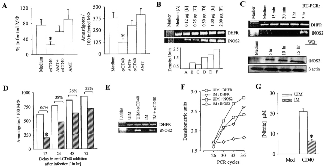
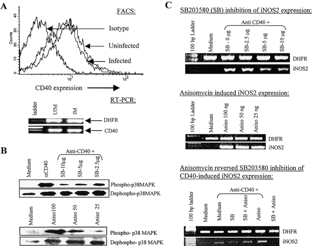
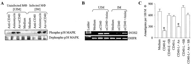
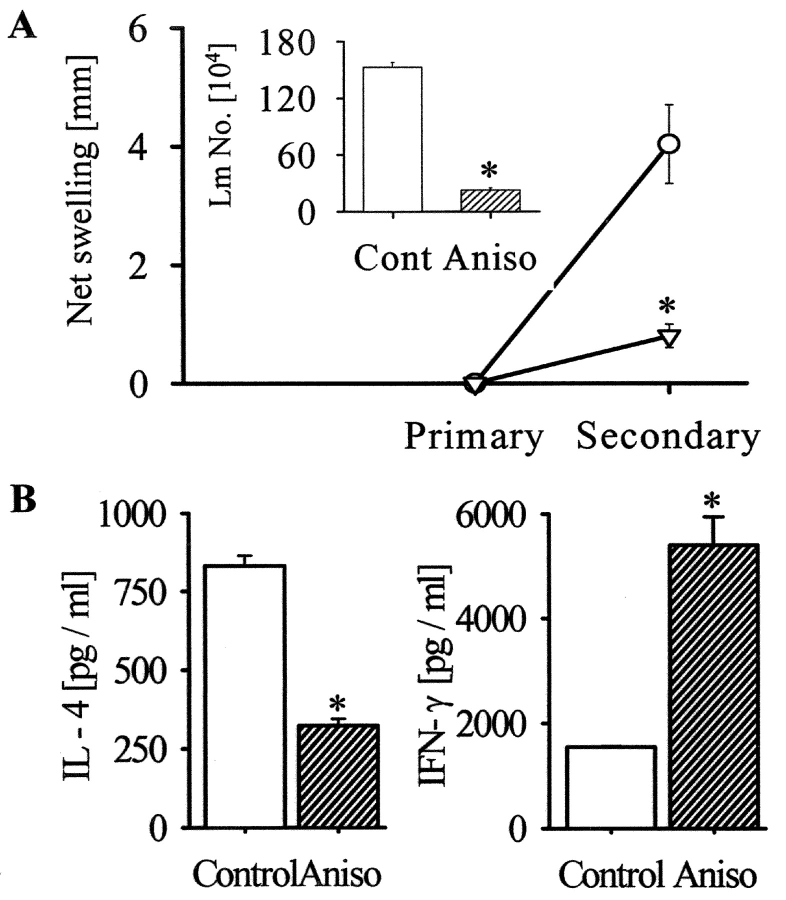
Leishmania, a protozoan parasite, lives and multiplies as amastigote within macrophages. It is proposed that the macrophage expressed CD40 interacts with CD40 ligand on T cells to induce IFN-gamma, a Th1-type cytokine that restricts the amastigote growth. Here, we demonstrate that CD40 cross-linking early after infection resulted in inducible nitric oxide synthetase type-2 (iNOS2) induction and iNOS2-dependent amastigote elimination. Although CD40 expression remained unaltered on L. major-infected macrophages, delay in the treatment of macrophages or of mice with anti-CD40 antibody resulted in significant reduction in iNOS2 expression and leishmanicidal function suggesting impaired CD40 signaling in Leishmania infection. The inhibition of CD40-induced iNOS2 expression by SB203580, a p38-mitogen activated protein kinase (p38MAPK)-specific inhibitor, and the reversal of the inhibition by anisomycin, a p38MAPK activator, suggested a crucial role of p38MAPK in CD40 signaling. Indeed, the CD40-induced p38MAPK phosphorylation, iNOS2 expression and anti-leishmanial function were impaired in Leishmania-infected macrophages but were restored by anisomycin. Anisomycin's effects were reversed by SB203580 emphasizing the role of p38MAPK in CD40-induced iNOS2-dependent leishmanicidal function. Anisomycin administration in L. major-infected BALB/c mice resulted in significant reduction in the parasite load and established a host-protective Th1-type memory response. Also implicated in these findings is a scientific rationale to define novel anti-parasite drug targets and to bypass the problem of drug resistance.


Figures

Figure 1.
Figure 1.
CD40 stimulation results in the induction of iNOS2 expression and iNOS2-dependent amastigote elimination. (A) CD40 induces leishmanicidal activity in BALB/c-derived peritoneal macrophages. The macrophages were infected with L. major promastigotes at a ratio of 1:10 for 6 h at 33°C and anti-CD40 antibody (clone 3/23; 2 μg/ml), with or without AMT, were added to the cultures. After 72 h, the macrophages were fixed, Giemsa-stained, and the percent-infected macrophages and the number of amastigotes per 100 infected macrophages were counted under a microscope. Values are expressed in mean ± SD. Anti-CD40 antibody treatment reduced infection significantly (*, P < 0.01). (B) CD40 induces iNOS2 expression. The BALB/c-derived peritoneal macrophages were incubated with different doses of the anti-CD40 antibody as indicated. RNA was extracted 3 h after stimulation and the RT-PCR for iNOS2 expression was performed as described in Materials and Methods. The density units shown in the bar diagram are arbitrary units obtained from the densitometric analysis using the Quantity-One software. (C) Time-kinetics of CD40-induced iNOS2 expression. Macrophages were stimulated with anti-CD40 antibody (2 μg/ml) as described above. Cells were lysed at different time points after anti-CD40 antibody treatment to obtain RNA (RT-PCR) or protein (WB). RNA was used for iNOS2 RT-PCR and the protein was used for iNOS2 Western blot (WB) analysis as described in Materials and Methods. (D) Delay in anti-CD40 antibody treatment results in the loss of its anti-leishmanial function. BALB/c-derived macrophages were infected with promastigotes at a 1:10 ratio in 16-well tissue culture slides for the indicated period. The cultures were treated with anti-CD40 or left untreated for another 24 h. The macrophages were fixed, Giemsa-stained, and 500 cells were counted as described under Materials and Methods. Values are expressed in mean ± SD (*, P < 0.01). (E) iNOS2 induction by CD40 is impaired in Leishmania-infected macrophages. Macrophages were cultured uninfected or Leishmania-infected for 72 h, followed by anti-CD40 antibody (2 μg/ml) treatment for 3 h. RNA was isolated from these macrophages and RT-PCR for DHFR and iNOS was performed. (F) RNA was isolated from the macrophages as described in the panel (E). RT-PCR for iNOS2 was performed for the indicated cycles and densitometric analysis was done. (G) CD40-induced nitrite production is impaired in Leishmania-infected macrophages. The macrophage cultures, as stated above, were maintained for 72 h. Culture supernatants were collected and nitrite concentration was measured using Griess reagent and calculated by comparison to NaNO2 as a standard. The data (mean ± SD) showing significant reduction of CD40-induced nitrite production (*, P < 0.005) in Leishmania infection is from one of three experiments.
Figure 2.
Figure 2.
CD40 signals through p38MAPK in macrophages and its expression remain unchanged in Leishmania-infected macrophages. (A) CD40 expression is not down-regulated in the Leishmania-infected macrophages. BALB/c-derived macrophages were cultured uninfected or Leishmania-infected for 72 h. The macrophages were stained with anti-CD40-PE antibody and CD40 expression was analyzed with a FACSVantage™ flow cytometer. CD40 expression by RT-PCR was performed as described in Materials and Methods. (B) Anisomycin (Aniso), a p38MAPK activator, and CD40 cross-linking induces p38MAPK phosphorylation and SB203580 (SB), an inhibitor of p38MAPK, inhibits the CD40-triggered p38MAPK phosphorylation. Macrophages were stimulated with anti-CD40 (2 μg/ml) alone or with the indicated doses of SB203580 or anisomycin for 15 min. Cell extracts were prepared and Western blot was performed to detect phospho- and dephospho-p38MAPK as described earlier. (C) SB203580 inhibits while anisomycin induces the CD40-triggered iNOS2 expression. RNA was isolated from BALB/c macrophages, which were stimulated with anti-CD40 antibody alone (2 μg/ml) or with the indicated doses of SB203580 or anisomycin alone or in combination, as indicated, for 3 h and RT-PCR for iNOS2 message was performed as described earlier. The data shown is from one of three individual experiments.
Figure 3.
Figure 3.
CD40-induced p38MAPK phosphorylation, iNOS2 induction, and anti-leishmanial function are impaired during Leishmania infection but restored by anisomycin. The uninfected and the L. major–infected macrophages, treated as indicated, were lysed to obtain protein, for studying p38MAPK phosphorylation (A) or RNA, for studying iNOS2 expression (B), as described in Materials and Methods. In some experiments (C), macrophages were treated with anti-CD40 antibody either at the beginning of infection (CD40-E) with or without SB203580 or 72 h after infection (CD40-L) with or without anisomycin. Macrophages were fixed, stained and counted to record the number amastigotes per 100 macrophages. The data (mean ± SD) showed that anisomycin rescued the CD40-induced anti-leishmanial effect (CD40-L+An) significantly (*, P < 0.01).
Figure 4.
Figure 4.
Anisomycin treatment results in significant decrease in infection (A and B) and is associated with a Th1-type response (C). BALB/c mice were infected with L. major promastigotes (2 × 10⁶) and were treated either at the beginning of infection with saline (Control) or anti-CD40 (CD40-E) or 7 d after infection with anti-CD40 antibody alone (CD40-L) or in combination with anisomycin (Aniso). The footpad swelling was measured weekly (A) and the parasite load was measured on day 35 post-infection (B). CD4+ T cells from lymph nodes of naive or infected mice treated with saline, anti-CD40 antibody or anisomycin were stimulated in vitro with anti-CD3 + anti-CD28 for 36 h. The cell culture supernatants were assayed for IL-4 and IFN-γ (C). The data (mean ± SD) represents one of three individual experiments. Anti-CD40 or anisomycin treatment reduced infection significantly (*, P < 0.001), decreased IL-4 and increased IFN-γ production significantly (P < 0.01).
Figure 5.
Figure 5.
Priming of BALB/c mice with ultra-low dose of L. major plus anisomycin establishes a host-protective Th1-type memory response. (A) BALB/c mice, primed with 5 × 10² Leishmania promastigotes alone (Circle) or with anisomycin (inverted triangle), showed no demonstrable infection after 6 wk (Primary). The mice were then challenged with L. major promastigotes (2 × 10⁶). The net footpad swelling in the control (open bar) or anisomycin-treated (hatched bar) mice was measured 6 wk after infection (Secondary). Limiting dilution assay assessed parasite load in the footpad (Inset). Values are expressed in mean ± SD. Anisomycin priming reduced the infection significantly (*, P < 0.001). (B) CD4+ T cells from popliteal lymph nodes of the control (open bar) or anisomycin-treated (hatched bar) mice were stimulated with anti-CD3+anti-CD28 for 36 h. IL-4 and IFN-γ were measured in the supernatants by ELISA. The data (mean ± SD) representing one of three experiments shows that anisomycin priming reduced IL-4 production but increased IFN-γ production significantly (*, P < 0.005).

References

    1. Murray, H.W., G.L. Spitalny, and C.F. Nathan. 1985. Activation of mouse peritoneal macrophages in vitro and in vivo by interferon-gamma. J. Immunol. 134:1619–1622. - PubMed
    1. Russell, D.G., and P. Talamas-Rohana. 1989. Leishmania and the macrophage: a marriage of inconvenience. Immunol. Today. 10:328–333. - PubMed
    1. Olivier, M., B.J. Romero-Gallo, C. Matte, J. Blanchette, B.I. Posner, M.J. Tremblay, and R. Faure. 1998. Modulation of interferon-gamma-induced macrophage activation by phosphotyrosine phosphatases inhibition. Effect on murine leishmaniasis progression. J. Biol. Chem. 273:13944–13949. - PubMed
    1. Reiner, S.L., and R.M. Locksley. 1995. The regulation of immunity to Leishmania major. Annu. Rev. Immunol. 13:151–177. - PubMed
    1. Miga, A., S. Master, M. Gonzalez, and R.J. Noelle. 2000. The role of CD40-CD154 interactions in the regulation of cell-mediated immunity. Immunol. Invest. 29:111–114. - PubMed

Publication types

MeSH terms