. 2003 Apr 29;100(9):5324-9.
doi: 10.1073/pnas.0831037100. Epub 2003 Apr 14.

An important regulatory role for CD4+CD8 alpha alpha T cells in the intestinal epithelial layer in the prevention of inflammatory bowel disease

Affiliations

An important regulatory role for CD4+CD8 alpha alpha T cells in the intestinal epithelial layer in the prevention of inflammatory bowel disease

Gobardhan Das et al. Proc Natl Acad Sci U S A. .

Abstract

The normal immunoregulatory mechanisms that maintain homeostasis in the intestinal mucosa, despite continuous provocation by environmental antigens, are jeopardized in inflammatory bowel diseases. Although previous studies have suggested that intestinal intraepithelial lymphocytes prevent spontaneous intestinal inflammation, there is limited knowledge about the characteristics of regulatory cells in the intestinal intraepithelial lymphocytes population. Here we show that CD4(+)CD8 alpha alpha(+) double-positive cells present in the intestinal intraepithelial lymphocytes population can suppress T helper 1-induced intestinal inflammation in an IL-10-dependent fashion. CD4(+) T cells stimulated along the Th2 but not the Th1 lineage, when transferred to RAG-1-/- mice, acquire CD8 alpha alpha expression on reaching the intestinal epithelium, and on arrival there, augment their production of IL-10. We show that a precursor CD4(+) T cell after limited, but not repeated, stimulation by IL-4 is able to become a double-positive-regulatory cell on exposure to the intestinal microenvironment in mice. Both CD8 alpha alpha acquisition and IL-10 production depend critically on the NF-kappa B-GATA-3-axis that we have previously shown is essential for differentiation to the Th2 phenotype and for the induction of airway inflammation. Our studies identify a mechanism for the generation of regulatory T cells in the intestine that may play an important role in controlling inflammatory bowel disease.


Figures
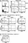
Figure 1
Figure 1
CD4 T cells in the iIEL compartment acquire CD8α. (A) iIELs were harvested from C57BL/6 mice and stained for TCR and coreceptors. Gating on TCRαβ showed that only a small number of CD4 T cells is found in the iIEL population in comparison to that in the spleen. Gating on CD4 showed that a large number of CD4 cells also express CD8α in iIELs but not in the spleen. However, none of these CD4+CD8α+ DP cells express CD8β. (B) TCRαβ CD4+ T cells were sorted from AND TCR Tg mice, activated with a combination of anti-CD3 and anti-CD28, and adoptively transferred to syngeneic RAG-1−/− mice. CD4 T cells were harvested from the iIEL compartment on the indicated days and examined for coreceptor expression. A large number of CD4 cells acquired CD8α but not CD8β. (C) Splenic TCRαβ CD4+ T cells were isolated from non-Tg mice, adoptively transferred to RAG-1−/− mice and analyzed as in B.
Figure 2
Figure 2
Transcription factors NF-κB and GATA-3 are involved in the acquisition of CD8α on CD4 T cells in the iIEL compartment. (A) iIELs were isolated from GATA-3DN mice and stained for their coreceptors. Gating on CD4 shows that very few CD4 T cells express CD8α+. (B) iIELs isolated from NF-κB p50−/− contain no CD4+CD8αα+ DP cells. (C) AND TCR Tg cells were cultured under Th1 and Th2 polarizing conditions for 3 days. They were then adoptively transferred to syngeneic RAG-1−/− mice. Four weeks posttransfer, they were harvested from iIEL compartment and stained for the expression of CD8α. Th2-polarized cells were able to acquire CD8α on their surface, whereas Th1-polarized cells failed to do so. (D) IL-4-producing cells either were enriched by positive selection after 4 days in culture under Th2-differentiating conditions (>80% enrichment) or were repeatedly stimulated under Th2 conditions and transferred to syngeneic RAG-1−/− mice. Thirty days after transfer, CD4 T cells were recovered from the iIEL compartment and analyzed for the expression of CD8α. IL-4-producing T cells from the primary culture yielded a considerable number of DP cells, whereas repeatedly stimulated cells could not generate DP cells in the iIELs. (E) CD4 T cells were cultured under either Th1 or Th2 polarizing conditions for 4 days. Cells were washed and restimulated under neutral conditions, and 48 h later they were stained with anti-CD30 antibody. Th2 cells showed a higher expression of CD30 and a single peak, whereas addition of anti-IL-4 and IFN-γ yielded essentially CD30-negative cells.
Figure 3
Figure 3
CD4+CD8αα+ DP iIELs possess a cytokine profile similar to regulatory (Treg) or suppressor T cells (Ts). (A) CD4+CD8αα+ DP cells were sorted by using a FACSVantage sorter. RNA was isolated from 0.5 × 10⁶ cells and used for RT-PCR for indicated cytokines by using specific primer pairs. (B) AND TCR Tg Th2-polarized cells were generated by culturing under Th2 conditions and their cytokine profile was measured by ELISA. They were transferred to RAG-1−/− mice. Four weeks after transfer, Tg cells were harvested from the iIEL compartment and CD4+CD8αα+ DP cells were sorted by using FACSVantage. They were activated with anti-CD3 + anti-CD28 along with IL-2. Forty-eight hours later, supernatants were harvested tested for their cytokine profile by ELISA.
Figure 4
Figure 4
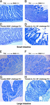
Reciprocal relationship of Th1 and CD4+CD8αα+ DP cells in the induction of intestinal inflammation. A total of 1 × 10⁶ Th2 (A and E) and Th1 (B and F) cells were transferred to RAG-1−/− mice. Four weeks later, the intestines were harvested and fixed in periodate fixing buffer. Tissues were embedded in paraffin blocks and sectioned. H&E-stained sections were examined for inflammation. (C and G) CD4+CD8αα+ DP cells were sorted from mice previously reconstituted with Th2 cells, and 1 × 10⁶ cells were transferred to RAG-1−/− mice. Four days later, they were challenged with 3 × 10⁶ Th1 cells. (D and H) Cells that were unable to produce IL-10 lacked protective ability. After 4 weeks, the intestines were harvested and sections were subjected to H&E (×100). Four animals were used per group, and the experiments were repeated twice with similar results. Histologic data shown are representative of two independent experiments.
Figure 5
Figure 5
CD4+CD8αα+ DP cells are found in the iIELs in IL-10−/− mice. iIELs were harvested from IL-10−/− mice and their control littermates. Cells were stained for their expression of CD8α. Gating on CD4 shows that IL-10−/− and its control littermates both contain CD4+CD8αα+ DP cells.
Figure 6
Figure 6
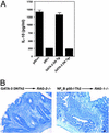
GATA-3 DN and NF-κB p50−/− CD4 T cells produce less IL-10 and can induce intestinal inflammation. (A) CD4 T cells from GATA-3 DN or NF-κB p50−/− mice were cultured under Th2 polarizing conditions, and IL-10 secreted in the culture supernatant was measured by ELISA. (B) Th2 cells derived from CD4 T cells isolated from GATA-3 DN and NF-κB p50−/− mice were adoptively transferred to syngeneic RAG-2−/− and RAG-1−/− mice. Four weeks later, the intestines were harvested, fixed, and paraffin-embedded, and sections were stained with H&E and scored for inflammation (×100).

References

    1. Mombaerts P, Mizoguchi E, Grusby M J, Glimcher L H, Bhan A K, Tonegawa S. Cell. 1993;75:274–282. - PubMed
    1. Powrie F, Correa-Oliveira R, Mauze S, Coffman R L. J Exp Med. 1994;179:589–600. - PMC - PubMed
    1. Simpson S J, Shah S, Comiskey M, de Jong Y P, Wang B, Mizoguchi E, Bhan A K, Terhorst C. J Exp Med. 1998;187:1225–1234. - PMC - PubMed
    1. Bhan A K, Mizoguchi E, Smith R N, Mizoguchi A. Immunol Rev. 1999;169:195–207. - PubMed
    1. Iijima H, Takahashi I, Kishi D, Kim J K, Kawano S, Hori M, Kiyono H. J Exp Med. 1999;190:607–615. - PMC - PubMed

Publication types

MeSH terms

LinkOut - more resources