. 2003 Apr 15;187(8):1165-77.
doi: 10.1086/368133. Epub 2003 Apr 2.

Intracellular and interstitial expression of Helicobacter pylori virulence genes in gastric precancerous intestinal metaplasia and adenocarcinoma

Affiliations

Intracellular and interstitial expression of Helicobacter pylori virulence genes in gastric precancerous intestinal metaplasia and adenocarcinoma

Cristina Semino-Mora et al. J Infect Dis. .

Abstract

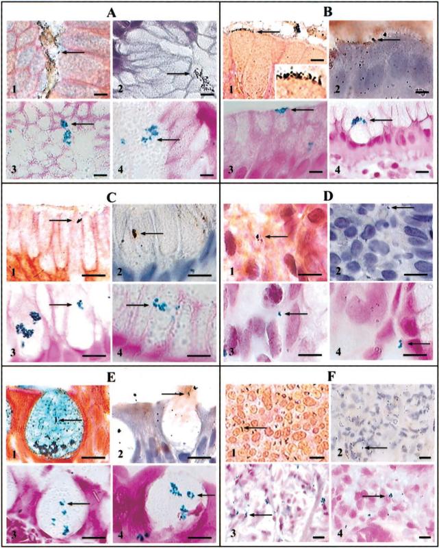
Gastric intestinal metaplasia (IM) and gastric cancer are associated with Helicobacter pylori, but the bacterium often is undetectable in these lesions. To unravel this apparent paradox, IM, H. pylori presence, and the expression of H. pylori virulence genes were quantified concurrently using histologic testing, in situ hybridization, and immunohistochemistry. H. pylori was detected inside metaplastic, dysplastic, and neoplastic epithelial cells, and cagA and babA2 expression was colocalized. Importantly, expression of cagA was significantly higher in patients with IM and adenocarcinoma than in control subjects. The preneoplastic "acidic" MUC2 mucin was detected only in the presence of H. pylori, and MUC2 expression was higher in patients with IM, dysplasia, and cancer. These novel findings are compatible with the hypothesis that all stages of gastric carcinogenesis are fostered by persistent intracellular expression of H. pylori virulence genes, especially cagA inside MUC2-producing precancerous gastric cells and pleomorphic cancer cells.


Figures

Figure 1
Figure 1
Luminal, intracellular, and interstitial colonization of the gastric mucosa by Helicobacter pylori in 4 groups of patients (bars, 10 μm). Bacteria are visualized in the lumen of antral gastric glands (A), in close proximity to the luminal surface of columnar epithelial cells (B; insert, details of H. pylori), within mucus-secreting cells (C), in the areolar connective tissue (D), within goblet cells (E), and in pleomorphic cells of gastric adenocarcinoma (F). Within each group of 4 pictures, picture 1 depicts Genta stain, picture 2 depicts anti–H. pylori immunohistochemistry, picture 3 depicts cagA expression by in situ hybridization (ISH), and picture 4 depicts babA2 expression by ISH.
Figure 2
Figure 2
A, (pictures 1 and 3), Dual-fluorescent localization of 16S rRNA–positive Helicobacter pylori (red). In picture 2, the 16S rRNA–positive bacterial cluster expresses cagA (green), whereas in picture 4, there is no colocalization of 16S rRNA and cagA (bars, 10 μm). B, H. pylori and mucin detection using high-iron diamine/Alcian blue/Steiner stain (bars, 10 μm) [23]. High-iron diamine stains sulfomucin black, Alcian blue stains sialomucin blue, and silver stains H. pylori. Picture 1, adhesion of 1 H. pylori to the apical membrane of a goblet cell in an area containing sulfomucin; picture 2, 4 H. pylori sectioned under different angles and surrounded by a clear halo within grey-stained sulfomucin; picture 3, H. pylori surrounded by an area stained blue only (no sulfomucin) inside a goblet cell in the process of discharging mucus into the gastric lumen (note that the halo surrounding H. pylori stains blue); and picture 4, 1 H. pylori sectioned transversely and surrounded by a clear halo within gray-stained sulfomucin. C, Apomucin expression in 3 serial sections of normal and precancerous mucosa by dual fluorescence in situ hybridization (FISH; pictures 1−4) and immunoperoxidase immunohistochemistry (IHC; pictures 5−8; bars, 10 μm). In normal epithelial cells (pictures 1 and 2 and pictures 5 and 6), MUC5AC is expressed (pictures 1 and 5; arrows) but MUC2 is not (pictures 2 and 6). In goblet cells (pictures 3 and 4 and pictures 7 and 8), MUC5AC is not expressed (pictures 3 and 7) but MUC2 is expressed (pictures 4 and 8; arrows). D, MUC5AC and MUC2 expression in 3 serial sections of adenocarcinoma matrix by dual FISH (pictures 1 and 2) and immunoperoxidase IHC (pictures 3 and 4; bars, 10 μm). Pictures 1 and 2 (serial section 2): dual-fluorescent reaction for MUC5AC (picture 1, negative) and MUC2 (picture 2, positive; arrow) expression by FISH; picture 3 (serial section 1): anti–MUC5AC IHC (negative); and picture 4 (serial section 3): anti-MUC2 IHC (positive; arrow).
Figure 3
Figure 3
Fluorescence in situ hybridization (FISH) laser confocal microscopic illustration of Helicobacter pylori intracellular localization. Right, pseudo–3-dimensional reconstruction of 10 serial 0.3-μm scans recorded by observing a FISH preparation of H. pylori–specific 16S rRNA at 1000× magnification (bar, 10 μm). Left, schematic representation of cell contours and of some of the most prominent intracellular clusters of H. pylori. A, absorptive columnar cell; G, goblet cell.
Figure 4
Figure 4
Transmission electron micrographs of a metaplastic gland containing columnar absorptive cells with surface microvilli and associated terminal web. A, Cell containing intracytoplasmic Helicobacter pylori (arrows) that are not surrounded by specialized cellular interface (magnification, ×17,000). B, Detail of the surface of a cell illustrating the presence of an H. pylori in the gastric gland lumen that is close to the surface microvilli and to the associated terminal web. No specialized pedestal-like formation is seen. A low-electron-density circular spot is present in one of the extremities of the bacteria, suggesting the accumulation of polyphosphate granule (magnification, ×33,000; bar, 1 μm).
Figure 5
Figure 5
Goblet cell mucus granules of different densities creating a “checkerboard” appearance [29]. Long arrow, structure that closely resembles a late-stage coccoidal form of Helicobacter pylori [30]. Arrowheads, 3 structures resembling flagellar filaments that appear to emerge from a single pole of the cell and are in close contact with the mucus granules (magnification, ×24,000; bar, 1 μm).

References

    1. Anonymous . IARC Monograph on the evaluation of carcinogenic risks to humans: schistosomes, liver flukes and Helicobacter pylori. International Agency for Research on Cancer; Lyon, France: 1994. - PMC - PubMed
    1. Uemura N, Mukai T, Okamoto S, et al. Effect of Helicobacter pylori eradication on subsequent development of cancer after endoscopic resection of early gastric cancer. Cancer Epidemiol Biomarkers Prev. 1997;6:639–42. - PubMed
    1. Helicobacter and Cancer Collaborative Group Gastric cancer and Helicobacter pylori: a combined analysis of 12 case control studies nested within prospective cohorts. Gut. 2001;49:347–53. - PMC - PubMed
    1. Blaser MJ, Perez-Perez GI, Kleanthous H, et al. Infection with Helicobacter pylori strains possessing cagA is associated with an increased risk of developing adenocarcinoma of the stomach. Cancer Res. 1995;55:2111–5. - PubMed
    1. Censini S, Lange C, Xiang Z, et al. cag, a pathogenicity island of Helicobacter pylori, encodes type I–specific and disease-associated virulence factors. Proc Natl Acad Sci USA. 1996;93:14648–53. - PMC - PubMed

Publication types

MeSH terms