. 2003 May;72(5):1300-7.
doi: 10.1086/375116. Epub 2003 Apr 14.

2-Methyl-3-hydroxybutyryl-CoA dehydrogenase deficiency is caused by mutations in the HADH2 gene

Affiliations

2-Methyl-3-hydroxybutyryl-CoA dehydrogenase deficiency is caused by mutations in the HADH2 gene

Rob Ofman et al. Am J Hum Genet. 2003 May.

Abstract

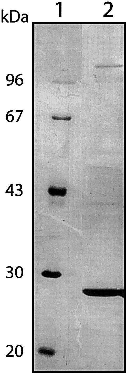
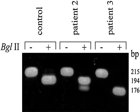
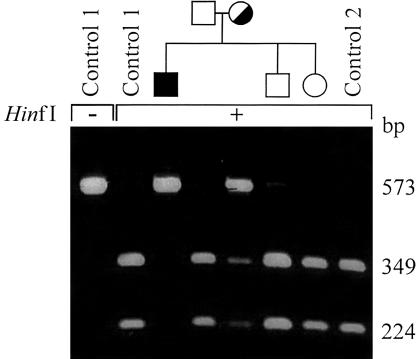
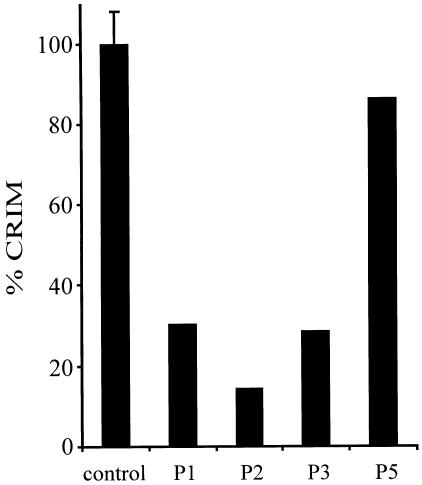
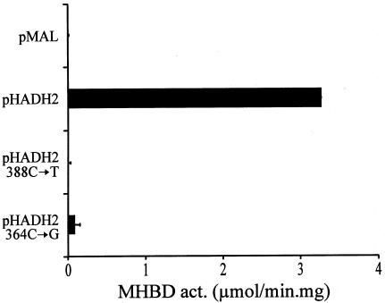
2-methyl-3-hydroxybutyryl-CoA dehydrogenase (MHBD) deficiency is a novel inborn error of isoleucine degradation. In this article, we report the elucidation of the molecular basis of MHBD deficiency. To this end, we purified the enzyme from bovine liver. MALDI-TOF mass spectrometry analysis revealed that the purified protein was identical to bovine 3-hydroxyacyl-CoA dehydrogenase type II. The human homolog of this bovine enzyme is a short-chain 3-hydroxyacyl-CoA dehydrogenase, also known as the "endoplasmic reticulum-associated amyloid-beta binding protein" (ERAB). This led to the identification of the X-chromosomal gene involved, which previously had been denoted "HADH2." Sequence analysis of the HADH2 gene from patients with MHBD deficiency revealed the presence of two missense mutations (R130C and L122V). Heterologous expression of the mutant cDNAs in Escherichia coli showed that both mutations almost completely abolish enzyme activity. This confirms that MHBD deficiency is caused by mutations in the HADH2 gene.


Figures

Figure  1
Figure 1
Sodium dodecyl sulfate-polyacrylamide gel electrophoresis (SDS-PAGE) analysis of bovine liver MHBD. MHBD was purified from bovine liver and analyzed using SDS-PAGE (Laemmli 1970) followed by silver staining (Rabilloud et al. 1988). Lane 1, molecular weight markers; Lane 2, purified bovine liver MHBD.
Figure  2
Figure 2
Detection of HADH2 388C→T mutation by RFLP analysis. The zygosity of the 388C→T mutation found in patients 2 and 3 (table 1) was studied using RFLP analysis. Genomic DNA was amplified using the primers 2284HADH2frw plus “M13 reverse”-tagged Ex3/4rev. PCR conditions were as described for the other genomic DNA amplifications. After amplification, the PCR products were incubated for 2 h at 37°C in the presence (+) or absence (−) of 10 units of BglII, and, afterward, the restriction products were separated on a 2% agarose gel.
Figure  3
Figure 3
Detection of the HADH2 364C→G mutation by RFLP analysis in a family with MHBD deficiency. The presence of the 364C→G mutation found in patient 4 (table 1) was studied in other family members (father, mother, brother, and sister) and two unrelated control subjects. Therefore, fragment 2 was amplified from genomic DNA, followed by an incubation in the presence (+) or absence (−) of the restriction enzyme HinfI. Finally, the restriction fragments were separated on a 2% agarose gel.
Figure  4
Figure 4
Immunological detection of MHBD in cultured skin fibroblasts from patients with MHBD deficiency. For immunoblot analysis, 50 μg protein of cultured skin fibroblasts from control subjects and patients with MHBD deficiency was applied on a 10% polyacrylamide gel and separated, as described elsewhere (Laemmli 1970). After electrophoresis, proteins were transferred to a nitrocellulose membrane by semidry blotting, probed with the polyclonal antibody raised against recombinant human MHBD, and developed (Wanders et al. 1995). After staining of the blot with NBT/BCIP, the result was digitalized by use of a desktop scanner and the amount of MHBD protein quantified, using the National Insitutes of Health Image 1.62 software program. CRIM = cross reactive immunological material. Control-mean value of three control subjects represented as 100% (±SD); 1 = patient 1; 2 = patient 2; 3 = patient 3, and 5 = patient 5 (see table 1).
Figure  5
Figure 5
Heterologous expression of human MHBD in E. coli. The complete ORF of HADH2 from a control subject and from patients with MHBD deficiency were amplified and ligated into the bacterial expression vector pMAL-C2X, as described. Each ORF was sequenced to exclude sequence errors introduced during PCR. Transformed bacteria (INVα) were grown in 20-ml LB medium supplemented with 100 μg/ml ampicillin to an OD600 of ∼0.5, and IPTG was added to a final concentration of 1 mM to induce protein expression. After 4 h at 37°C, cells were pelleted and resuspended in 1 ml 10 mM sodium phosphate buffer pH 7.4, containing 140 mM NaCl, 0.1% (wt/vol) Triton X-100 and protease inhibitors (1 tablet Completemini [Boehringer Mannheim] in 10-ml solution). Lysis was achieved by sonication at 9 W for 10 s. The bacterial lysate was centrifuged for 10 min at 14,000×gav, and the pellet was discarded. The supernatant was used for protein measurement and MHBD activity, as described. Expression levels of the fusion protein in the bacterial extracts were equal, on the basis of immunoblot analysis (data not shown). pMAL = expression with empty pMAL-c2x vector; pHADH2 = expression with vector containing the wild-type human MHBD; pHADH2 388C→T = expression of human MHBD mutant with R130C; pHADH2 364C→G = expression of human MHBD mutant with L122V.

References

Electronic-Database Information

    1. GenBank and National Center for Biotechnology Information (NCBI), http://www.ncbi.nlm.nih.gov/ (for ERAB [accession numbers U96132 and AF037438] and HADH2 [NM_004493])
    1. Online Mendelian Inheritance in Man (OMIM), http://www.ncbi.nlm.nih.gov/Omim/ (for 2-methylacetoacetyl-CoA thiolase deficiency, 2-methylbutyryl-CoA dehydrogenase deficiency, 2-methyl-3-hydroxybutyryl-CoA dehydrogenase deficiency, and Alzheimer disease)
    1. Swiss-Prot Protein Knowledgebase, http://www.ebi.ac.uk/swissprot/ (for a protein sequence database)

References

    1. Bradford MM (1976) A rapid and sensitive method for the quantitation of microgram quantities of protein utilizing the principle of protein-dye binding. Anal Biochem 72:248–254 - PubMed
    1. Ensenauer R, Niederhoff H, Ruiter JP, Wanders RJ, Schwab KO, Brandis M, Lehnert W (2002) Clinical variability in 3-hydroxy-2-methylbutyryl-CoA dehydrogenase deficiency. Ann Neurol 51:656–659 - PubMed
    1. Frackowiak J, Mazur-Kolecka B, Kaczmarski W, Dickson D (2001) Deposition of Alzheimer’s vascular amyloid-β is associated with decreased expression of brain L-3-hydroxyacyl-coenzyme A dehydrogenase (ERAB). Brain Res 907:44–53 - PubMed
    1. Furuta S, Kobayashi A, Miyazawa S, Hashimoto T (1997) Cloning and expression of cDNA for a newly identified isozyme of bovine liver 3-hydroxyacyl-CoA dehydrogenase and its import into mitochondria. Biochim Biophys Acta 1350:317–324 - PubMed
    1. Ghaedi K, Itagaki A, Toyama R, Tamura S, Matsumura T, Kawai A, Shimozawa N, Suzuki Y, Kondo N, Fujiki Y (1999) Newly identified Chinese hamster ovary cell mutants defective in peroxisome assembly represent complementation group A of human peroxisome biogenesis disorders and one novel group in mammals. Exp Cell Res 248:482–488 - PubMed

Substances

Associated data