. 2003 Apr 2:4:6.
doi: 10.1186/1471-2202-4-6.

Discrete nuclear structures in actively growing neuroblastoma cells are revealed by antibodies raised against phosphorylated neurofilament proteins

Affiliations

Discrete nuclear structures in actively growing neuroblastoma cells are revealed by antibodies raised against phosphorylated neurofilament proteins

Shannon E Weigum et al. BMC Neurosci. .

Abstract

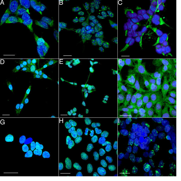
Background: Nuclear objects that have in common the property of being recognized by monoclonal antibodies specific for phosphoprotein epitopes and cytoplasmic intermediate filaments (in particular, SMI-31 and RT-97) have been reported in glial and neuronal cells, in situ and in vitro. Since neurofilament and glial filaments are generally considered to be restricted to the cytoplasm, we were interested in exploring the identity of the structures labeled in the nucleus as well as the conditions under which they could be found there.

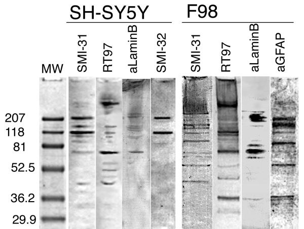
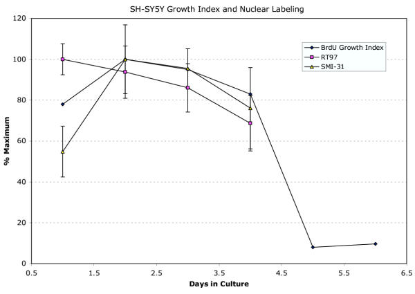
Results: Using confocal microscopy and western analysis techniques, we determined 1) the immunolabeled structures are truly within the nucleus; 2) the phosphoepitope labeled by SMI-31 and RT-97 is not specific to neurofilaments (NFs) and it can be identified on other intermediate filament proteins (IFs) in other cell types; and 3) there is a close relationship between DNA synthesis and the amount of nuclear staining by these antibodies thought to be specific for cytoplasmic proteins. Searches of protein data bases for putative phosphorylation motifs revealed that lamins, NF-H, and GFAP each contain a single tyrosine phosphorylation motif with nearly identical amino acid sequence.

Conclusion: We therefore suggest that this sequence may be the epitope recognized by SMI-31 and RT-97 mABs, and that the nuclear structures previously reported and shown here are likely phosphorylated lamin intermediate filaments, while the cytoplasmic labeling revealed by the same mABs indicates phosphorylated NFs in neurons or GFAP in glia.


Figures

Figure 1
Figure 1
Immunolocalization of mABs SMI-31, SMI-32, anti-BrdU, and pAB anti-GFAP. A. A micrograph showing a single, confocal image plane of SH-SY5Y cells labeled with SMI-31 mAB followed by an Alexa-Fluor-488 conjugated secondary antibody (green). Nuclei were revealed by the TO-PRO-3 DNA probe (blue). The green SMI-31 mAB labeling appears as white dots within the blue nuclei, and as filamentous green structures in the cytoplasm. B. A single image plane of SH-SY5Y cells grown on glass coverslips and labeled with RT97 mAB followed by an Alexa-Fluor-488 conjugated secondary antibody (green). Nuclei were revealed by TO-PRO-3 probe (blue). RT97 mAB labeling appears as white dots in nuclei and as filamentous or clumpy green structures in the cytoplasm C. A confocal projection of SH-SY5Y cells grown on glass coverslips and labeled with SMI-32 mAB followed by an Alexa-Fluor-488 conjugated secondary antibody (green). Nuclei were revealed by TO-PRO-3 probe (blue). SMI-32 mAB did not label nuclei but did reveal filamentous green structures in the cytoplasm. D. A confocal projection of F98 rat glioma cells grown on glass coverslips and labeled with SMI-31 mAB followed by an Alexa-Fluor-488 conjugated secondary antibody (green). Nuclei were revealed by TO-PRO-3 probe (blue). SMI-31 mAB labeling appears as white dots in nuclei and as filamentous green structures in the cytoplasm. Labeling in these cells was so intense that the nuclei appeared completely full of the labeled epitope. E. A confocal projection of F98 rat glioma cells grown on glass coverslips and labeled with RT97 mAB followed by an Alexa-Fluor-488 conjugated secondary antibody (green). Nuclei were revealed by TO-PRO-3 probe (blue). SMI-31 mAB labeling appears as white dots in nuclei. RT97 staining of nuclear and cytoplasmic structures in F98 rat glioma cells was similar but less intense than seen with SMI-31 on F98 cells. F. A confocal projection of F98 cells grown on glass coverslips and labeled with anti-GFAP polyclonal AB followed by an Alexa-Fluor-488 conjugated secondary antibody (green). Nuclei were revealed by TO-PRO-3 probe (blue). The anti-GFAP revealed the expected filamentous green structures in the cytoplasm but did not label structures in nuclei. The white areas that look as if they are in the nucleus are actually green staining in the cytoplasm below (seen through) the blue nucleus. G, H, I. Confocal projections of SH-SY5Y cells showing nuclei labeled with TO-PRO-3 (blue) and anti-BrdU (green, white when co-localized with blue). These panels show nuclei of cells in culture for 1, 3, and 6 days, respectively. Labeling intensity appeared to decline as the culture became confluent, suggesting a corresponding decline in DNA synthesis and cells exiting the cell cycle. See also Figure 4. *the calibration bar in each panel represents 5 μm.
Figure 2
Figure 2
Western analysis with SMI-31, RT97, anti-lamin B, SMI-32 on SH-SY5Y, and SMI-31, RT97, anti-lamin B, anti-GFAP on F98 cell extracts subjected to SDS-PAGE. On SH-SY5Y, the SMI-31 and SMI-32 lanes show strong labeling of bands corresponding to the MW of NF-H, and NF-M. Additional bands, not seen in the SMI-32, are present in the SMI-31 and RT97 lanes between and above the NF-H and NF-M, and probably represent phosphorylated forms of NF-M and NF-H. SMI-31, RT-97 and anti-lamin B also revealed a band or closely spaced bands at approximately 70 kD which corresponds approximately to the MW of lamin B. Western analysis with anti-lamin A/C revealed no bands in either SH-SY5Y or F98 extracts (not shown). On F98 cells, SMI-31 labeled several well defined bands, including those of MW corresponding to GFAP and lamin B. The RT97 mAB also strongly labeled several F98 bands, including bands corresponding to GFAP and lamin B. The anti-lamin B mAB strongly labeled bands at the appropriate MW (~70 kD) and also a cluster of bands in the range of the heavy NF MW (~207 kD). The anti-GFAP pAB labeled a band at the appropriate MW (~52 kD) and also band higher of MW appropriate for multimers of GFAP (~100, 150, 200 kD). Labeled bands lower than 65 kD may represent degradation products produced during cell extraction. Negative controls (control blots stained with appropriate normal serum) showed no labeling (data not shown).
Figure 3
Figure 3
A portion of the amino acid sequences for NF-H, GFAP, and lamin A, B, and C. The Match-Box server prints aligned sequences in lower case, an unaligned as uppercase. The numbers printed below each column of aligned amino acids indicates the reliability of the alignment, where 1 = 10%, 3 = 20%, and 5 = 50% chance of equal occurrence in related or unrelated sequences (low scores are best, Depiereux et al, 1997). While we found many phosphorylation motifs in the sequences examined, the indicated tyrosine kinase motif was the only one shared with a high degree of identity among all 5 sequences. As it occurs only once, the remainder of the sequences are not shown. The darker gray shading indicates the predicted motif for NF-H (the reference sequence), while the light gray shows regions of GFAP and lamins that include the tyrosine (bold-face "Y") in a motif with very high sequence identity.
Figure 4
Figure 4
Graph showing a strong correlation between the culture proliferation index (percent nuclei showing BrdU incorporation) and the extent of nuclear labeling as a function of days in culture for SH-SY5Y cells immuno-stained for SMI-31, RT97. Values for SMI-31 and RT-97 labeling represent averages (± SEM) of the percentage of nuclear area labeled as determined by confocal projection. Proliferation index data is shown through day 5 and 6 to illustrate that the culture enters a confluent, stationary phase. See also Figure 1G,1H and 1I.

References

    1. Schilling K, Duvernoy C, Keck S, Pilgrim C. Detection and partial characterization of a developmentally regulated nuclear antigen in neural cells in vitro and in vivo. J Histochem Cytochem. 1989;37:241–247. - PubMed
    1. Singh MV, Price KJ, Bhatnagar R, Johnson R, Malhotra SK. J1-31 antigen of astrocytes: cytoplasmic and nuclear localization. Dendron. 1992;1:91–108.
    1. Singh MV, Price KJ, Bhatnagar R, Malhotra SK. Novel rod-shaped structures identified in glioma cell nuclei by immunolabeling and confocal laser fluorescence microscopy. Biomedical Letters. 1994;50:163–172.
    1. Malhotra SK, Bhatnagar R, Shnitka TK, Herrera JJ, Koke JR, Singh MV. Rat glioma cell line as a model for astrogliosis. Cytobios. 1995;82:39–51. - PubMed
    1. Sanford Jeremy. Nuclear Compartments: Nuclear Splicing Speckles. In: Wendy Bickmore, editor. Nuclear Protein Database (NDP) UK, Medical Research Council Human Genetics Unit; 2002. http://npd.hgu.mrc.ac.uk/compartments/speckles.html

Publication types

MeSH terms

Associated data