. 2003 Apr;111(8):1125-32.
doi: 10.1172/JCI17170.

EGF amplifies the replacement of parvalbumin-expressing striatal interneurons after ischemia

Affiliations

EGF amplifies the replacement of parvalbumin-expressing striatal interneurons after ischemia

Tetsuyuki Teramoto et al. J Clin Invest. 2003 Apr.

Abstract

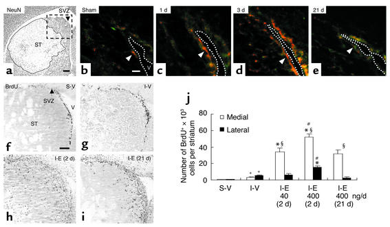
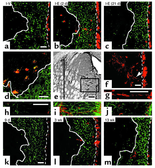
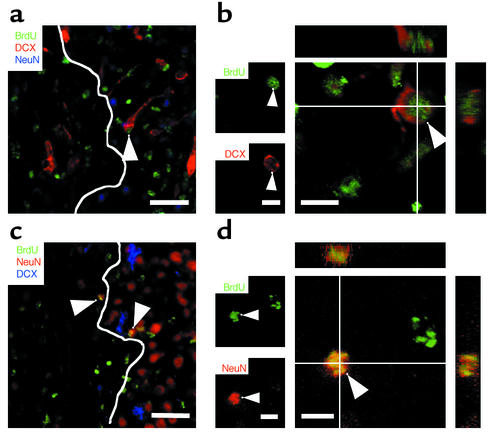
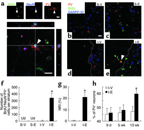
EGF promotes proliferation and migration of stem/progenitor cells in the normal adult brain. The effect of epidermal growth factor on neurogenesis in ischemic brain is unknown, however. Here we show that intraventricular administration of EGF and albumin augments 100-fold neuronal replacement in the injured adult mouse striatum after cerebral ischemia. Newly born immature neurons migrate into the ischemic lesion and differentiate into mature parvalbumin-expressing neurons, replacing more than 20% of the interneurons lost by 13 weeks after ischemia and representing 2% of the total BrdU-labeled cells. These data suggest that administration of EGF and albumin could be used to manipulate endogenous neurogenesis in the injured brain and to promote brain self-repair.


Figures

Figure 1
Figure 1
EGF-enhanced proliferation after ischemia. (a) Ischemia reperfusion caused a marked loss of NeuN+ neurons (outlined area) confined to the striatum (ST). SVZ, subventricular zone. Scale bar is 200 μm. (be) Without ischemia (b), few nestin+ cells (green) expressing EGF receptors (red) surrounded the lateral ventricle (V). After ischemia, the intensity of EGF receptor immunoreactivity in nestin+ SVZ cells (arrowheads) and the number of nestin+ cells expressing EGF receptor increased at 1 day (c), reached maximum at 3 days (d), and returned to basal levels at 21 days (e). The dotted line delineates lateral ventricle. Scale bar is 20 μm (n = 3–4/time-point). (fi) BrdU+ cells at 9 days after sham or ischemia in an area corresponding to the box (a). After vehicle, the number of BrdU+ cells was higher after ischemia (g) than sham (f) (quantification in j). Ischemia followed by EGF (I-E; 400 ng/day) increased BrdU+ cells and was greater when EGF infusion started 2 days (h) rather than 21 days (i) after ischemia. (f) Scale bar is 100 μm. Bregma, 0.8 mm. (j) Quantification. S-V, sham-operated; I-V, ischemic group with vehicle; I-E (2d), ischemic groups with EGF (400 ng/day) initiated at day 2; I-E (21d), day 21 after ischemia. The effects of EGF (40 and 400 ng/day) initiated at day 2 were compared. +Significant difference between S-V and I-V; *between vehicle- and EGF-treated ischemic groups; #between groups treated with EGF 2 days or 21 days; §between medial and lateral striatum within the same group; n = 3–4/group, unpaired Student t test, P < 0.05.
Figure 2
Figure 2
Migrating neuroblasts after EGF treatment. (a) Using confocal microscopy, 5 weeks after ischemia, very few DCX+ cells (red) were observed in the striatal lesion (white solid line) after vehicle. DCX+ cells dramatically increased by EGF infusion initiated on day 2 (b) but not on day 21 (c) (n = 3–4 per group; for quantification see Table 1), revealing the importance of EGF signaling for cell migration early after ischemia. (d) Colocalization of DCX (red) and Tuj1 (green) confirmed their phenotype. (e) DCX+ cells were predominantly within medial (M) relative to lateral (L) striatum. (f and g) Chains or clusters of DCX+ (red) cells (arrowhead, magnified in g) connecting SVZ and the striatal lesion were observed in medial striatum (e). (hj) GFAP+ glial cells (green) and DCX+ neuroblasts (red) between SVZ (right end) and the damaged striatum (left side) were rare in vehicle (h) or with EGF administration initiated at day 21 (j), compared with EGF at day 2 post-ischemia (i). Note the orientation of GFAP+ processes in (i). (km) The spatial distribution and number of DCX+ (red) cells changed with time after EGF. DCX+ cells were few at the end of the infusion period (k) and progressively increased in the area devoid of NeuN+ cells (green) at 3 weeks (l) and 5 weeks (b). By 13 weeks, DCX+ were mostly outside the lesion (m), suggesting that DCX+ cells may have differentiated into mature neurons. Scale bar is 50 μm except for (e), 100 μm. Bregma, 0.8 mm. Dotted line, the border between SVZ and striatum.
Figure 3
Figure 3
Neuronal differentiation in the ischemic striatum. (a and c) Representative confocal microscopic images of BrdU, DCX, and NeuN colabeling showing that BrdU-labeled neurons (arrowheads) were located in the boundary zone of the ischemic lesion at 5 (a) and 13 (c) weeks. A white solid line delineates the lesion area. Scale bar is 50 μm. (b and d) Confocal 3D analyses of BrdU+DCX+ cells at 5 weeks (b) and BrdU+NeuN+ cells at 13 weeks (d) after ischemia. Reconstructed orthogonal images are presented as viewed from the sides in both the x-z (top) and y-z (right) planes. Arrowheads indicate double-labeled cells. Scale bar is 10 μm. n = 5–6 per group (for quantification, see Table 1).
Figure 4
Figure 4
Neuronal replacement in the ischemic striatum. (a) Confocal 3D reconstruction revealed newborn PV+ neurons (arrowhead) triple-labeled with BrdU, NeuN, and PV in the medial striatum of EGF-treated mice at 13 weeks after ischemia. (be) The triple-labeling with BrdU, PV, and DARPP-32 revealed that newborn PV+ neurons (e, arrowhead) were located in the boundary zone of the ischemic lesion. White solid line delineates the lesion area. Scale bars are 20 μm. (f) Numbers of BrdU+NeuN+PV+ cells in the medial striatum at 13 weeks after ischemia. Ud, undetected in VAS-V and S-E groups. (g) NRI (see text) indicates ratio between newborn neurons and the neurons lost after injury. (h) Progressive repopulation of PV+ neurons in the ischemic striatum measured as a percentage of PV+ neurons in ischemic striatum relative to the number in the nonischemic contralateral striatum. *Significant difference between ischemic group administered with vehicle (I-V) or EGF (I-E) (P < 0.01); n = 5-6/group, unpaired Student t test.

References

    1. Betarbet R, Zigova T, Bakay RA, Luskin MB. Dopaminergic and GABAergic interneurons of the olfactory bulb are derived from the neonatal subventricular zone. Int. J. Dev. Neurosci. 1996;14:921–930. - PubMed
    1. Chiasson BJ, Tropepe V, Morshead CM, van der Kooy D. Adult mammalian forebrain ependymal and subependymal cells demonstrate proliferative potential, but only subependymal cells have neural stem cell characteristics. J. Neurosci. 1999;19:4462–4471. - PMC - PubMed
    1. Alvarez-Buylla A, Garcia-Verdugo JM. Neurogenesis in adult subventricular zone. J. Neurosci. 2002;22:629–634. - PMC - PubMed
    1. Reynolds BA, Tetzlaff W, Weiss S. A multipotent EGF-responsive striatal embryonic progenitor cell produces neurons and astrocytes. J. Neurosci. 1992;12:4565–4574. - PMC - PubMed
    1. Reynolds BA, Weiss S. Generation of neurons and astrocytes from isolated cells of the adult mammalian central nervous system. Science. 1992;255:1707–1710. - PubMed

Publication types

MeSH terms