. 2003 Apr;111(8):1133-45.
doi: 10.1172/JCI16432.

CD28-dependent Rac1 activation is the molecular target of azathioprine in primary human CD4+ T lymphocytes

Affiliations

CD28-dependent Rac1 activation is the molecular target of azathioprine in primary human CD4+ T lymphocytes

Imke Tiede et al. J Clin Invest. 2003 Apr.

Abstract

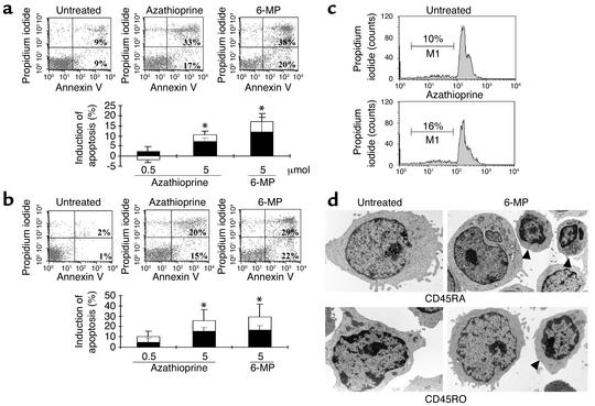
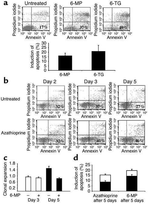
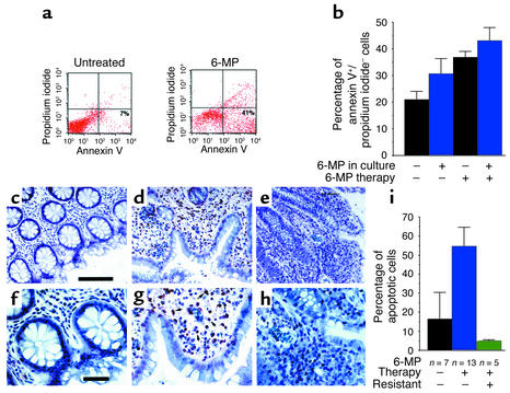
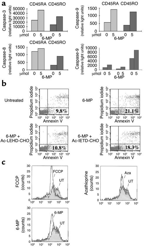
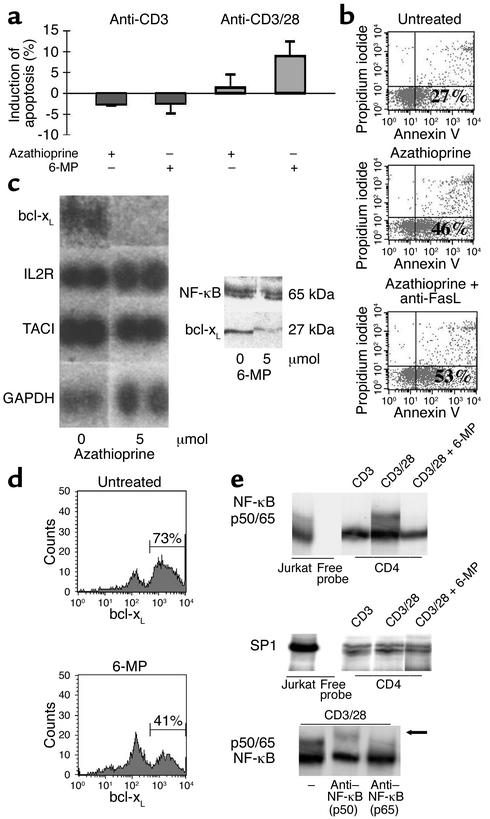
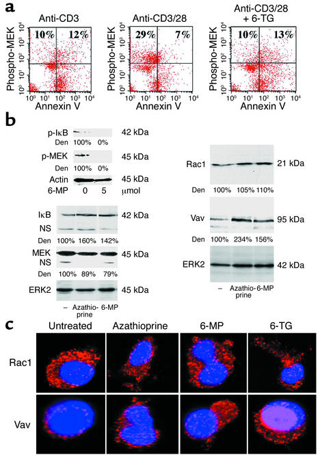
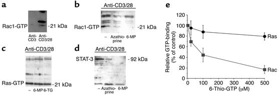
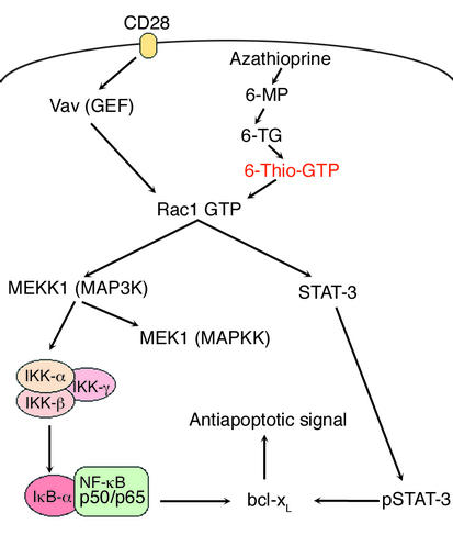
Azathioprine and its metabolite 6-mercaptopurine (6-MP) are immunosuppressive drugs that are used in organ transplantation and autoimmune and chronic inflammatory diseases such as Crohn disease. However, their molecular mechanism of action is unknown. In the present study, we have identified a unique and unexpected role for azathioprine and its metabolites in the control of T cell apoptosis by modulation of Rac1 activation upon CD28 costimulation. We found that azathioprine and its metabolites induced apoptosis of T cells from patients with Crohn disease and control patients. Apoptosis induction required costimulation with CD28 and was mediated by specific blockade of Rac1 activation through binding of azathioprine-generated 6-thioguanine triphosphate (6-Thio-GTP) to Rac1 instead of GTP. The activation of Rac1 target genes such as mitogen-activated protein kinase kinase (MEK), NF-kappaB, and bcl-x(L) was suppressed by azathioprine, leading to a mitochondrial pathway of apoptosis. Azathioprine thus converts a costimulatory signal into an apoptotic signal by modulating Rac1 activity. These findings explain the immunosuppressive effects of azathioprine and suggest that 6-Thio-GTP derivates may be useful as potent immunosuppressive agents in autoimmune diseases and organ transplantation.


Figures

Figure 1
Figure 1
Azathioprine and its metabolites induce T cell apoptosis. CD45RA (a) and CD45RO (b) T cell subsets were isolated from peripheral blood of healthy volunteers and stimulated with antibodies to CD3 and CD28 and recombinant IL-2 in the presence or absence of indicated concentrations of azathioprine and 6-MP. T cell apoptosis was assessed by FACS analysis after 5 days of cell culture. Azathioprine and 6-MP led to an induction of annexin-positive, propidium iodide–negative T cells, suggesting that they induced T cell apoptosis. The FACS data is representative of 6–10 independent experiments per group. The average induction of specific apoptosis from 6–10 patients per group (induction of annexin-positive, propidium iodide–negative cells compared with untreated cells indicated by black sections of bars, induction of annexin-positive, propidium iodide–positive cells compared with untreated cells indicated by white sections) by azathioprine and 6-MP ± SEM is shown in the lower panels. Statistically significant changes are indicated (*P < 0.01). (c) Determination of apoptosis and cell cycle distribution by the Nicoletti technique. Peripheral blood CD4+ T cells from healthy volunteers were stimulated as above for 5 days, followed by analysis of DNA content by the Nicoletti technique. The percentage of apoptotic cells in the subdiploid peak is indicated by M1. (d) Azathioprine and 6-MP induce morphologic changes of CD45RA CD4+ T cells (upper panels) and CD45RO CD4+ T cells (lower panels) indicative of apoptosis, as assessed by transmission electron microscopy. Peripheral blood CD4+ T cells from healthy volunteers were stimulated as above for 5 days. Upon 6-MP treatment, T cells exhibited typical signs of apoptosis (arrowheads), with dense nuclear condensation and degeneration of organelles. Magnification, ×7200.
Figure 2
Figure 2
(a) Peripheral blood CD4+ T cells from healthy volunteers were stimulated with antibodies to CD3 and CD28 and recombinant IL-2 and cultured in the presence or absence of 6-MP and 6-TG for 4–5 days. T cell apoptosis was assessed by FACS analysis (upper panels). The average level of 6-MP– and 6-TG–specific apoptosis (induction of annexin-positive, propidium iodide–negative T cells compared with untreated cells) ± SEM from four independent experiments is shown in the lower panel. (b) Kinetics of azathioprine-induced apoptosis in primary CD4+ T lymphocytes. CD4+ T cells were cultured in the presence or absence of azathioprine for 2–5 days as indicated. T cell apoptosis was assessed by FACS analysis at the indicated time points. (c) 6-MP suppresses clonal expansion of activated primary CD4+ T lymphocytes in cell culture. CD4+ T cells were cultured in the presence (+) or absence (–) of 6-MP for 3–5 days. The clonal expansion of T cells during cell culture was calculated as specified in Methods. (d) CD4+ T cells were cultured in the absence of azathioprine or 6-MP for 5 days, followed by addition of azathioprine or 6-MP to the cell culture for an additional 5 days. The percentage of annexin-positive, propidium iodide–negative cells was then determined at day 10 by FACS analysis. The average percentage of azathioprine- and 6-MP–specific apoptosis (induction of annexin-positive, propidium iodide–negative cells compared with untreated cells indicated by black sections of bars, induction of annexin-positive, propidium iodide–positive cells compared with untreated cells indicated by white sections) ± SEM is shown in the lower panel.
Figure 3
Figure 3
Azathioprine treatment induces T cell apoptosis in IBD. (a) LPMCs were isolated and stimulated with anti-CD2/CD28 plus IL-2 in the presence or absence of 6-MP for 5 days followed by FACS analysis. (b) Peripheral blood CD4+ T cells were isolated from patients with IBD receiving azathioprine/6-MP and patients with IBD before therapy and stimulated with anti-CD3/CD28 plus IL-2 in the presence or absence of 6-MP for 5 days. The number of apoptotic cells (annexin-positive, propidium iodide–negative cells) was determined by FACS analysis after 5 days and stratified according to clinical data on azathioprine therapy. Data represent mean values ± SEM from eight patients with IBD per group. (ch) TUNEL assays for the detection of apoptotic lamina propria cells in patients with IBD under azathioprine therapy. Successful azathioprine treatment associated with low-level gut inflammation was characterized by a high number of apoptotic cells (shown at low [d] and high [g] magnification) as compared with control patients with low-level gut inflammation not receiving azathioprine (c and f). In contrast, unsuccessful azathioprine treatment associated with high-level gut inflammation was characterized by a very low percentage of apoptotic cells (e and h). Data are representative of 5–13 patients per group. Magnification, ×20 (bar = 100 μm) (ce) and ×40 (bar = 50 μm) (fh). (i) Quantification of the percentage of apoptotic, TUNEL-positive LPMCs in patients with IBD receiving 6-MP as compared with untreated control patients. Clinical 6-MP responsiveness was associated with a marked, significant (P < 0.01) increase in the percentage of apoptotic LPMCs as compared with control patients with IBD not receiving 6-MP. Data are mean values ± SEM of 5–13 patients per group.
Figure 4
Figure 4
Azathioprine induces a mitochondrial pathway of apoptosis. (a) Activity of caspase-3, -8, and -9 upon treatment of T cells with 6-MP. CD45RA and CD45RO T cell subsets were stimulated with antibodies to CD3 and CD28 and recombinant IL-2 for 5 days in the presence or absence of 6-MP, as indicated. There was a marked induction of caspase-9 activity upon azathioprine treatment. A second independent experiment showed similar results (data not shown). Data on caspase-9 activity from three independent healthy blood donors are shown in the right lower panel. (b) Specific blockade of caspase-9 by acetyl-LEHD-CHO (Ac-LEHD-CHO) suppresses 6-MP–induced apoptosis. CD4+ T lymphocytes from the peripheral blood of healthy volunteers were stimulated with antibodies to CD3 and CD28 in the presence or absence of 6-MP, 10 μM acetyl-LEHD-CHO, and 10 μm acetyl-IETD-CHO. Although acetyl-IETD-CHO had little effect, 6-MP–induced T cell apoptosis could be suppressed by acetyl-LEHD-CHO. (c) Measurement of ΔΨm in primary CD4+ T lymphocytes upon treatment with azathioprine, 6-MP, and FCCP (positive control). Peripheral blood CD4+ T cells from healthy volunteers were stimulated with antibodies to CD3 and CD28 and recombinant IL-2 and cultured in the presence or absence of azathioprine or 6-MP for 5 days as indicated. Cells were then loaded with JC-1 for 20 minutes followed by FACS analysis to determine ΔΨm. Both azathioprine and 6-MP as well as FCCP led to a marked reduction of ΔΨm as compared with untreated primary CD4+ T cells. One representative experiment of two is shown. Aza, azathioprine; UT, untreated.
Figure 5
Figure 5
(a) Azathioprine-induced apoptosis is critically dependent on costimulation with CD28. CD4+ T lymphocytes were stimulated in the presence or absence of azathioprine and 6-MP, as indicated. T cell apoptosis was assessed by FACS analysis using annexin V/propidium iodide staining at day 5 of cell culture. (b) Azathioprine-induced apoptosis is independent of the CD95/CD95L system. Primary CD4+ T lymphocytes were stimulated as above in the presence or absence of azathioprine and a neutralizing CD95L antibody. T cell apoptosis was assessed by FACS analysis at day 5 of cell culture. (c) The left panel shows a gene array for apoptosis-related genes in T lymphocytes. CD4+ T lymphocytes were stimulated as above in the presence or absence of azathioprine. The right panel shows that 6-MP suppresses bcl-xL protein expression. Cellular proteins were isolated after 3 days of cell culture and assessed for bcl-xL or cellular NF-κB expression by Western blot analysis. (d) FACS analysis for intracellular bcl-xL expression in permeabilized lymphocytes upon 6-MP treatment. Purified CD4+ T lymphocytes were stimulated in the presence or absence of 6-MP. FACS analysis for bcl-xL in permeabilized cells was performed after 5 days of cell culture. (e) 6-MP suppresses nuclear NF-κB activation. CD4+ T lymphocytes were stimulated in the presence or absence of 6-MP, as indicated. Nuclear proteins were isolated after 3 days and analyzed for NF-κB (upper panel) or SP-1 (middle panel) activity by gel retardation assays (EMSAs). Nuclear extracts from PMA-stimulated Jurkat T cells served as positive controls. The lower panel represents a supershift analysis of the upper complex using extracts from anti-CD3– plus anti-CD28–stimulated primary T cells. The addition of antibodies to p50 or p65 to the EMSA reaction is indicated. 6-MP treatment led to downregulation of the NF-κB p50/p65 complex.
Figure 6
Figure 6
Azathioprine blocks the Rac1/MEK kinase pathway. (a) Analysis of phosphorylation of MEK, a MAP kinase kinase that can be activated by MEKK (see Figure 8). Purified CD4+ T lymphocytes were stimulated in the presence or absence of 6-TG. Intracellular staining for phospho-MEK by FACS analysis was made after 3 days. (b) 6-MP suppresses CD28-induced MEK and IκB phosphorylation. Purified CD4+ T lymphocytes were stimulated in the presence or absence of 6-MP. Cellular proteins were isolated after 3 days and analyzed by Western blotting. The upper left panels show phospho-IκB (p-IκB) or phospho-MEK (p-MEK) activity upon 6-MP treatment. Band intensity was quantified by densitometry and normalized to actin levels. The lower left panels show IκB or MEK protein expression upon azathioprine and 6-MP treatment. Band intensity was normalized to ERK2 levels. The right panels show Rac1 and vav protein expression upon azathioprine and 6-MP treatment. Azathioprine and 6-MP treatment had little effect on Rac1 protein levels, whereas vav levels were increased in cellular extracts. Band intensity was quantified by densitometry and normalized to ERK2 levels. Den, densitometry. (c) 6-MP induces vav accumulation. CD4+ T lymphocytes were stimulated in the presence or absence of azathioprine, 6-MP, or 6-TG for 5 days. Cells were immunostained with Rac1-specific antibodies and vav-specific antibodies and Cy3-labeled secondary antibodies (red). Nuclei were counterstained with Hoechst blue. Confocal microscopy showed that the expression of the Rac1-associated guanosine exchange factor vav was increased upon treatment with azathioprine and its metabolites, whereas Rac1 levels were nearly unchanged.
Figure 7
Figure 7
(a) Rac1 activation assay in activated primary T cells. Purified CD4+ T lymphocytes were stimulated with IL-2 and antibodies to CD3 or IL-2 plus antibodies to CD3 and CD28 for 3 days. GTP-bound Rac1 (Rac1-GTP) was analyzed using PAK to determine Rac1 activation. CD28 costimulation led to induction of Rac1 activation in primary T cells. (b) Azathioprine and 6-MP suppress Rac1 activation. Purified CD4+ T lymphocytes were stimulated in the presence or absence of azathioprine or 6-MP for 3 days, as indicated. GTP-bound Rac1 was analyzed using PAK to determine Rac1 activation. Azathioprine treatment led to a reduction of CD28-dependent Rac1 activation. One representative experiment out of five is shown. (c) 6-MP and 6-TG fail to modulate Ras activation. GTP-bound Ras (Ras-GTP) was analyzed using Raf RGD to determine Ras activation. Azathioprine and its metabolites did not affect Ras activation. One representative experiment of three is shown. (d) Downregulation of STAT-3 in primary T cells upon treatment with azathioprine or 6-MP. Purified CD4+ T lymphocytes were stimulated in the presence or absence of azathioprine and 6-MP for 3 days, as indicated. Since Rac1 binds and activates STAT-3 (53), GTP-bound Rac1 was obtained using PAK, and STAT-3 levels were determined by immunoblotting. (e) Competition of GTP binding to Rac1 or Ras by 6-Thio-GTP. Recombinant Rac1 or Ras was incubated with radiolabeled GTP ([3H]GTP) and increasing amounts of chemically synthesized 6-Thio-GTP (0–500 μM). Next, Rac1 was obtained using PAK-1 agarose, followed by analysis of [3H]GTP-bound Rac1 by scintillation counting. Similarly, Ras was obtained using Raf RGD agarose followed by analysis of [3H]GTP-bound Ras by scintillation counting. 6-Thio-GTP led to a concentration-dependent suppression of [3H]GTP-bound Rac1 but had little effect on [3H]GTP-bound Ras.
Figure 8
Figure 8
A model for azathioprine-mediated immunosuppression. Hypothetical mechanism of the action of azathioprine in primary CD4+ T cells upon CD28 costimulation. In normal T cells, CD28 costimulation leads to vav activation, causing replacement of the Rac1-bound GDP with GTP (62). Activated Rac1 in turn leads to activation of the MEKK/IκB/NF-κB pathway and STAT-3 activation, both of which result in enhanced bcl-xL levels. Augmented bcl-xL levels then provide an important antiapoptotic signal. Azathioprine and its metabolites 6-MP and 6-TG specifically target Rac1 activation by the generation of 6-Thio-GTP, which binds to Rac1. Blockade of Rac1 activation leads to suppression of bcl-xL expression through inhibition of STAT-3 and NF-κB activation, followed by a mitochondrial pathway of apoptosis. MAP, mitrogen-activated protein; IκB, inhibitor of NF-κB. IKK, IκB kinase; MAPKK; MAPK kinase; MAP3K, MAPKK kinase.

Comment in

  • Azathioprine: old drug, new actions.
    Maltzman JS, Koretzky GA. Maltzman JS, et al. J Clin Invest. 2003 Apr;111(8):1122-4. doi: 10.1172/JCI18384. J Clin Invest. 2003. PMID: 12697731 Free PMC article. No abstract available.

Similar articles

Cited by

References

    1. McGeown M, et al. Ten-year results of renal transplantation with azathioprine and prednisolone as only immunosuppression. Lancet. 1988;1:983–991. - PubMed
    1. Andreone PA, et al. Reduction of infectious complications following heart transplantation with triple-drug immunotherapy. J. Heart Transplant. 1986;5:13–19. - PubMed
    1. BritishDutch Multiple Sclerosis Azathioprine Trial Group. Double-masked trial of azathioprine in multiple sclerosis. Lancet. 1988;2:179–186. - PubMed
    1. DeSilva M, Hazleman BL. Long-term azathioprine in rheumatoid arthritis. A double-blind study. Ann. Rheum. Dis. 1981;40:560–568. - PMC - PubMed
    1. Ginzler E, Sharon E, Diamond H, Kaplan D. Long term maintenance therapy with azathioprine in systemic lupus erythematosus. Arthritis Rheum. 1975;18:27–35. - PubMed

Publication types

MeSH terms