. 2003 Apr;111(8):1181-90.
doi: 10.1172/JCI16651.

Mutations in factor H reduce binding affinity to C3b and heparin and surface attachment to endothelial cells in hemolytic uremic syndrome

Affiliations

Mutations in factor H reduce binding affinity to C3b and heparin and surface attachment to endothelial cells in hemolytic uremic syndrome

Tamara Manuelian et al. J Clin Invest. 2003 Apr.

Abstract

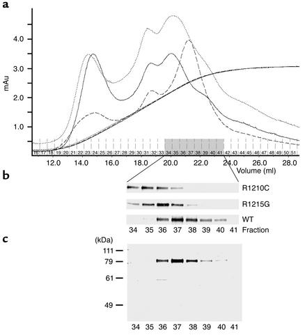
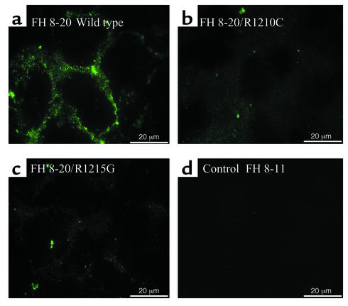
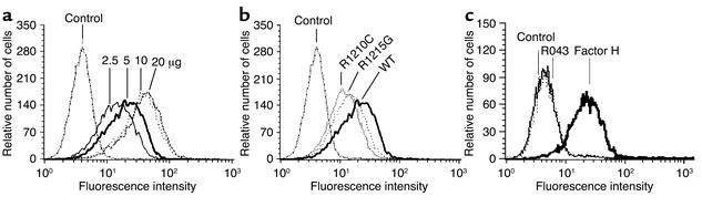
Hemolytic uremic syndrome (HUS) is a disease characterized by microangiopathic hemolytic anemia, thrombocytopenia, and acute renal failure. Recent studies have identified a factor H-associated form of HUS, caused by gene mutations that cluster in the C-terminal region of the complement regulator factor H. Here we report how three mutations (E1172Stop, R1210C, and R1215G; each of the latter two identified in three independent cases from different, unrelated families) affect protein function. All three mutations cause reduced binding to the central complement component C3b/C3d to heparin, as well as to endothelial cells. These defective features of the mutant factor H proteins explain progression of endothelial cell and microvascular damage in factor H-associated genetic HUS and indicate a protective role of factor H for tissue integrity during thrombus formation.


Figures

Figure 1
Figure 1
Identification of mutant factor H protein in serum of HUS patients. (a) Sera from normal individual (control) (lane 1) and HUS patients with the R1210C (F106, lane 2), the R1215G (F34, lane 3), and the E1172Stop mutation (R043, lane 4) were separated by SDS-PAGE and assayed by Western blotting using anti–factor H antiserum. Note the band with 175 kDa depicted by the arrow in serum of patient F106 (lane 2). Serum derived from patient F34 with the R1215G mutation showed normal factor H, and no additional band was detected. In serum from patient with the E1172Stop mutation (R043), a band of higher mobility with a reduced molecular weight is identified. (b) Reactivity of the normal plasma factor H and the 175 band with antisera and mAb’s specific for factor H. Sera from a healthy individual (lanes 1–3) and patient F106 with the R1210C mutation (lanes 4–10) were separated by SDS-PAGE, and after Western blotting, reacted with the indicated polyclonal and monoclonal Ab’s (lanes 1, 4). (c) Western blotting and (d) silver staining of reduced and deglycosylated (N-glycosidase F; GF) control and patient samples.
Figure 2
Figure 2
The mutant form of factor H binds less efficiently to heparin. Sera from a healthy control (a) and from HUS patient F106 with the R1210C mutation (b) were applied to heparin affinity chromatography, and after washing, bound proteins were eluted with the linear NaCl gradient. Individual fractions of 500 μl were collected starting at an NaCl concentration of 200 mM. The fractions were assayed by SDS-PAGE in combination with Western blotting. The 175-kDa mutant factor H eluted at a lower salt concentration (fraction 35) prior to the wild-type factor H protein (fraction 41). The arrow indicates the position of the mutant factor H protein.
Figure 3
Figure 3
Recombinant expression of wild-type factor H SCRs 8–20 and HUS mutations of factor H. (a) Culture supernatants of insect cells infected with the recombinant viruses were harvested 5 days after infection, separated by SDS-PAGE, and used for Western blot analysis. (b) Purified recombinant proteins representing the wild-type factor H protein (SCRs 8–20) or the proteins with the indicated mutations were separated by SDS-PAGE and detected by silver staining. The mobility of the marker is indicated on the left.
Figure 4
Figure 4
Binding of recombinant and purified mutant factor H protein to C3d analyzed by surface plasmon resonance. (a) The wild-type FH8-20 protein (solid line), mutant proteins FH8-20/R1210C (dotted line), and FH8-20/R1215G (dashed line) were injected at a concentration of 2–3 μg/ml into a flow cell coupled with C3d. The binding of the wild-type protein is apparent from the distinct association and dissociation phases of the binding curve, whereas the binding of the mutants is decreased. All recombinant proteins were tested on two different chip surfaces coated with C3d, and representative figures are shown. (b) Binding of purified mutant factor H (FH) (E1172Stop) to C3b was assayed and compared with that of purified native factor H.
Figure 5
Figure 5
Heparin affinity chromatography binding of recombinant wild-type deletion mutant factor H (SCRs 8-20). (a) Culture supernatant of insect cells infected with recombinant baculovirus coding for the recombinant wild-type protein (FH 8-20) and the two mutant forms, R1210C mutant (i.e., FH 8-20/R1210C) or the R1215G mutant (i.e., FH 8-20/R1215G) was applied to heparin affinity chromatography. After loading, the column was thoroughly washed, and bound proteins were eluted by an NaCl gradient. Absorbencies are indicated for recombinant wild-type protein by the dashed line, the R1210C mutant by the solid line, and the R1215 mutant by the dotted line. The identical conductivity graphs show that elution was performed under identical conditions. mAu, milliampere units. (b) SDS-PAGE and Western blot analysis of fractions 34–41 of the individual proteins. (c) The separation yields pure protein as shown for wild-type protein fractions 34–41 after SDS-PAGE separation in combination with silver staining. The mobility of the marker proteins is indicated on the left.
Figure 6
Figure 6
Binding of recombinant wild-type and mutant factor H proteins to HUVECs: immunofluorescence. HUVECs cultivated in serum-free medium were incubated with cell culture supernatant containing the indicated recombinant proteins, i.e., the recombinant wild-type protein (FH 8-20; WT) (a), the R1210C mutant (i.e., FH 8-20/R1210C) (b), and the R1215G mutant (i.e., FH 8-20/R1215G) (c), and FH 8-11 (d) was used as a control. Unfixed cells were used directly for immunofluorescence analysis by staining with factor H antiserum in combination with an FITC-labeled secondary antiserum. The bars in the lower corner show the length of 20 μm.
Figure 7
Figure 7
Binding of recombinant mutant, mutant factor H (E1172Stop), and wild-type proteins to endothelial cells. (a) Dose-dependent binding of recombinant wild-type factor H (FH 8-20). HUVECs incubated in serum-free medium were incubated with the indicated amounts of purified recombinant wild-type factor H 8-20, and binding was assayed by FACS analysis. (b) Comparison of recombinant mutant R1210C (i.e., FH 8-20/R1210C), or R1215G (i.e., FH 8-20/R1215G), or wild-type factor H (FH 8-20). HUVECs cultivated in serum-free medium were incubated with the indicated proteins, and binding was assayed by FACS analysis. (c) Binding of purified factor H protein with the E1172Stop mutation isolated from serum of patient R043 and of purified wild-type factor H to HUVECs. HUVECs were treated with either 5 μg of mutant factor H protein purified from plasma of patient R043 or with 5 μg purified intact wild-type factor H, and binding was assayed by FACS.

References

    1. Kaplan BS, Meyers KE, Schulman SL. The pathogenesis and treatment of hemolytic uremic syndrome. J. Am. Soc. Nephrol. 1998;9:1126–1133. - PubMed
    1. Remuzzi G, Ruggenenti P. The hemolytic uremic syndrome. Kidney Int. Suppl. 1998;66:S54–S57. - PubMed
    1. Kaplan BS, Drummond KN. The hemolytic-uremic syndrome is a syndrome. N. Engl. J. Med. 1978;298:964–966. - PubMed
    1. Thompson RA, Winterborn MH. Hypocomplementaemia due to a genetic deficiency of beta 1H globulin. Clin. Exp. Immunol. 1981;46:110–119. - PMC - PubMed
    1. Buddles MR, Donne RL, Richards A, Goodship J, Goodship TH. Complement factor H gene mutation associated with autosomal recessive atypical hemolytic uremic syndrome. Am. J. Hum. Genet. 2000;66:1721–1722. - PMC - PubMed

Publication types