. 2003 Apr;111(8):1221-30.
doi: 10.1172/JCI17215.

Role of RANK ligand in mediating increased bone resorption in early postmenopausal women

Affiliations

Role of RANK ligand in mediating increased bone resorption in early postmenopausal women

Guitty Eghbali-Fatourechi et al. J Clin Invest. 2003 Apr.

Abstract

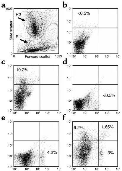
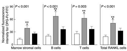
Studies in rodents have implicated various cytokines as paracrine mediators of increased osteoclastogenesis during estrogen deficiency, but increases in RANKL, the final effector of osteoclastogenesis, have not been demonstrated. Thus, we isolated bone marrow mononuclear cells expressing RANKL on their surfaces by two-color flow cytometry using FITC-conjugated osteoprotegerin-Fc (OPG-Fc-FITC) as a probe. The cells were characterized as preosteoblastic marrow stromal cells (MSCs), T lymphocytes, or B lymphocytes by using Ab's against bone alkaline phosphatase (BAP), CD3, and CD20, respectively, in 12 premenopausal women (Group A), 12 early postmenopausal women (Group B), and 12 age-matched, estrogen-treated postmenopausal women (Group C). Fluorescence intensity of OPG-Fc-FITC, an index of the surface concentration of RANKL per cell, was increased in Group B over Groups A and C by two- to threefold for MSCs, T cells, B cells, and total RANKL-expressing cells. Moreover, in the merged groups, RANKL expression per cell correlated directly with the bone resorption markers, serum C-terminal telopeptide of type I collagen and urine N-telopeptide of type I collagen, in all three cell types and inversely with serum 17beta-estradiol for total RANKL-expressing cells. The data suggest that upregulation of RANKL on bone marrow cells is an important determinant of increased bone resorption induced by estrogen deficiency.


Figures

Figure 1
Figure 1
Representative flow cytometry dot plots sorted for BAP+/RANKL+ cells from bone marrow aspirates of an untreated postmenopausal woman. (a) Forward/side light scatter profile of unstained cells following Ficoll-Paque density centrifugation. The R1 gate represents the mononuclear cells that include the MSCs. The R2 gate represents granulocyte precursor cells and these are excluded from analysis. (b) The dot plot profile of the autofluorescence from the irrelevant mouse IgG1 isotype that was used as a control for anti-BAP Ab. (c) The dot plot profile of the BAP+ cells (left upper quadrants in af) as assessed by single-color flow cytometry using the monoclonal anti-BAP (B4-78) stained with a PE-conjugated secondary Ab. The quadrant limits were set so that the left upper quadrant that defines the specific fluorescence for the BAP+ cells contains less than 5% of the nonspecific fluorescence from the irrelevant mouse IgG1 isotype control. (d and e) The analogous single-color flow cytometry using the human Fc-FITC control and OPG-Fc-FITC probe for RANKL+ sorted cells, respectively. (f) The dual-color dot plot profile for BAP+/RANKL sorted cells (left upper quadrant), BAP/RANKL+ sorted cells (right lower quadrant), and for combined BAP+ and RANKL+ sorted cells (right upper quadrant). The numerical percentages represent the stained cells per quadrant expressed as a proportion of total mononuclear cells in the R1 gate.
Figure 2
Figure 2
Formation of mineralized nodules during culture. BAP+/RANKL+ cells were cultured in either nutrient medium (a and c) or in osteoblast differentiation medium (b and d) for 21 days. When stained using von Kossa stain (a and b) or with alizarin red-S (c and d), mineralized nodules were apparent when cells were cultured in osteoblast differentiation medium. The BAP+/RANKL and BAP/RANKL+ cells did not proliferate sufficiently in culture to allow mineralization to be assessed.
Figure 3
Figure 3
Demonstration of the lack of TRAIL expression in cells sorted by FACS using RT-PCR analysis. In ethidium bromide–stained agarose gel of PCR products, BAP+/RANKL cells sorted by dual-color flow cytometry are shown in lane 1 and BAP+/RANKL+ cells are shown in lane 2. Size markers are given in lane 3. Note that the BAP+ cells binding the OPG-Fc-FITC probe (lane 2) express the RANKL, but not TRAIL mRNA. Conversely, the BAP+ cells not binding the OPG-Fc-FITC probe (lane 1) express TRAIL mRNA, but do not express RANKL.
Figure 4
Figure 4
Changes in OPG-Fc-FITC fluorescence as an index of mean RANKL surface concentration per cell (mean ± SEM). The premenopausal women (Group A) are shown by the white bars, the untreated postmenopausal women (Group B) by gray bars, and the estrogen-treated postmenopausal women (Group C) by black bars. There is a highly significant (ANOVA P values as indicated) difference among three groups. There is a two- to threefold significant increase of the mean RANKL fluorescence intensity per cell in Group B over Group A (*) and a somewhat smaller significant increase in Group B over Group C (**) as analyzed by the Student-Newman-Keuls test.

Comment in

References

    1. Riggs BL, Khosla S, Melton LJ. Sex steroids and the construction and conservation of the adult skeleton. Endocr. Rev. 2002;23:279–302. - PubMed
    1. Khosla S, Melton LJ, Riggs BL. Estrogen and the male skeleton. J. Clin. Endocrinol. Metab. 2002;87:1443–1450. - PubMed
    1. Manolagas SC. Birth and death of bone cells: basic regulatory mechanisms and implications for the pathogenesis and treatment of osteoporosis. Endocr. Rev. 2000;21:115–137. - PubMed
    1. Pacifici R. Estrogen, cytokines, and pathogenesis of postmenopausal osteoporosis. J. Bone Miner. Res. 1996;11:1043–1051. - PubMed
    1. Tanaka S, et al. Macrophage colony-stimulating factor is indispensable for both proliferation and differentiation of osteoclast progenitors. J. Clin. Invest. 1993;91:257–263. - PMC - PubMed

Publication types

MeSH terms