. 2003 May;23(9):3067-78.
doi: 10.1128/MCB.23.9.3067-3078.2003.

Autoinhibition of the kit receptor tyrosine kinase by the cytosolic juxtamembrane region

Affiliations

Autoinhibition of the kit receptor tyrosine kinase by the cytosolic juxtamembrane region

Perry M Chan et al. Mol Cell Biol. 2003 May.

Abstract

Genetic studies have implicated the cytosolic juxtamembrane region of the Kit receptor tyrosine kinase as an autoinhibitory regulatory domain. Mutations in the juxtamembrane domain are associated with cancers, such as gastrointestinal stromal tumors and mastocytosis, and result in constitutive activation of Kit. Here we elucidate the biochemical mechanism of this regulation. A synthetic peptide encompassing the juxtamembrane region demonstrates cooperative thermal denaturation, suggesting that it folds as an autonomous domain. The juxtamembrane peptide directly interacted with the N-terminal ATP-binding lobe of the kinase domain. A mutation in the juxtamembrane region corresponding to an oncogenic form of Kit or a tyrosine-phosphorylated form of the juxtamembrane peptide disrupted the stability of this domain and its interaction with the N-terminal kinase lobe. Kinetic analysis of the Kit kinase harboring oncogenic mutations in the juxtamembrane region displayed faster activation times than the wild-type kinase. Addition of exogenous wild-type juxtamembrane peptide to active forms of Kit inhibited its kinase activity in trans, whereas the mutant peptide and a phosphorylated form of the wild-type peptide were less effective inhibitors. Lastly, expression of the Kit juxtamembrane peptide in cells which harbor an oncogenic form of Kit inhibited cell growth in a Kit-specific manner. Together, these results show the Kit kinase is autoinhibited through an intramolecular interaction with the juxtamembrane domain, and tyrosine phosphorylation and oncogenic mutations relieved the regulatory function of the juxtamembrane domain.


Figures

FIG. 1.
FIG. 1.
CD analyses of wild-type, mutant, and diphosphorylated forms of Kit JM peptide. (A) CD spectra of 100 μM wild-type pep-tide (GPMYEVQWKVVEEINGNNYVYIDPTQLPYDHKWEFPRN) in 20 mM Tris-Cl, pH 7.4 (−gdn), or in 20 mM Tris-Cl, 4 M guanidine, pH 7.4 (+gdn), at 4°C. Similar results were obtained in other aqueous buffers. deg, degrees. (B) Direct comparison of the CD spectra of wild-type, mutant (GPMYEVQWKEEINGNNYVYIDPTQLPYDHKWEFPRN), and diphosphorylated (GPMYEVQWKVVEEINGNNpYVpYIDPTQLPYDHKWEFPRN) forms of Kit JM peptides (all 100 μM in 20 mM Tris-Cl, pH 7.4). (C) Thermal denaturation of wild-type or phospho-wild-type Kit JM peptides (CD measurement at 220 nm). (D) Calculation of the unfolded fraction of the wild-type Kit JM peptide as a function of temperature. The melting temperature (Tm) was calculated to be 39°C.
FIG. 2.
FIG. 2.
Binding analyses of wild-type, mutant, and phosphorylated forms of the Kit JM peptide. (A) Differential affinities of wild-type (WT), mutant (M), and phosphorylated (pWT) peptides for intracellular Kit (KitΔjux). The amount of Kit bound to immobilized peptides or to BSA-blocked streptavidin beads (lane b) was detected by α-histidine blotting (output). One out of 20 of the input kinases was loaded for comparison. (B) Mapping of the JM-catalytic domain interaction to the N-terminal kinase lobe. The top portion is a list of GST-Kit fusion proteins made, and the bottom portion is an analysis of GST-Kit fusion proteins (0.4 mg/ml) binding to immobilized wild-type JM peptides as detected by silver staining. Lane numbers correspond to the GST construct assigned above. Arrows point to the relevant protein band. (C) Wild-type and mutant peptides were directly compared in binding to GST-TK1 (increasing concentrations of 22, 27.5, 36.7, 55, and 110 μg/ml) by α-GST blotting. (D) The top portion shows gel filtration of intracellular Kit kinase alone, kinase with wild-type JM peptide, and kinase with control aprotinin peptide. Molecular size standards are indicated in the first graph. The shoulder peak in the middle graph represents the kinase-peptide complex. No complex formation was observed for the aprotinin peptide. The bottom portion shows α-histidine (KitΔjux) and avidin (Kit JM peptide) blotting of gel filtration fractions which demonstrate that the Kit JM forms a stable complex with the entire kinase domain in solution.
FIG. 3.
FIG. 3.
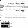
Expression and activity of recombinant Kit proteins. (A) The top portion shows wild-type, JM mutant, and inactive mutant intracellular Kit kinases expressed by using the baculovirus system. The wild-type kinase comprises the entire intracellular portion. The JM mutant kinases are deletions of the entire JM region, Y544-H579 (Δjux), a double valine (ΔV558V559-jux), or a tyrosine valine (ΔY567V568-jux). The catalytically inactive D790A mutant carries a mutation in the catalytic loop of the kinase domain. The bottom portion shows sequence alignment of the JM regions of type III RTKs (Kit, PDGFRα and PDGFRβ, Fms, and Flt3). (B) α-Histidine and 4G10 Western blot analyses of Kit kinases. Wild-type and JM mutant Kit kinases were found to be tyrosine phosphorylated after expression and purification, whereas the D790A mutant kinase had no detectable phosphotyrosine. The D790A mutant also exhibited no activity toward exogenous peptide substrates (data not shown). Lane numbers correspond to the Kit kinase assigned in panel A.
FIG. 4.
FIG. 4.
Exogenous substrate specificities and ATP-binding measurement for wild-type and mutant forms of Kit. (A) The x axis corresponds to the peptide substrate as follows: lane a, src-related peptide; lane b, angiotensin I; lane c, dynorphin A; lane d, gastrin; lane e, KitY719. One optical density at 340 nm (OD340) unit = 65.2 nmol of NADH. Results represent the mean values of triplicate assays and are presented with standard error bars. The background NADH oxidation rate of 0.12 mOD/min in the absence of kinase was subtracted from the final velocities. (B) Km ATP was measured for the wild type (0.4 μM), KitΔjux (0.3 μM), and KitΔYVjux (0.3 μM). Spectrophotometric assays of Kit kinases were performed in triplicate by using the peptide substrate angiotensin I with various ATP concentrations. The mean average velocities, after subtracting the background NADH oxidation, were plotted and fitted into the Michaelis-Menten equation, and Km values were obtained by nonlinear regression analysis.
FIG. 5.
FIG. 5.
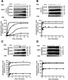
Activation kinetics of the recombinant dephosphorylated Kit kinase. Autophosphorylation of Kit and phosphorylation of the substrate enolase (en) was analyzed by 4G10 blotting. y-axis units were generated by densitometry. These analyses were performed for wild-type (A), Δjux (B), ΔVV-jux (C), and ΔYV-jux (D) kinases. For wild-type Kit, 20, 40, 80, and 120 nM concentrations are represented by filled circles, open circles, filled triangles, and open squares, respectively. For all the mutant kinases, 40, 80, and 120 nM are represented by filled circles, open circles, and filled triangles, respectively. The 50-s time point at the 40 nM kinase concentration (*) of panel C was an outlying value and was not used in generating the progress curve. Results are representative of three independent experiments.
FIG. 6.
FIG. 6.
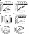
Inhibition of full-length intracellular Kit kinase activity by JM peptides in trans. (A) Inhibition of autophosphorylation. Dephosphorylated recombinant Kit kinase was incubated with various concentrations of wild-type or mutant JM peptide. Kit autophosphorylation was assayed at the indicated time points by 4G10 blotting. No inhibition of Kit kinase activity was observed with the control aprotinin peptide at 120 μM (right panel). Peptide concentrations of 0, 10, 20, 40, and 60 μM are represented by open circles, filled circles, open squares, filled diamonds, and open triangles, respectively. These results are representative of two independent experiments. (B) Percent inhibition is calculated from the phosphorylation units shown in panel A at 4 min. Kit autophosphorylation in the absence of the JM peptide was normalized to 100%. (C) Kit autophosphorylation in the presence of phospho-JM peptide. The conditions used were the same as those described for panel A. Phosphopeptide concentrations of 0, 10, 20, 40, and 60 μM are represented by open squares, open diamonds, open triangles, crosses, and filled diamonds, respectively. (D) In trans inhibition of substrate phosphorylation by Kit JM peptides. The phosphorylation of the exogenous peptide substrate dynorphin A (1 mM) by preactivated Kit was measured in the presence of [γ-32P]ATP and JM wild-type and mutant peptides at 0 (open circles), 0.1 (filled circles), 0.2 (filled triangles), 0.4 (open squares), and 0.6 μM (open diamonds) concentrations. 32P incorporation was converted to picomoles of phosphate. The values are the means of triplicate assays and are presented with standard error bars.
FIG. 7.
FIG. 7.
Growth suppression of a Kit-transformed cell line by ectopic expression of the Kit JM domain. Rat 2 cells expressing either the vector alone (pBMN Lyt2) or a constitutively active form of Kit (pBMN Lyt2; Kit Δ27) were selected by cell sorting. Vector control and Kit Δ27 Rat 2 cells were subsequently transfected with either GFP alone or with the Kit JM domain fused to GFP at its carboxy terminus, selected in G418, and verified for GFP expression by fluorescence microscopy. Known numbers of the selected cells were seeded, and cell growth was estimated after 40, 60, or 90 h of culture. Data shown are percent growth inhibition in cells expressing Kit JM relative to that of GFP-transfected controls.
FIG. 8.
FIG. 8.
Model of Kit activation through JM-mediated autoinhibition. The Kit JM domain is portrayed as a cylinder. P represents phosphotyrosine residues. Monomeric Kit is autoinhibited by the interaction of the JM domain with the N-terminal kinase lobe prior to ligand stimulation. Ligand-induced dimerization drives autophosphorylation at the slow rate of the repressed kinase. Activation of Kit involves phosphorylation of the JM domain and activation segment. Once phosphorylated, the JM domain loses its secondary structure and is released from the N-terminal lobe. The kinase then undergoes rapid autophosphorylation characteristic of the fully activated state.

References

    1. Barker, S. C., D. B. Kassel, D. Weigl, X. Huang, M. A. Luther, and W. B. Knight. 1995. Characterization of pp60c-src tyrosine kinase activities using a continuous assay: autoactivation of the enzyme is an intermolecular autophosphorylation process. Biochemistry 34:14843-14851. - PubMed
    1. Baxter, R. M., J. P. Secrist, R. R. Vaillancourt, and A. Kazlauskas. 1998. Full activation of the platelet-derived growth factor beta-receptor kinase involves multiple events. J. Biol. Chem. 273:17050-17055. - PubMed
    1. Besmer, P., J. E. Murphy, P. C. George, F. H. Qiu, P. J. Bergold, L. Lederman, H. W. Snyder, Jr., D. Brodeur, E. E. Zuckerman, and W. D. Hardy. 1986. A new acute transforming feline retrovirus and relationship of its oncogene v-kit with the protein kinase gene family. Nature 320:415-421. - PubMed
    1. Binns, K. L., P. P. Taylor, F. Sicheri, T. Pawson, and S. J. Holland. 2000. Phosphorylation of tyrosine residues in the kinase domain and juxtamembrane region regulates the biological and catalytic activities of Eph receptors. Mol. Cell. Biol. 20:4791-4805. - PMC - PubMed
    1. Buchwald, G., E. Hostinova, M. G. Rudolph, A. Kraemer, A. Sickmann, H. E. Meyer, K. Scheffzek, and A. Wittinghofer. 2001. Conformational switch and role of phosphorylation in PAK activation. Mol. Cell. Biol. 21:5179-5189. - PMC - PubMed

Publication types

MeSH terms

LinkOut - more resources