. 2003 May;23(9):3079-90.
doi: 10.1128/MCB.23.9.3079-3090.2003.

p38alpha isoform Mxi2 binds to extracellular signal-regulated kinase 1 and 2 mitogen-activated protein kinase and regulates its nuclear activity by sustaining its phosphorylation levels

Affiliations

p38alpha isoform Mxi2 binds to extracellular signal-regulated kinase 1 and 2 mitogen-activated protein kinase and regulates its nuclear activity by sustaining its phosphorylation levels

Victoria Sanz-Moreno et al. Mol Cell Biol. 2003 May.

Abstract

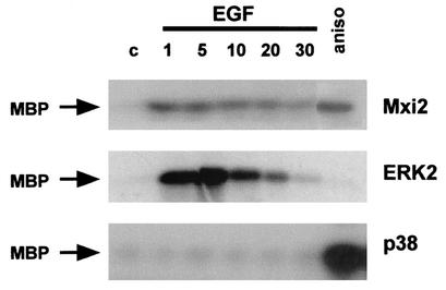
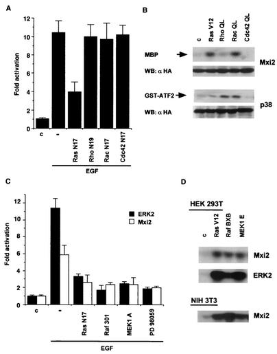
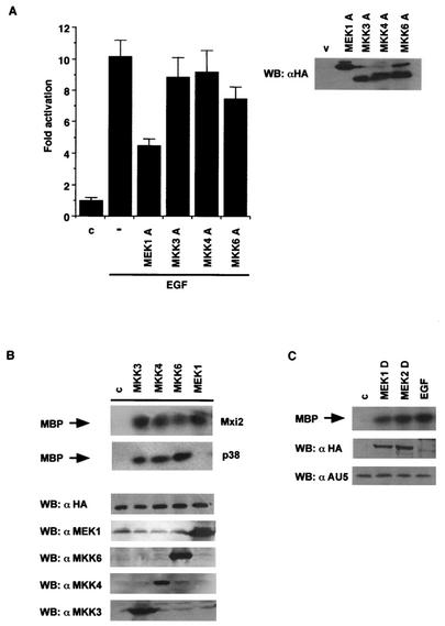
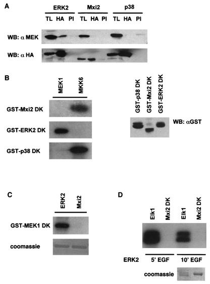
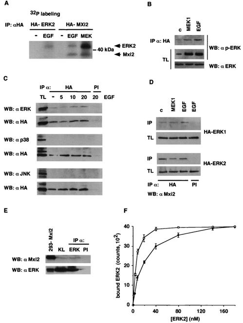
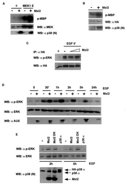
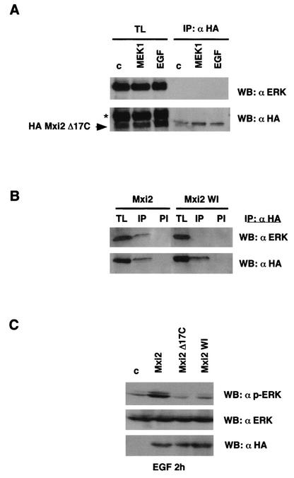
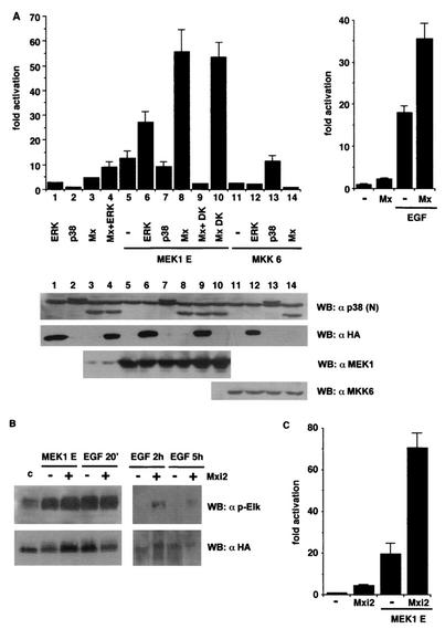
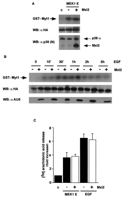
Mxi2 is a p38alpha splice isoform that is distinctively activated by mitogenic stimuli. Here we show that Mxi2 immunoprecipitates carry a kinase activity that is persistently activated by epidermal growth factor in a fashion regulated by Ras, Raf, and MEK. We demonstrate that this kinase activity can be attributed not to Mxi2 but rather to extracellular signal-regulated kinases 1 and 2 (ERK1/2), which coimmunoprecipitated with Mxi2 both by ectopic expression and in a physiological environment like the kidney. Furthermore, we provide evidence that Mxi2-ERK interaction has profound effects on ERK function, demonstrating that Mxi2 prolongs the duration of the ERK signal by sustaining its phosphorylation levels. Interestingly, we show that the effects of Mxi2 on ERK are restricted to nuclear events. Mxi2 potently up-regulates ERK-mediated activation of the transcription factors Elk1 and HIF1alpha but has no effect on the activity of ERK cytoplasmic substrates RSK2 and cPLA(2), induced by epidermal growth factor or by MEK. Overall, our findings point to Mxi2 as a unique member of the p38 family that may have an unprecedented role in the regulation of the functions of ERK mitogen-activated protein kinases.


Figures

FIG. 1.
FIG. 1.
Time-dependent activation of Mxi2. 293T cells transfected with HA-tagged Mxi2, ERK2, and p38 (1 μg) were starved for 12 h and stimulated with 10 μg of anisomycin (aniso) per ml for 20 min or with EGF (100 ng/ml). At the indicated times (minutes), the phosphotransfer activities in the immunoprecipitated kinases were determined by an in vitro kinase assay with MBP as the substrate. c, control.
FIG. 2.
FIG. 2.
Mediation of Mxi2 activation by the components of the ERK pathway. (A) Effects of Ras GTPase dominant inhibitory mutant forms on Mxi2 activation induced by EGF. Kinase activities in 293T cells transfected with HA-Mxi2 and the indicated dominant inhibitory mutant forms (250 ng) are shown. After starvation, the indicated cells were stimulated with EGF (100 ng/ml) for 5 min. The data shown are the average ± the standard error of the mean of three independent experiments, relative to the activity levels in control cells. (B) Effects of activated Ras family GTPases on Mxi2. Kinase activities in cells transfected with HA-Mxi2 or HA-p38 (1 μg) and the indicated GTPase (0.5 μg). (Bottom) Expression of HA-Mxi2 and HA-p38. (C) Effects of the inhibition of the proteins composing the ERK pathway on Mxi2 activation induced by EGF. Cells were cotransfected with HA-tagged Mxi2 or ERK2 and the indicated mutant forms (250 ng). Upon starvation and pretreatment for 30 min with 10 μM PD98059 where indicated, cells were stimulated with EGF and kinase assays were performed. The data shown are the average ± the standard error of the mean of three independent experiments, relative to the activity levels in control cells. (D) Effects of the constitutive activation of the components of the ERK pathway on Mxi2. Kinase activities in 293T or NIH 3T3 cells cotransfected with HA-Mxi2 or HA-ERK2 and expression vectors (0.5 μg) encoding the indicated constructs. WB, Western blot; c, control.
FIG. 3.
FIG. 3.
MAPKKs as mediators of Mxi2 activation. (A) Effects of MAPKK dominant inhibitory mutant forms on Mxi2 activation induced by EGF. Cells were transfected with AU5-tagged Mxi2 and the indicated HA-tagged inhibitory mutant forms (250 ng). The indicated cells were stimulated with EGF (100 ng/ml) for 5 min. The data shown are the average ± the standard error of the mean of three independent experiments relative to the activity detected in control cells. (Right side) Expression levels of the MAPKKs in total lysates. (B) Activation of Mxi2 by MAPKKs. Kinase activities in cells transfected with HA-tagged Mxi2 or p38, together with the indicated MAPKKs (1 μg). (Bottom) Expression levels of HA-Mxi2 and of the different MAPKKs. (C) Activation of Mxi2 by constitutively active MEK isoforms. Kinase activities detected in cells transfected with AU5-Mxi2 and activated, HA-tagged MEK1 and -2 (1 μg) or stimulated with EGF. (Bottom) Expression levels of AU5-Mxi2 and HA-MEK1 and -2. WB, Western blot; c, control; v, vector.
FIG. 4.
FIG. 4.
Interactions of Mxi2 with MEK1 and ERK2. (A) Coimmunoprecipitation of MEK with MAPKs. 293T cells were transfected with 1 μg of HA-tagged Mxi2, ERK2, and p38. After 12 h of starvation, anti-HA (HA) and preimmune (PI) immunoprecipitates were immunoblotted with anti-MEK. TL, total lysates. (B) Analysis of MEK substrates. Kinase assays were performed with immunoprecipitated, constitutively active, HA-tagged MEK1 and MKK6 by using as substrates equal amounts (0.5 μg; right side) of fusions of GST with Mxi2, ERK2, and p38 kinase-inactive mutant forms (DK). (C) Analysis of the phosphorylation of MEK1 by Mxi2. Kinase assays of HA-tagged ERK2 and Mxi2 immunoprecipitated from cells treated with EGF for 5 min by using as the substrate 0.5 μg of GST-MEK1 DK. (D) Analysis of the phosphorylation of Mxi2 by ERK2. Kinase assays of HA-tagged ERK2 immunoprecipitated from cells stimulated with EGF for the indicated times by using as substrates fusions of GST with Mxi2 DK and Elk1. WB, Western blot.
FIG. 5.
FIG. 5.
Association of ERK1 and -2 with Mxi2. (A) Detection of phosphoproteins in Mxi2 immunoprecipitates (IP). 293T cells transfected with HA-ERK2 or HA-Mxi2 and MEK1E (1 μg) were labeled with 32P and stimulated with EGF (100 ng/ml) for 5 min where indicated. After anti-HA immunoprecipitation, phosphoproteins were detected in dried gels. The positions of Mxi2 and ERKs are indicated. (B) Detection of phosphorylated ERKs in association with Mxi2. Cells transfected with HA-Mxi2 and stimulated with cotransfected MEK1E or with EGF for 5 min were probed for phospho-ERK in anti-HA immunoprecipitates and their respective total lysates (TL). (C) Association of MAPKs with Mxi2. Cells transfected with HA-Mxi2 were stimulated with EGF for the indicated times, and anti-HA and preimmune (PI) immunoprecipitates were probed for ERK, JNK, and p38. (Bottom) Expression of immunoprecipitated HA-Mxi2. (D) Association of Mxi2 with ERK1 and -2. Cells transfected with HA-ERK1 and -2, in addition to AU5-Mxi2, were stimulated with cotransfected MEK1E or EGF for 5 min and anti-HA and preimmune immunoprecipitates, and their respective total lysates were probed for Mxi2. (E) Association of Mxi2 with ERKs in rat kidney. Lysates from homogenized kidneys (KL) were immunoprecipitated with anti-ERK or preimmune antibodies and probed for coimmunoprecipitating Mxi2. Total lysate from Mxi2-transfected 293T cells was run alongside as a control. (F) Affinity of ERK2 binding to Mxi2. Increasing concentrations of [35S]methionine-labeled ERK2 were allowed to bind in vitro to GST-Mxi2 (open squares) or to GST-p38 (black circles) bound to glutathione-Sepharose beads. Beads were washed, bound proteins were electrophoresed by SDS-PAGE, and bound ERK2 was counted by phosphorimager. The data shown are the average ± the standard error of the mean of three independent experiments. WB, Western blot; c, control.
FIG. 6.
FIG. 6.
Effects of Mxi2 on ERK phosphorylation and activation levels. (A) Effects of Mxi2 on the activation of ERK2 by MEK. 293T cells were transfected with HA-ERK2 cotransfected with vector or with MEK1E (1 μg) in the presence (+) or absence (−) of Mxi2 (1 μg). Phosphorylated MBP (p-MBP) levels indicate ERK activity levels. (Bottom) Expression levels of MEK and Mxi2. (B) Effect of Mxi2 on ERK2 basal activity levels. Cells were transfected with HA-ERK2 (1 μg) in the presence (+) or absence (−) of Mxi2 (1 μg). After 18 h of starvation, ERK kinase activity was determined as described previously. Phospho-MBP levels became apparent after prolonged exposure at −70°C. (Bottom) Expression levels of HA-ERK2 and Mxi2. (C) Effects of Mxi2 on EGF-induced phosphorylation of ERK2. Cells transfected with HA-ERK2 (1 μg) and increasing amounts of Mxi2 (2 and 5 μg) were stimulated with EGF for 5 min and probed for phospho-ERK in anti-HA immunoprecipitates. (Bottom) Levels of immunoprecipitated HA-ERK2. (D) Effects of Mxi2 on the dephosphorylation rate of ERK2. Cells were transfected with (+) or without (−) AU5-Mxi2 and stimulated with EGF (100 ng/ml), and phospho-ERK levels in total lysates were examined at the indicated times. (Bottom) Expression of ERK1/2 and AU5-Mxi2. (E) Effects of Mxi2 dead kinase (DK) on ERK2 dephosphorylation. Cells transfected with the indicated constructs were stimulated with EGF, and phospho-ERK levels were determined after 2 and 5 h. (Bottom) Expression of p38 proteins detected by immunoblotting with anti-p38 N terminus antibody. WB, Western blot; c, control.
FIG. 7.
FIG. 7.
Effects of Mxi2 on ERKs require the C terminus of Mxi2. (A) ERKs cannot bind to a deletion mutant form of Mxi2 lacking the C terminus. 293T cells were transfected with HA-Mxi2 Δ17C (1 μg) and stimulated with EGF (100 ng/ml) for 5 min or with cotransfected MEK1 E where indicated. Anti-HA immunoprecipitates (IP) and their respective total lysates (TL) were probed for the presence of ERK. (Bottom) Levels of HA-Mxi2 Δ17C protein. The position of HA-Mxi2 Δ17C is indicated. The asterisk shows the remaining anti-ERK signal from the previous blot. (B) ERKs cannot bind to the Mxi2 WI mutant form. Cells were transfected with HA-Mxi2 or with HA-Mxi2 WI (1 μg). Lysates were immunoprecipitated with an anti-HA antibody or with preimmune (PI) serum and probed for associated ERK. (Bottom) Expression of the HA-tagged proteins. (C) Effect of the mutant form of Mxi2 lacking the C terminus on ERK2 dephosphorylation. Cells transfected with the indicated constructs (1 μg) were stimulated with EGF (100 ng/ml), and phospho-ERK levels were determined after 2 h. (Bottom) Total ERK protein levels and expression of the Mxi2 HA-tagged proteins detected by anti-ERK and anti-HA immunoblotting, respectively. WB, Western blot; c, control.
FIG. 8.
FIG. 8.
Regulation of ERK-dependent nuclear events by Mxi2. (A, left side) Effects of Mxi2 on Elk1-dependent gene expression. Elk1transactivation was examined in NIH 3T3 cells cotransfected with GAL4-Elk1 (TAD) and the indicated plasmids. ERKs were HA tagged; p38 and Mxi2s were AU5 tagged (Mx + DK = Mxi2 + ERK2 DK). The results shown are the average ± the standard error of the mean of at least five independent experiments. (Bottom) Expression levels of the different kinases. (Right side) Effects of Mxi2 on Elk1transactivation induced by EGF in 293T cells. The results shown are the average ± the standard error of the mean of three independent experiments. In both parts, values are expressed relative to the activity detected in vector-transfected cells. Luciferase activities were normalized to the β-galactosidase activity. (B) Effect of Mxi2 on Elk1 phosphorylation. 293T cells were transfected with HA-Elk1 with (+) or without (−) AU5-Mxi2 and stimulated with cotransfected MEK1E or with EGF (100 ng/ml) for 20 min, 2 h, and 5 h as shown. Phospho-Elk1 levels were determined by immunoblotting. (Bottom) Expression of HA-Elk1. (C) Effects of Mxi2 on HIF1α-dependent expression. HIF1α transactivation was determined in NIH 3T3 cells cotransfected with GAL4-HIF1α (TAD) and the indicated plasmids. Luciferase activities were normalized to the β-galactosidase activity. Values are expressed relative to the activity detected in vector-transfected cells. The results are the average ± the standard error of the mean of three independent experiments. WB, Western blot; c, control.
FIG. 9.
FIG. 9.
Effect of Mxi2 on the activation of ERK cytoplasmic substrates. (A) Effect of Mxi2 on MEK-induced activation of RSK2. RSK2 kinase activities with GST-Myt1 as the substrate in 293T cells transfected with MEK1E with (+) or without (−) AU5-Mxi2 in addition to HA-RSK2. (Bottom) Expression of HA-RSK2 and p38 proteins. (B) Effect of Mxi2 on the long-term activation of RSK2 induced by EGF. Cells were transfected with (+) or without (−) AU5-Mxi2 and stimulated with EGF (100 ng/ml), and RSK2 activation levels were determined after the indicated times. (Middle) Expression of HA-RSK2 in total lysates. (Bottom) AU5-Mxi2 expression levels. (C) Effect of Mxi2 on the activation of cPLA2. Arachidonic acid release was measured in 293T cells labeled with [3H]arachidonic acid previously transfected with MEK1E or stimulated for 30 min with EGF in the presence or absence of transfected Mxi2 as indicated. The data shown are the average ± the standard error of the mean of three independent experiments expressed relative to the values found in unstimulated, vector-transfected cells. WB, Western blot; c, control.

Similar articles

Cited by

References

    1. Ajenjo, N., D. S. Aaronson, E. Ceballos, C. Richard, J. León, and P. Crespo. 2000. Myeloid leukemia cell growth and differentiation are independent of mitogen-activated protein kinase ERK1/2 activation. J. Biol. Chem. 275:7189-7197. - PubMed
    1. Brunet, A., G. Pages, and J. Pouyssegur. 1994. Growth factor-stimulated MAP kinase induces rapid retrophosphorylation and inhibition of MAP kinase kinase (MEK1). FEBS Lett. 346:299-303. - PubMed
    1. Catling, A. D., H.-J. Schaeffer, C. W. M. Reuter, G. R. Reddy, and M. J. Weber. 1995. A proline-rich sequence unique to MEK1 and MEK2 is required for Raf binding and regulates MEK function. Mol. Cell. Biol. 15:5214-5225. - PMC - PubMed
    1. Chen, G., M. Hitom, J. Han, and D. W. Stacey. 2000. The p38 pathway provides negative feedback for Ras proliferative signaling. J. Biol. Chem. 275:38973-38980. - PubMed
    1. Coso, O. A., M. Chiariello, J. C. Yu, H. Teramoto, P. Crespo, N. Xu, T. Miki, and J. S. Gutkind. 1995. Rac-1 and cdc42 control the activity of JNK (SAPK) signaling pathway. Cell 81:1137-1146. - PubMed

Publication types

MeSH terms

Substances