. 2003 May;23(9):3091-102.
doi: 10.1128/MCB.23.9.3091-3102.2003.

Histone deacetylase 1/mSin3A disrupts gamma interferon-induced CIITA function and major histocompatibility complex class II enhanceosome formation

Affiliations

Histone deacetylase 1/mSin3A disrupts gamma interferon-induced CIITA function and major histocompatibility complex class II enhanceosome formation

Eleni Zika et al. Mol Cell Biol. 2003 May.

Abstract

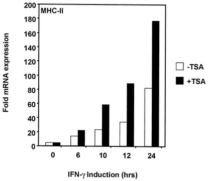
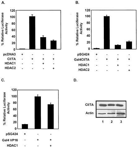
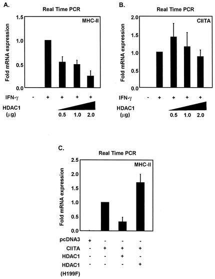
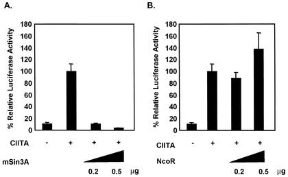
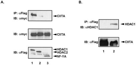
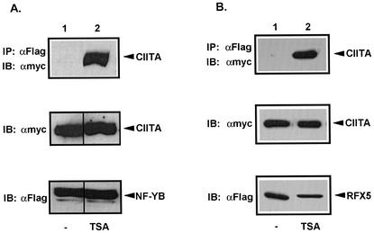
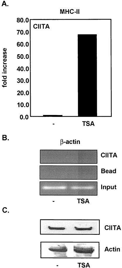
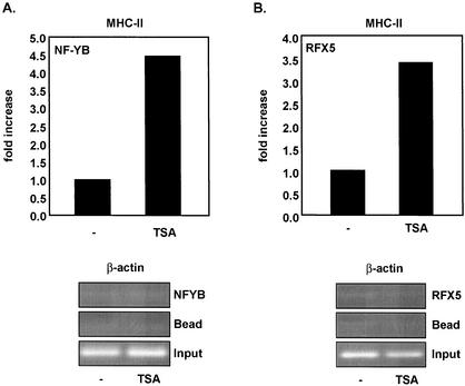
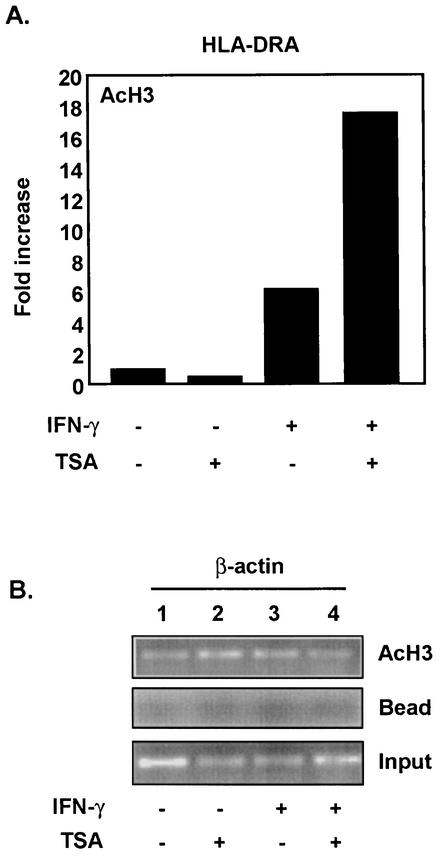
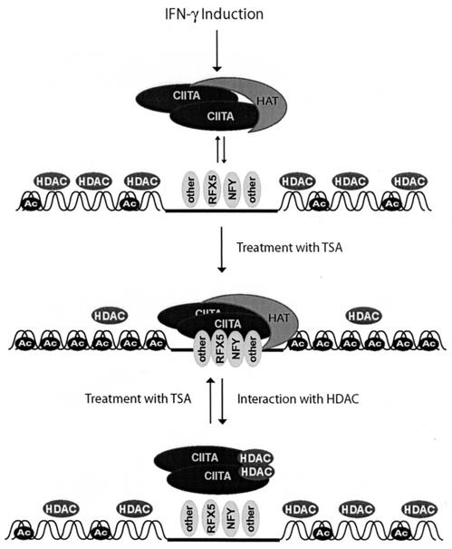
The class II transactivator (CIITA) is a master transcriptional regulator of major histocompatibility complex class II (MHC-II) promoters. CIITA does not bind DNA, but it interacts with the transcription factors RFX5, NF-Y, and CREB and associated chromatin-modifying enzymes to form an enhanceosome. This report examines the effects of histone deacetylases 1 and 2 (HDAC1/HDAC2) on MHC-II gene induction by gamma interferon (IFN-gamma) and CIITA. The results show that an inhibitor of HDACs, trichostatin A, enhances IFN-gamma-induced MHC-II expression, while HDAC1/HDAC2 inhibits IFN-gamma- and CIITA-induced MHC-II gene expression. mSin3A, a corepressor of HDAC1/HDAC2, is important for this inhibition, while NcoR, a corepressor of HDAC3, is not. The effect of this inhibition is directed at CIITA, since HDAC1/HDAC2 reduces transactivation by a GAL4-CIITA fusion protein. CIITA binds to overexpressed and endogenous HDAC1, suggesting that HDAC and CIITA may affect each other by direct or indirect association. Inhibition of HDAC activity dramatically increases the association of NF-YB and RFX5 with CIITA, the assembly of CIITA, NF-YB, and RFX5 enhanceosome, and the extent of H3 acetylation at the MHC-II promoter. These results suggest a model where HDAC1/HDAC2 affect the function of CIITA through a disruption of MHC-II enhanceosome and relevant coactivator-transcription factor association and provide evidence that CIITA may act as a molecular switch to modulate MHC-II transcription by coordinating the functions of both histone acetylases and HDACs.


Figures

FIG. 1.
FIG. 1.
Global inhibition of deacetylation by TSA enhances the inducible expression of MHC-II. Real-time PCR analysis was performed to measure endogenous mRNA levels of MHC-II after IFN-γ induction. HeLa cells were induced with 500 U of IFN-γ/ml for a time course of 24 h and treated with 100 nM TSA. Values represent the averages for three experiments. Samples were normalized to number of GAPDH copies.
FIG. 2.
FIG. 2.
HDAC1 and HDAC2 repress CIITA transactivation function. (A) HDAC1 and HDAC2 repress MHC-II promoter activation. COS 7 cells were cotransfected with 100 ng of CIITA, 1 μg of either HDAC1 or HDAC2, and 0.5 μg of MHC-II-luciferase reporter. Luciferase activity is reported as percent activation relative to that by CIITA alone. (B) HDAC1 and HDAC2 repress a Gal4-CIITA construct. Transfection was performed as described for panel A. (C) Overexpression of HDAC1 does not affect Gal4-VP16 activation. Transfection was performed as described for panel A. Values are shown as mean percent relative luciferase activity ± standard error of the mean for three experiments, each of which was repeated in triplicate. pSG424 is the empty vector control for Gal4 CIITA and Gal4-VP16. (D) HDAC1 or HDAC2 overexpression does not affect CIITA protein levels. Equal amounts of Fg-CIITA and pcDNA3 (top panel, lane 1) or HDAC1 (top panel, lane 2) and HDAC2 (top panel, lane 3) were transfected in COS 7 cells, and Western analysis was performed using anti-Fg antibodies. As a loading control we also immunoblotted with antibodies against actin (bottom panel).
FIG. 3.
FIG. 3.
HDAC1 specifically represses inducible expression of endogenous MHC-II. (A) Real-time PCR analysis was performed to measure endogenous mRNA levels of MHC-II in the presence of increasing amounts of HDAC1. HeLa cells were induced with IFN-γ (500 U/ml) for 24 h. (B) Overexpression of HDAC1 does not inhibit CIITA mRNA expression. CIITA promoter IV mRNA was measured by real-time PCR. (C) HDAC1 deacetylase activity is required for inhibition of CIITA-mediated activation of endogenous MHC-II. Real-time PCR analysis was performed to measure endogenous mRNA levels of MHC-II in the presence of HDAC1. Equal amounts of CIITA and HDAC1 were transfected into HeLa cells, and mRNA was isolated 24 h posttransfection. An HDAC1 deacetylase-defective mutant (H199F) failed to inhibit MHC-II transcription. Values are means ± standard errors of the means for three experiments. Samples were normalized to the number of 18S rRNA copies.
FIG. 4.
FIG. 4.
mSin3A mediates repression of CIITA transactivation function. (A) COS 7 cells were cotransfected with 100 ng of CIITA, increasing amounts of mSin3A, and 0.5 μg of DRA-luciferase reporter. Luciferase activity is reported as percent activation relative to that by CIITA alone. (B) NcoR is not required for repression of MHC-II promoter activity. Transfection was performed as described for panel A. Values are shown as mean percent relative luciferase activity ± standard error of the mean for three experiments, each of which was repeated in triplicate.
FIG. 5.
FIG. 5.
CIITA associates with HDAC1. (A) Myc-CIITA coimmunoprecipitates with Fg-HDAC1 and Fg-HDAC2. COS 7 cells were transfected with equal amounts of Myc-CIITA and Fg-HDAC1 or Fg-HDAC2. The top panel shows the results for immunoprecipitation (IP) with anti-Fg M5 antibody, followed by immunoblotting with anti-Myc 9E10. CIITA interacted strongly with HDAC1 (lane 1) but only weakly with HDAC2 (lane 2). Association with NF-YA was also tested as a negative control (lane 3). Expression of Myc-CIITA was confirmed in the middle panel, and expression levels of Fg-HDAC1, Fg-HDAC2, and Fg-NF-YA were confirmed in the bottom panels. (B) CIITA coimmunoprecipitates with endogenous HDAC1 in 293T cells. Fg-CIITA was immunoprecipitated from 293T whole-cell lysates with anti-Fg M5 antibody. Endogenous HDAC1 was detected in the top panel (lane 2) by immunoblotting with mouse anti-HDAC1 antibody (Santa Cruz Biotechnology). As a negative control, a bead-only immunoprecipitation was also performed (lane 1). Expression levels of Fg-CIITA were confirmed in the bottom panel.
FIG. 6.
FIG. 6.
Overexpression of HDAC1 or HDAC2 does not change the nuclear localization of CIITA. COS 7 cells were transfected with 1 μg of GFP-CIITA and 3 μg of HDAC1, HDAC2, or empty vector (pcDNA3). Immunofluorescence was detected 24 h posttransfection.
FIG. 7.
FIG. 7.
TSA promotes association of CIITA with NF-YB and RFX5. (A) TSA enhances interaction of CIITA with NF-YB (compare lanes 1 and 2). COS 7 cells were transfected with equal amounts of Myc-CIITA and Fg-NF-YB and treated with 300 nM TSA. The top panel shows the results for immunoprecipitation (IP) with anti-Fg M5 antibody, followed by immunoblotting with anti-Myc 9E10. Expression of Myc-CIITA in lysates was confirmed in the middle panel, whereas expression levels of Fg-NF-YB in lysates were confirmed in the bottom panel. (B) TSA enhances interaction of CIITA with RFX5 (compare lanes 1 and 2). Expression and detection of Myc-CIITA interaction with Fg-RFX5 was performed using the same procedure as described for panel A. Expression levels of Myc-CIITA and Fg-RFX5 were confirmed in the middle and bottom panels, respectively.
FIG. 8.
FIG. 8.
(A) TSA enhances CIITA recruitment to the MHC-II promoter. 293T cells were transfected with Fg-CIITA and treated with 300 nM TSA. Chromatin immunoprecipitation was performed using anti-Fg M5. MHC-II promoter DNA was detected by quantitative real-time PCR. Data are presented as increases compared to results with untreated cells. Real-time PCR values were determined by subtracting values obtained from bead-only immunoprecipitations and normalizing to the total amount of MHC-II promoter DNA added to the immunoprecipitation reaction (input). Data shown are representative of three independent experiments. (B) TSA does not promote association of CIITA with the β-actin promoter. Chromatin was prepared from transiently transfected 293T cells as was done for panel A. PCR was performed to detect β-actin promoter DNA sequences. Input represents 1% of the total chromatin introduced into each immunoprecipitation reaction. (C) TSA does not affect CIITA protein levels. 293T cells were transfected with Fg-CIITA and treated with TSA as for panel A. CIITA was detected by Western analysis anti-Fg antibody. As a loading control we also immunoblotted with antibodies against actin (bottom panel).
FIG. 9.
FIG. 9.
TSA promotes a stable association of RFX5 and NF-YB with the MHC-II promoter. (A) TSA enhances NF-YB association with the MHC-II promoter (top panel) but not the β-actin promoter (bottom panel). 293T cells were transiently transfected with Fg-NF-YB and treated with TSA (300 nM). Chromatin immunoprecipitation was performed using anti-Fg M5. MHC-II promoter sequences were detected by quantitative real-time PCR, and β-actin promoter was detected by PCR. Data are presented as increases compared to results with untreated cells. Real-time PCR values were determined by subtracting values obtained from bead-only immunoprecipitations and normalizing to the total amount of MHC-II promoter DNA added to the immunoprecipitation reaction (Input). (B) TSA enhances RFX5 association with the MHC-II promoter (top panel). 293T cells were transfected with Fg-RFX5 and treated with TSA as described for panel A. Chromatin immunoprecipitation was performed as for panel A. Real-time PCR values were determined as for panel A. Association of RFX5 with the β-actin promoter was not detected (bottom panel).
FIG. 10.
FIG. 10.
(A) TSA enhances IFN-γ-dependent acetylation of histone H3 (AcH3) at the MHC-II promoter. HeLa cells were induced with IFN-γ (500 U/ml) for 24 h and treated with 100 nM TSA. Chromatin immunoprecipitation was performed using anti-acetyl H3. Immunoprecipitated DNA was analyzed by real-time PCR, and values were determined as described in the legend for Fig. 8A. (B) IFN-γ does not affect H3 acetylation at the β-actin promoter. Chromatin immunoprecipitation was performed as for panel A, and DNA was analyzed by PCR. Input represents 1% of the total chromatin introduced into each immunoprecipitation reaction.
FIG. 11.
FIG. 11.
Model for the role of HDACs in MHC-II regulation. In the absence of inducing signals, such as IFN-γ signals, histones are hypoacetylated at the MHC-II promoter due to the presence of HDAC and absence of HAT activity. Association of MHC-II DNA-binding factors with the MHC-II promoter is observed at a low level. When CIITA is induced by IFN-γ, it associates with MHC-II transcription factors, such as RFX5 and NF-YB, and HATs. These interactions open chromatin and correlate with increased acetylated H3. If HDAC activity is inhibited by TSA, CIITA-NF-YB-RFX5 interactions are further stabilized and MHC-II enhanceosome formation is enhanced. Histones also become hyperacetylated, and maximal activation is achieved. At the end of the induction phase, HDAC may interact with CIITA, resulting in the disassembly of the entire enhanceosome complex.

Similar articles

Cited by

References

    1. Ashburner, B. P., S. D. Westerheide, and A. S. Baldwin, Jr. 2001. The p65 (RelA) subunit of NF-êB interacts with the histone deacetylase (HDAC) corepressors HDAC1 and HDAC2 to negatively regulate gene expression. Mol. Cell. Biol. 21:7065-7077. - PMC - PubMed
    1. Ayer, D. E. 1999. Histone deacetylases: transcriptional repression with SINers and NuRDs. Trends Cell Biol. 9:193-198. - PubMed
    1. Beresford, G. W., and J. M. Boss. 2001. CIITA coordinates multiple histone acetylation modifications at the HLA-DRA promoter. Nat. Immunol. 2:652-657. - PubMed
    1. Cressman, D. E., K. C. Chin, D. J. Taxman, and J. P. Ting. 1999. A defect in the nuclear translocation of CIITA causes a form of type II bare lymphocyte syndrome. Immunity 10:163-171. - PubMed
    1. Cressman, D. E., W. J. O'Connor, S. F. Greer, X. S. Zhu, and J. P. Ting. 2001. Mechanisms of nuclear import and export that control the subcellular localization of class II transactivator. J. Immunol. 167:3626-3634. - PubMed

Publication types

MeSH terms

Substances